Mitochondria are the only semi-autonomous organelles in cells with a double-membrane structure and are widely present in all types of eukaryotic cells. They serve as the central hub for cellular energy metabolism. In addition to driving key energy conversion processes such as oxidative phosphorylation and ATP synthesis, mitochondria also play essential roles in fatty acid metabolism, regulation of apoptosis, and calcium signaling. As the executors of these biological functions, mitochondrial proteins play an important role in regulating life processes and disease mechanisms through their expression levels, localization status, structural conformation, and post-translational modifications. With increasing attention on mitochondria in the contexts of metabolic disorders, neurodegenerative diseases, cancer development, and aging, obtaining high-quality and highly purified mitochondrial proteins has become a foundational requirement in functional omics, structural biology, and translational medicine research. However, mitochondrial protein extraction presents numerous technical challenges due to the organelle's complex structure, difficulties in layer separation, diverse protein composition with highly variable properties, low abundance, susceptibility to cytoplasmic contamination, and the tendency of mitochondria to undergo autophagy or degradation.
To address the above challenges, MtoZ Biolabs has developed a mitochondrial protein extraction kit that has been systematically optimized for key parameters including extraction efficiency, preservation of protein activity, and removal of contaminants. By integrating a refined separation workflow with highly effective buffer systems, the kit enables researchers to reliably isolate high-quality mitochondrial proteins from a variety of cell types. It provides a robust and dependable sample preparation solution for studies in metabolomics, proteomics, and disease mechanism research.
Product Overview
The mitochondrial protein extraction kit is specifically designed for the efficient extraction of mitochondrial proteins and enables rapid isolation of high-quality mitochondrial proteins from a variety of animal and plant cells. The unique lysis buffer formulation effectively disrupts the plasma membrane and separates mitochondria from intracellular components, while preserving the native conformation and biological activity of mitochondrial proteins to the greatest extent. The kit includes carefully optimized components such as cell lysis buffer, protein extraction solution, and protease inhibitors, which efficiently remove intracellular impurities and other membrane-associated elements, ensuring high-quality and high-purity protein yields.
In addition to its robust performance, the mitochondrial protein extraction kit is user-friendly and allows for fast sample processing, significantly reducing preparation time and effort. The extracted mitochondrial proteins are compatible with a wide range of downstream applications, including Western blotting, immunoprecipitation, enzymatic activity assays, and mass spectrometry (LC-MS/MS). The kit provides a powerful experimental foundation for mitochondrial biology research, disease mechanism studies, and drug screening.
Product Details
|
Product Details |
Size |
Storage Condition |
|
Mitochondria Isolation Reagent |
125 mL |
Store at -20ºC for one year
|
|
Trypan Blue Staining Solution |
10 mL |
|
|
Mitochondria Storage Buffer |
15 mL |
|
|
Mitochondria Lysis Buffer |
15 mL |
|
|
PMSF (crystals) |
1.5 mL (100 mM) |
|
|
PMSF (solvent) |
1.5 mL |
Protocol
1. Solution Preparation
Remove all reagents from the -20℃ freezer and thaw at room temperature. Keep all solutions on ice. Add 1.5 mL of PMSF solvent to the PMSF crystals and mix thoroughly to prepare 1.5 mL of 100 mM PMSF solution. Store the prepared 100 mM PMSF solution at -20℃. Before adding the Mitochondria Separation Reagent or Mitochondria Lysis Buffer to the sample, supplement with 100 mM PMSF to a final concentration of 1 mM.
2. Cell Collection
Harvest the required number of cells and gently resuspend the cell pellet in pre-chilled PBS. Centrifuge at 600 ×g, 4℃ for 5 minutes. Discard the supernatant.
3. Pretreatment
Add 1-2.5 mL of mitochondria separation reagent containing PMSF to 2-5 × 10⁷ cells. Gently resuspend and incubate on ice for 10-15 minutes.
4. Homogenization
Transfer the cell suspension to a homogenizer and perform 10-30 strokes.
Note:
a. Optimize the number of strokes based on cell type and homogenizer.
b. After 10 strokes, take 2 μL of homogenate and mix with 30-50 μL of Trypan Blue. If <80% of the cells are stained blue (indicating membrane rupture), increase strokes by 5 and repeat until >80% are positive. Do not over-homogenize to avoid mitochondrial damage.
5. Debris Removal
Centrifuge the homogenate at 600 ×g, 4℃ for 10 minutes.
Note: This step removes nuclei, cell debris, and unbroken cells. Centrifuging at 1,000 ×g increases purity but may reduce mitochondrial yield.
6. Mitochondria Isolation
Transfer the supernatant to a clean centrifuge tube and centrifuge at 11,000 ×g, 4℃ for 10 minutes.
Note: This step pellets the mitochondria. Using 3,500 ×g will yield purer mitochondria but with lower recovery.
7. Mitochondria Harvesting
Discard the supernatant; the pellet contains mitochondria.
Note: To collect cytoplasmic proteins free of mitochondria, take the supernatant from this step and centrifuge at 12,000 ×g, 4℃ for 10 minutes. The resulting supernatant contains cytoplasmic proteins. Protein concentration can be measured using the BCA assay.
8. Applications of Isolated Mitochondria
a. Functional and enzymatic studies of intact mitochondria: Resuspend mitochondria from 2-5 × 10⁷ cells in 150-200 μL of mitochondrial storage buffer.
b. Mitochondrial protein analysis: Add 150-200 μL of mitochondria lysis buffer (pre-supplemented with PMSF) to the mitochondria pellet from 2-5 × 10⁷ cells. After lysis, centrifuge at 12,000 ×g, 4℃ for 3-5 minutes. The resulting supernatant can be used for PAGE, Western blotting, IP, enzymatic assays, or BCA quantification.
Figures
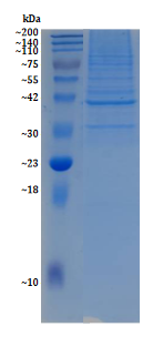
Figure 1. SDS-PAGE Gel Map of Mitochondrial Protein (Hela Cell) Extracted by the Kit.
Features and Benefits
1. Efficient Mitochondrial Protein Extraction
The mitochondrial protein extraction kit utilizes an optimized lysis buffer formulation that enables rapid and efficient extraction of mitochondrial proteins from cells while preserving their native conformation and biological activity to the greatest extent possible.
2. Preservation of Native Protein Activity
Protease inhibitors are included in the extraction process to effectively prevent protein degradation. The entire procedure is carried out under low-temperature conditions to ensure that mitochondrial proteins remain intact and biologically active.
3. Easy Operation and Short Processing Time
The simplified workflow allows users to complete mitochondrial protein extraction and purification in a short time, significantly saving experimental time and effort.
4. High-Purity Protein Recovery
Through multiple rounds of centrifugation and impurity removal steps, it is possible to ensure the extraction of mitochondrial proteins with high purity, suitable for downstream analyses requiring sensitive and accurate data.
5. Compatibility with Multiple Downstream Applications
The extracted mitochondrial proteins are compatible with various downstream applications, including Western blotting, mass spectrometry, co-immunoprecipitation, and enzyme activity assays, providing reliable data for mitochondrial research.
Applications
1. Mitochondrial Function Research
Mitochondrial proteins extracted using the mitochondrial protein extraction kit can be applied to functional studies of mitochondria, including key processes such as energy metabolism, redox reactions, and ATP synthesis.
2. Mitochondrial Disease Research
The extracted mitochondrial proteins are suitable for investigating mitochondrial-related diseases, providing important insights for disease diagnosis and therapeutic development.
3. Fundamental Mitochondrial Biology Studies
Widely applicable to basic mitochondrial biology research, including studies on mitochondrial dynamics, membrane protein functions, and regulation of mitochondrial gene expression.
FAQs
Q1: What Types of Cells and Tissues Is the Kit Suitable for?
A1: The mitochondrial protein extraction kit is suitable for mitochondrial protein extraction from animal cells, plant cells, and certain microbial cells. For tissue samples such as liver, heart, or muscle, it is recommended to perform appropriate cutting and homogenization to better disrupt the cells and release mitochondria.
Q2: Will Limited Sample Quantity Affect the Extraction Efficiency?
A2: The kit supports low-input samples, requiring as few as 1×10⁷ cells or 50 mg of tissue. When working with smaller sample amounts, the reagent volumes can be scaled down proportionally, and incubation times can be shortened accordingly.
Q3: How to Resolve Mitochondrial Protein Degradation during the Experiment?
A3: Possible causes and solutions:1. Inactive protease inhibitors: Always add protease inhibitors fresh before use; avoid long-term storage that may lead to loss of activity. 2. Excessive operating temperature: Ensure the entire process is performed on ice, and pre-cool the centrifuge to 4°C.