


TEM-Based Exosome Identification Service
- End-to-end sample handling, including exosome isolation and enrichment (optional)
- Morphological analysis: vesicle size, membrane architecture, and structural homogeneity
- Quality assessment: purity, integrity, and contamination detection
- Aggregation and debris visualization
- Comprehensive image interpretation and structural annotation
Exosomes, small membrane-bound vesicles typically 30–150 nm in diameter, are secreted by cells via multivesicular body (MVB) pathways. As a key class of extracellular vesicles (EVs), exosomes play crucial roles in intercellular communication, immune modulation, and disease pathogenesis, making them an emerging focus in both basic research and precision medicine.
Due to their nanoscale size and complex biogenesis, exosomes require ultra-resolution techniques for accurate structural validation. Transmission Electron Microscopy (TEM), long regarded as the gold standard for exosome imaging, enables direct visualization of vesicle morphology, membrane integrity, and aggregation, providing irreplaceable insights into exosome quality and identity.
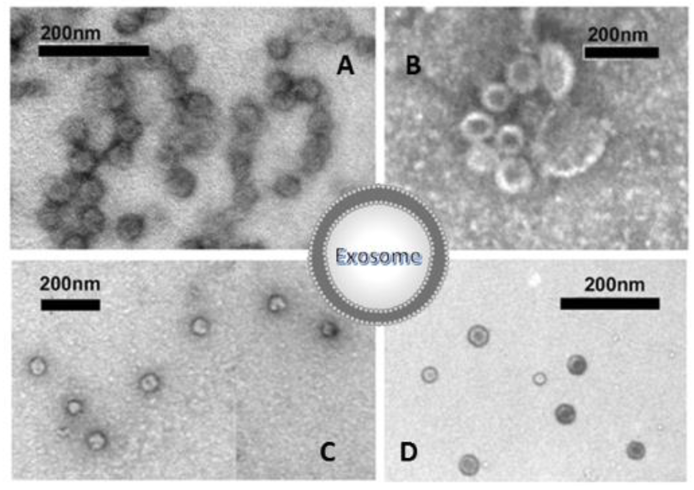
Di Bella, M. A. et al. Biology. 2022.
Figure 1. TEM-Based Exosome Morphology Validation
At MtoZ Biolabs, we offer comprehensive TEM-Based Exosome Identification Service that combines advanced imaging platforms with standardized protocols to support a wide array of biological samples—from cell culture media to clinical fluids and tissue extracts. Our service empowers researchers across exosome biology, diagnostics, and therapeutics with validated, publication-ready data.
Services at MtoZ Biolabs
To support diverse research workflows, MtoZ Biolabs provides not only high-resolution TEM imaging but also optional exosome extraction and pre-processing. Our TEM-Based Exosome Identification Service scope includes:
Analysis Workflow
Our TEM-Based Exosome Identification Service is built on a well-established workflow that ensures high reproducibility and data quality, delivering results that meet publication standards.
1. Sample Evaluation: We assess concentration, background, and quality upon receipt.
2. Fixation & Staining: Using glutaraldehyde fixation and phosphotungstic acid staining to enhance contrast and preserve vesicle ultrastructure.
3. Sectioning & Drying: Ultrathin sections are prepared to optimize resolution and minimize imaging artifacts.
4. TEM Acquisition: High-voltage TEM captures representative vesicle populations, enabling clear visualization of cup-shaped morphology and membrane structure.
5. Data Analysis & Delivery: Vesicle morphology, size distribution, and contamination levels are assessed. Clients receive high-resolution images, interpretation files, and a comprehensive report.
Why Choose MtoZ Biolabs?
✅ Nanoscale Precision: Capture the classical bilayer membrane and cup-shaped morphology of exosomes. Detect structural defects, aggregates, or contaminants at nanometer resolution.
✅ Multi-Matrix Compatibility: Effective with diverse sample types—no need for extensive pre-processing.
✅ Advanced Imaging Infrastructure: Access cutting-edge TEM platforms capable of resolving vesicles down to 30 nm.
✅ Integrated Quality Evaluation: Seamlessly combines with Western blotting, NTA, and protein marker analysis for multi-parameter exosome validation.
✅ End-to-End Support: From isolation to final images, MtoZ Biolabs provides a streamlined workflow that cuts hand-offs, lowers trial-and-error costs, and keeps your project moving.
✅ One-Time-Charge: Our pricing is transparent, no hidden fees or additional costs.
Sample Submission Suggestions
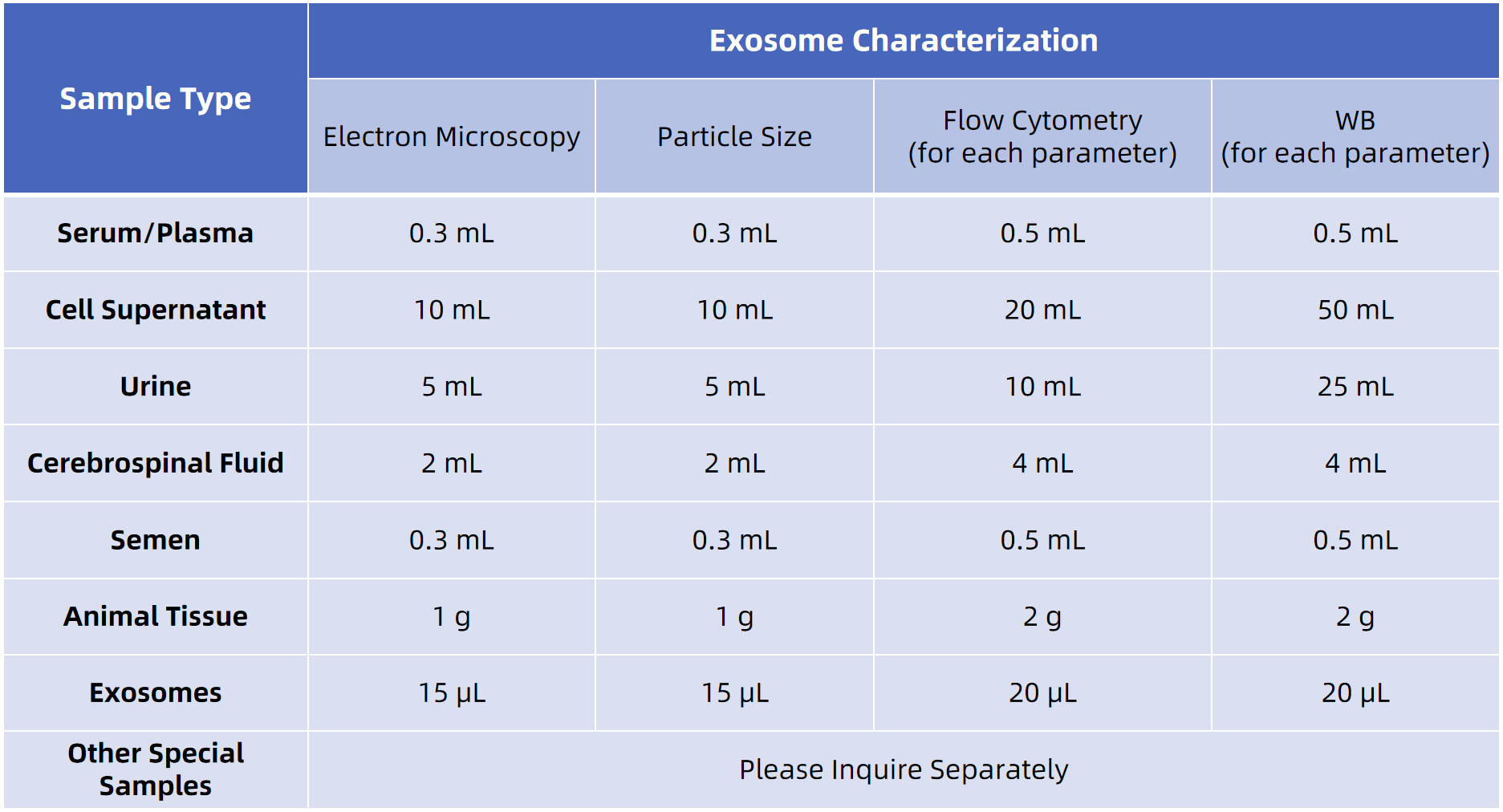
MtoZ Biolabs provides exosome isolation and protein analysis from a wide range of biological samples. We offer clear submission guidelines and can assist with sample processing to ensure consistent, high-quality data. For the best results, we recommend reaching out to our technical team before submission for personalized guidance.
What Could be Included in the Report?
1. High-resolution TEM images (TIFF/JPG ≥300 dpi)
2. Annotated vesicle morphology files
3. Comprehensive analysis report (PDF format)
4. Image interpretation: vesicle size, structural features, contaminants
5. Workflow summary and optional post-processing recommendations
FAQ
Q1: Can MtoZ Biolabs perform exosome isolation prior to TEM analysis?
Yes. We offer exosome extraction and enrichment services for cell culture media, tissues, and biological fluids (e.g., plasma, breast milk, saliva, urine). All isolation workflows are optimized for downstream TEM compatibility to ensure image quality and consistency.
Q2: What are the unique advantages of TEM for exosome research?
TEM is currently the only method capable of directly visualizing vesicle architecture with nanometer-level resolution. It is ideal for confirming the presence, shape, and integrity of exosomes, as well as detecting potential artifacts such as debris or aggregation. We recommend integrating TEM with Western blotting, NTA, and other orthogonal approaches to construct a reliable and comprehensive quality control system.
MtoZ Biolabs is committed to accelerating exosome-related research with integrated, high-quality solutions. In addition to our TEM-Based Exosome Identification Service, we offer exosome marker validation (Western blot), particle sizing and quantification (NTA), and custom workflow development. Contact us today to design a data-driven exosome analysis strategy tailored to your research needs.
Related Services
Exosome Separation & Purification Service
Exosomes Identification Service
Western Blotting based Exosome Characterization Service
Nanoparticle Tracking Analysis-based Exosome Characterization Service
How to order?

