




Tissue Proteomics: Sample Preparation, Workflow, Data Analysis, and Applications
Tissues such as liver, brain, muscle, and plant leaves differ in cellular composition, structure, and physiological function. Even within the same tissue, protein profiles may vary across sampling regions, developmental stages, and experimental conditions. Tissue proteins participate in metabolism, signaling, structural maintenance, and extracellular matrix formation. Changes in tissue protein composition and abundance therefore provide molecular clues for investigating tissue function and responses to experimental conditions.
What Is Tissue Proteomics?
Tissue proteomics is the systematic identification and relative quantification of proteins in animal or plant tissues using liquid chromatography-tandem mass spectrometry (LC-MS/MS). In a typical bottom-up proteomics workflow, tissues are disrupted to extract proteins, which are then enzymatically digested into peptides. Mass spectrometry data are searched against a protein sequence database to produce a protein identification list and relative quantification results.
The tissue proteome comprises proteins from tissue cells and the extracellular matrix. Bulk tissue proteomics examines the collected tissue as a whole, producing a protein profile that reflects contributions from different cell populations and tissue components. Compared with assays targeting a limited set of predefined proteins, tissue protein profiling provides a broader view of protein composition and a basis for comparing overall protein abundance across samples.
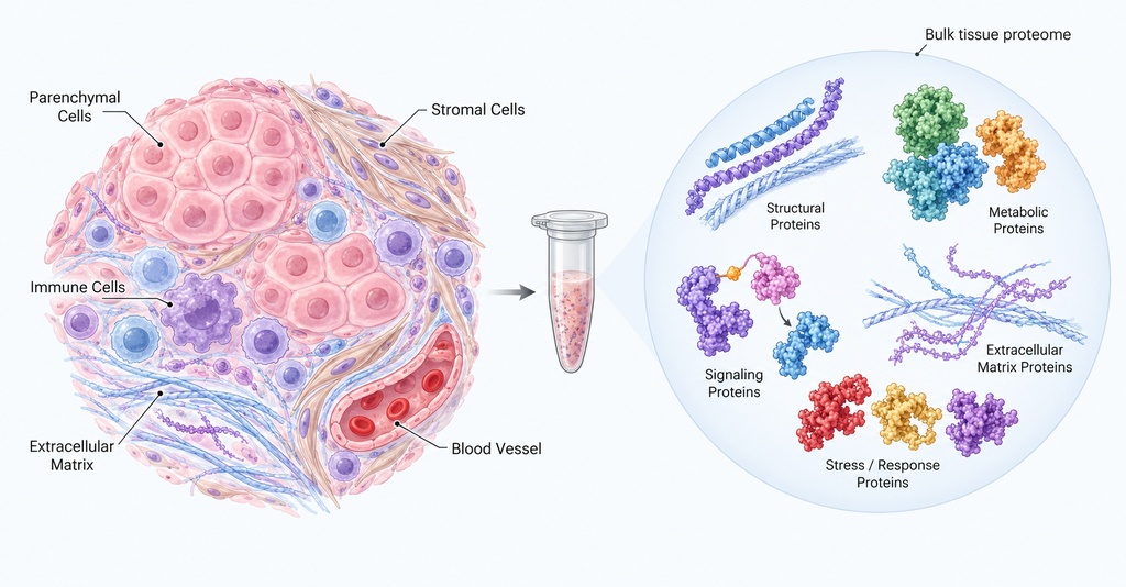
Figure 1. Tissue Composition and the Bulk Tissue Proteome.
What Research Information Can Tissue Proteomics Reveal?
Tissue proteomics connects protein-level characteristics with defined research conditions and provides three main types of information:
- Tissue protein composition: Generate a profile of identifiable tissue proteins, including proteins associated with tissue structure, metabolic activity, cellular functions, and the extracellular matrix.
- Changes in relative protein abundance: Compare abundance in the same tissue type across biological states, treatment conditions, or time points to prioritize proteins that show consistent trends.
- Functional and biological associations: Combine protein findings with functional annotations, metabolic pathways, and protein association information to identify potentially relevant biological processes and guide follow-up validation.
Tissue Sample Preparation and Analytical Considerations
Preparing tissue for proteomics involves selecting an appropriate submission format, checking the recommended input amount, documenting storage conditions, and verifying sample identity before submission.
1. Routine Tissue Sample Preparation
|
Sample item |
Basic requirements |
|
Supported tissues |
Supported tissues include animal tissues (e.g., liver, brain, heart, muscle, adipose tissue, and bone) and plant tissues. |
|
Submission formats |
Fresh tissue, snap-frozen or −80°C frozen tissue, tissue lysate, or protein extract; FFPE blocks, sections, and scrolls are also accepted. |
|
Recommended tissue amount |
Routine animal soft tissue: 20-50 mg per sample; plant tissue: 2-5 g of ground powder per sample. |
|
Minimum tissue amount |
Animal soft tissues: ≥10 mg; hard animal tissues: ≥75 mg; soft plant tissues: ≥2 g; hard plant tissues: ≥5 g. |
|
Extracted protein input |
At least 50 μg per sample, quantified by BCA or Bradford assay. |
|
Tissue collection and handling |
Clean tissue promptly and remove residual blood, necrotic areas, and non-target tissues. For routine tissues, cut into small pieces before snap-freezing; pancreatic tissue does not need to be cut into small pieces. |
|
Storage and shipping |
Snap-freeze in liquid nitrogen, store at −80°C, and ship on dry ice. Limit freeze-thaw cycles to 2-3 where possible and maintain a record. |
|
Sample information |
Provide species, tissue type, sampling location, sample ID, study group, and storage history. For lysates or protein extracts, specify reagent components. |
|
FFPE material |
FFPE blocks, sections, and scrolls may be submitted at room temperature. Protein extraction is required for FFPE workflows; fixation and embedding services are not provided. |
For detailed guidance on tissue collection, storage, and pre-submission checks, see How to Prepare Tissue Samples for Proteomics Analysis.
2. Special Tissue Types and Limited-Input Samples
Differences in tissue structure and matrix composition can change sample processing requirements. Common examples include:
- Dense or fibrous tissues: Bone, cartilage, muscle, connective, and fibrotic tissues may require tailored disruption, lysis, and protein extraction procedures.
- Tissues with complex matrices: Lipid-rich tissues, including adipose tissue, brain, and liver, as well as tissues rich in polysaccharides, polyphenols, or mucus, may require tailored cleanup procedures. Protein recovery may vary with sample composition.
- FFPE and limited-input materials: FFPE and long-stored tissues can be processed, and proteomics of laser-capture microdissection (LCM)-derived material is available. Repeatedly freeze-thawed or degraded tissues may require resampling.
For an explanation of how these tissue characteristics affect protein recovery and detection, see Challenges in Proteomics Analysis of Different Tissue Samples.
Tissue Sampling and Comparative Study Design
1. Sampling Regions and Tissue Comparability
Sampling regions may differ in cellular composition, extracellular matrix content, and tissue condition. Comparative tissue proteomics studies should consider three factors:
- Anatomical location: Select the same or comparable sampling locations for experimental and control groups wherever possible, and accurately record tissue origin and region.
- Region of interest: For injured, diseased, or experimentally treated tissues, define the region relevant to the research question and retain the corresponding histological information.
- Tissue composition: Consider differences in cell populations and matrix proportions. Samples with the same tissue weight may still have different bulk protein profiles.
2. Study Design
Once the sampling material is defined, research objectives should be translated into a practical comparison plan:
- Primary comparisons: Define the experimental and control groups and distinguish between tissue states, treatment factors, and sampling time points.
- Independent samples and paired relationships: Plan biological replicates based on biological variation, document tissues or regions collected from the same individual, and do not count technical replicates as independent samples.
- Sample and batch organization: Review submission formats and pre-analytical histories across groups, organize processing and analytical batches appropriately, and maintain essential experimental records.
For guidance on matching sampling regions, planning replicates, and aligning analytical methods with research objectives, see Experimental Design for Comparative Tissue Proteomics.
Tissue Proteomics Analysis Workflow
Intact tissues generally require disruption and protein extraction before bottom-up LC-MS/MS analysis. The process from laboratory sample assessment to protein-level results consists of six main stages.
1. Tissue Sample Assessment
Check tissue type, submission format, available amount, and storage condition to determine the appropriate processing route for intact tissue, FFPE material, or extracted protein.
2. Homogenization and Protein Extraction
Disrupt tissue and cellular structures to release and recover proteins, generating an extract suitable for subsequent preparation.
3. Protein Cleanup and Quality Control
Assess extract condition and protein recovery; when needed, reduce interference from lipids, lysis reagents, and other components before downstream analysis.
4. Protein Digestion and Peptide Preparation
Convert extracted proteins into peptides suitable for mass spectrometry and perform peptide cleanup where required.
5. LC-MS/MS Analysis
Separate complex peptide mixtures by liquid chromatography, then acquire mass spectrometry data on peptide ions and their fragments.
6. Protein Identification and Relative Quantification
Match peptides and proteins against an appropriate protein sequence database, calculate relative protein abundance, and generate identification lists, quantitative matrices, and basic quality-control information.
Tissue pretreatment, peptide preparation, and mass spectrometry acquisition form a connected process. For the purpose of each stage and the generation of protein-level results, see Tissue Proteomics Workflow from Protein Extraction to LC-MS/MS.
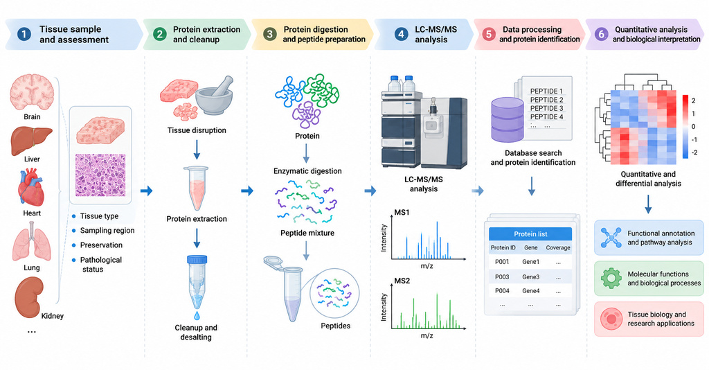
Figure 2. Overview of the LC-MS/MS-Based Tissue Proteomics Workflow.
Tissue Proteomics Results and Interpretation
A tissue proteomics report organizes protein identification, relative quantification, and predefined group comparisons into data modules for subsequent research.
|
Deliverable module |
Main results or files |
|
Protein identification |
Protein identification list and associated peptide information. |
|
Relative quantification |
Protein relative-abundance matrix mapped to individual sample IDs. |
|
Quality control |
QC information such as peptide length and identification scores. |
|
Sample-level and differential analysis |
Correlation, PCA, clustering, and differential-protein statistics as defined in the project plan. |
|
Functional and pathway analysis |
GO, KEGG, Reactome, and PPI analysis results according to project conditions. |
|
Project files |
Raw mass spectrometry data and analytical report. |
For a detailed evaluation of differential-protein evidence and interpretation in the context of tissue composition, see Why Tissue Proteomics Results Vary and How to Interpret Protein Changes.
Research Applications of Tissue Proteomics
1. Organ Function and Metabolic State
Comparing protein profiles from the same organ under different physiological or experimental conditions can reveal changes associated with liver metabolism, muscle energy utilization, and other organ functions, providing molecular leads for functional research.
2. Tissue Development and Maturation
As tissues develop and mature, protein composition and abundance change alongside tissue structure and functional state. Comparing developmental stages can identify candidate proteins associated with tissue differentiation, morphogenesis, and maturation.
3. Plant Tissues and Environmental Stress
Protein-profile comparisons in leaves, roots, and stems can be used to investigate responses to changes in temperature, water availability, or light, with particular attention to photosynthesis, metabolism, and defense-related proteins.
4. Tissue Injury and Repair
Animal tissue injury and repair studies can compare protein changes across experimental stages, focusing on the extracellular matrix, inflammation, and tissue-structure-associated proteins alongside tissue morphology and other experimental indicators.
5. Disease Models and Drug Interventions
Comparisons of disease-model tissues, drug-treated tissues, and matched controls can identify proteins associated with tissue state or experimental interventions, helping prioritize candidates for subsequent mechanistic studies. For specific research scenarios, see Tissue Proteomics for Disease and Drug Response Studies.
Frequently Asked Questions About Tissue Proteomics
Q1: Can multiple organs be analyzed within the same project?
Yes. Protein identification and relative quantification data can be generated for different organs, although their baseline protein compositions differ. When a study examines the effects of one treatment across multiple organs, comparisons should be defined within each organ, while individual-animal and tissue-origin information should be retained.
Q2: Should samples from different regions of the same tissue be pooled?
The decision depends on the research objective. To identify differences between regions, retain each region as a separate sample and record its origin. To obtain a combined protein profile from a predefined tissue area, pooling may be considered; however, pooled data cannot distinguish the individual changes occurring in each contributing region.
Q3: Can tissue proteins be identified when the study species lacks a well-developed protein database?
The available protein sequence references for the study species should first be reviewed. If a suitable UniProt database exists, or the client can provide a FASTA file for sequence matching, the protein identification strategy can be evaluated. When a client-supplied custom FASTA is used, GO, KEGG, Reactome, and PPI analyses are not included as default deliverables.
Q4: Can identified differential proteins undergo subsequent targeted validation?
Yes. PRM-based targeted protein analysis may be assessed for selected candidates, together with independent samples or other experimental methods to validate protein changes. Target selection should be based on the candidate proteins and sample conditions.
Tissue proteomics connects protein identification and relative abundance changes with histological observations, experimental phenotypes, and study context, providing molecular evidence about tissue states. Differential-protein and functional-analysis results can help prioritize candidate proteins and biological processes for validation using independent samples and follow-up functional experiments.
How to order?