




Tissue Proteomics for Disease and Drug Response Studies
Tissue proteomics characterizes protein abundance patterns in diseased and experimentally treated tissues. Comparisons with reference tissue identify proteins associated with disease status, while treatment comparisons reveal molecular responses to an intervention. Combined with histopathology and measurements of tissue function, these profiles provide a molecular perspective on disease-related alterations and experimental treatment effects.
In disease research, immune-associated proteins, metabolic enzymes, and extracellular matrix components can be examined alongside inflammatory lesions, altered tissue function, and fibrosis. In drug-response studies, changes in these protein groups can be related to measured differences in tissue injury, matrix deposition, or organ-specific function.
Disease-Associated Protein Changes in Tissue
1. Differential Protein Abundance Across Disease States
Comparisons of diseased and control tissues, including tumor and reference tissues, identify proteins with increased or decreased relative abundance. Disease-associated profiles may involve metabolic enzymes, immune-response proteins, stress-related factors, and components of tissue-specific function. In tumor tissue, coordinated changes among proliferation-, metabolism-, and immune-associated proteins characterize the molecular state of the sampled tissue. Liver and kidney disease studies may instead highlight metabolic or transport-related changes associated with altered organ function.
2. Protein Profiles in Disease Models
Disease models allow protein abundance profiles to be examined in the context of defined tissue pathology. The protein groups of interest vary with the biological features of the model:
- Liver injury models: Metabolic enzymes, inflammatory proteins, and stress-response factors characterize tissue injury and adaptation.
- Kidney disease models: Transport proteins and tissue-maintenance factors reflect renal functional and structural changes.
- Nervous-system disease models: Neuronal maintenance proteins and proteostasis components provide evidence of disease-associated molecular changes.
Across these models, the main application is to relate coordinated protein changes to a defined lesion or functional phenotype. For tissue-region selection and group comparability, see Experimental Design for Comparative Tissue Proteomics.
Protein Changes Associated with Tissue Pathology
1. Inflammation and Tissue Injury
Histopathology provides the morphological context for disease-associated protein changes. Inflammation and tissue injury often involve immune-response proteins, cellular stress factors, and tissue-repair components. In liver or kidney injury studies, enrichment of inflammatory proteins alongside histological lesions supports closer investigation of immune-associated processes. A broader pattern involving stress and repair proteins may characterize the accompanying injury response.
2. Extracellular Matrix Remodeling and Fibrosis
Fibrosis alters extracellular matrix (ECM) composition and tissue architecture. Tissue proteomics can identify coordinated differences in collagens, matrix-organizing proteins, and proteins associated with matrix turnover. When histological observations document matrix deposition, these protein changes help characterize the molecular features of remodeling and prioritize ECM-related candidates for further study.
3. Immune and Stromal Features in Tumor Tissue
Tumor tissue contains malignant cells, immune and stromal populations, and extracellular structures. Changes in immune-associated and stromal proteins characterize distinct components of the tumor-associated tissue environment. Combined with histological information on inflammatory infiltration and stromal architecture, these profiles help distinguish immune-associated patterns from matrix-related remodeling within the sampled tissue.
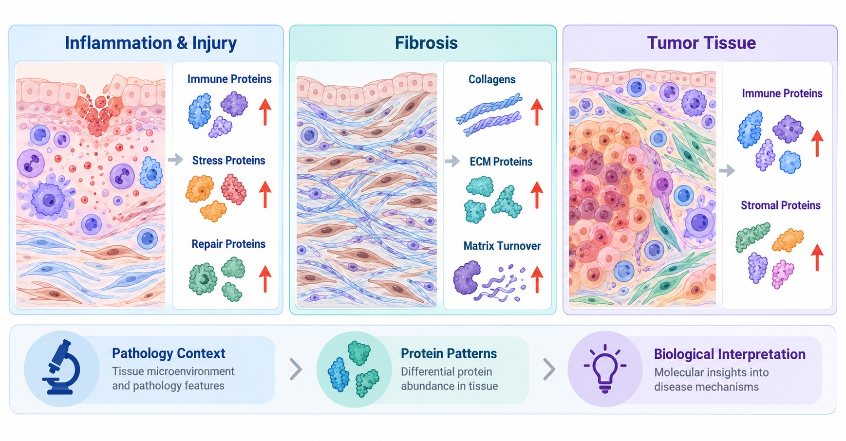
Figure 1. Disease-Associated Protein Patterns and Tissue Pathology.
Protein Responses to Drug Treatment
1. Metabolic Responses
Experimental drug treatment can change the abundance of proteins involved in energy production, nutrient utilization, lipid metabolism, and molecular transport. A coordinated response among mitochondrial enzymes directs attention to energy metabolism, whereas changes in lipid-processing proteins highlight lipid-handling pathways. Organ-specific protein composition provides context: metabolic enzymes may dominate one tissue response, while transport-associated proteins are more informative for another tissue.
2. Stress and Protein Homeostasis
Treatment-associated stress responses can involve molecular chaperones, antioxidant proteins, protein-folding factors, and components of protein degradation. Examining these proteins as functional groups helps distinguish coordinated proteostasis responses from isolated abundance changes. Histological injury and tissue-function measurements can then identify which stress-associated patterns accompany the experimental phenotype.
3. Inflammatory and Tissue-Function Responses
Immune-related proteins, extracellular components, transporters, and organ-specific proteins provide complementary information about tissue responses after treatment. For example, inflammatory protein changes can be evaluated with tissue inflammation, while transporter changes can be related to the measured function of the affected organ. Together, these findings characterize immune and tissue-function responses to the intervention.
Linking Protein Changes to Treatment Effects
1. Protein Patterns and Tissue Phenotypes
Treatment-associated protein changes become more informative when considered alongside tissue phenotypes measured in the same study. Immune- and repair-associated protein profiles can be compared with histological injury, while ECM changes can be evaluated with matrix deposition and tissue architecture. These associations can be analyzed across samples when paired molecular and phenotypic measurements are available. Molecular changes and tissue morphology may also evolve on different timescales, which is relevant when interpreting the timing of a response. For quantitative evidence and sample variation, see Why Tissue Proteomics Results Vary and How to Interpret Protein Changes.
2. Disease-Associated Patterns After Intervention
In disease models with an appropriate reference condition, post-treatment protein profiles can be examined in terms of three response patterns:
- Shift toward the reference profile: Disease-associated proteins move toward reference-tissue abundance after treatment.
- Persistent disease-associated changes: Part of the original disease protein signature remains altered after intervention.
- New treatment-associated changes: Additional proteins respond to the experimental intervention, revealing processes not prominent in the untreated disease group.
For example, inflammatory proteins may decrease after treatment while ECM proteins remain elevated. The contrast directs attention to different responses in inflammation and tissue remodeling, which can be assessed alongside histology and tissue-function measurements.
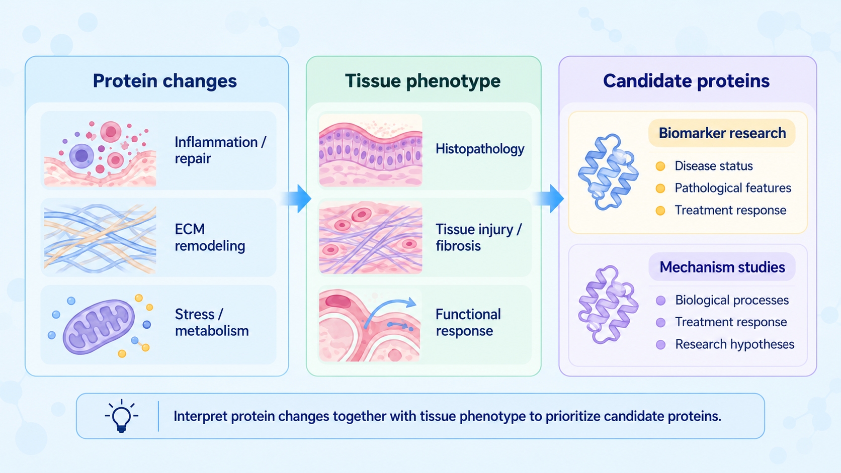
Figure 2. Linking protein changes with tissue phenotypes to prioritize candidates for biomarker and mechanism studies.
Candidate Proteins for Biomarker and Mechanism Studies
1. Candidates for Biomarker-Oriented Research
Proteins associated consistently with disease status, pathological features, or treatment response can be prioritized for biomarker-oriented research. A candidate linked to a defined lesion may be investigated as an indicator of tissue state; a protein that changes alongside a treatment-related phenotype may support studies of response monitoring in experimental models. Prioritization should consider reproducibility in independent samples, tissue context, and the intended research use.
2. Candidates for Mechanistic Research
Coordinated changes in ECM proteins can support hypotheses about matrix remodeling; metabolic and stress-response patterns can highlight processes associated with tissue injury or drug treatment. Candidates linked to defined tissue phenotypes offer more focused targets for functional studies than proteins selected solely by fold change. For example, a matrix-associated protein changing with both fibrosis-related histology and treatment response may be selected for independent measurement and downstream experiments.
Frequently Asked Questions
1. Can molecular changes appear before prominent tissue pathology?
Stress-, metabolism-, and immune-associated protein changes may be detectable before prominent morphological abnormalities appear. Comparing protein profiles with subsequent tissue observations or functional measurements helps establish how early molecular responses relate to the progression of the experimental model.
2. How can proteomics be used across stages of a disease model?
Proteomic comparisons across defined disease stages can identify proteins that emerge early, remain altered, or change as pathology develops. These temporal patterns distinguish stage-associated molecular processes and help select proteins for closer study of disease progression.
3. Can protein patterns be compared across different animal disease models?
Shared protein groups and functional themes can be compared across disease models when protein identifiers, tissue context, and study endpoints are aligned. Differences between models can also clarify which molecular responses are associated with distinct lesions or tissue compartments.
4. How should an unexpected stress-protein response after treatment be considered?
Assess unexpected stress-protein changes alongside injury-related proteins, tissue pathology, and functional measurements. A coordinated increase in stress and repair proteins, for example, provides a different interpretation from an isolated stress signature without corresponding tissue changes.
Conclusion
Tissue proteomics identifies protein patterns associated with disease state and experimental intervention. Relating these patterns to pathology and tissue function clarifies the molecular features of tissue injury, remodeling, and treatment response and supports the selection of candidates for biomarker-oriented and mechanistic research.
For the broader tissue-proteomics research pathway, see Tissue Proteomics: Sample Preparation, Workflow, Data Analysis, and Applications. Researchers can explore Tissue Proteomics Service at MtoZ Biolabs for tissue protein identification, relative quantification, differential analysis, and project-specific research objectives.
How to order?