



Tissue Proteomics Workflow from Protein Extraction to LC-MS/MS
Tissue proteomics analysis turns tissue material into protein-level data through a sequence of preparation, measurement, and data-processing steps. Because proteins are embedded within cells and tissue structures, intact tissue must first be disrupted and proteins extracted. The extract is then assessed and, when needed, cleaned up before enzymatic digestion converts proteins into peptides suitable for LC-MS/MS. LC-MS/MS records precursor-ion and fragment-ion information, which supports peptide identification, protein identification, and relative quantification.
The starting material determines where the workflow begins and which preparation steps need attention. Fresh or frozen intact tissue generally requires homogenization and protein extraction, while a tissue lysate or protein extract may enter at a later stage after concentration, solubility, and reagent compatibility are reviewed. Following the workflow from input to output clarifies the purpose of each stage and how the material produced at one step supports the next.
Tissue Sample Assessment
1. Sample Formats and Workflow Entry Points
The submitted material determines the first preparation stage and the information needed for assessment.
|
Sample Format |
Workflow Entry Point |
Information to Review |
|
Fresh or frozen intact tissue |
Homogenization and protein extraction |
Tissue type and source, sampling region, material condition, available amount |
|
FFPE block, section, or scroll |
FFPE-related preprocessing, then protein extraction |
Material format, fixation history, tissue source |
|
Biopsy or other low-input tissue |
Preparation matched to the available material |
Available amount and previous processing |
|
LCM-derived material |
Low-input proteomics preparation |
Recovered material and previous processing |
|
Tissue lysate |
Protein assessment; cleanup if needed before digestion |
Protein concentration, solubility, lysis-buffer composition |
|
Protein extract |
Protein assessment and downstream preparation |
Protein concentration, solubility, extraction reagents |
The table identifies typical entry points. FFPE material requires fixation- and paraffin-related preprocessing before protein extraction; lysates and protein extracts can enter later after their condition and reagent compatibility have been reviewed.
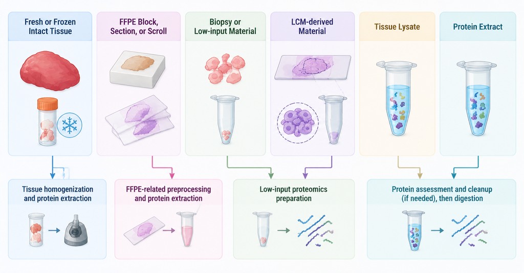
Figure 1. Sample Formats and Workflow Entry Points in Tissue Proteomics.
2. Pre-Processing Assessment
Review tissue type and source, material format, available amount, preservation status, previous processing, sample identifiers, and relevant group information. For tissue lysates or protein extracts, protein concentration, solubility, and lysis or extraction-reagent composition help determine the next preparation step. This initial review links the submitted material to the appropriate workflow entry point and identifies information that may affect downstream processing.
The assessment should distinguish an intact specimen from material that has already been lysed or extracted. That distinction determines which stages remain in the workflow and prevents unnecessary repetition of completed preparation steps.
Tissue Homogenization and Protein Extraction
1. Tissue Disruption and Protein Release
Intact tissue is disrupted so that proteins within cells and tissue structures become accessible to the extraction system. Homogenization improves contact between the material and the extraction environment and helps produce a more uniform preparation.
The degree of disruption depends on tissue architecture and composition. Dense or fibrous tissue and lipid-rich tissue may present different extraction considerations from softer tissue. The aim is to release proteins from the tissue matrix and create a preparation that can be handled consistently in later steps. For matrix-specific extraction challenges, see Challenges in Proteomics Analysis of Different Tissue Samples.
2. Protein Recovery and Downstream Compatibility
Protein extraction transfers released proteins into a recoverable preparation. Recovery depends on tissue disruption and extraction conditions, and different protein components may not be recovered equally. The resulting extract also needs suitable solubility and compatibility with subsequent cleanup and digestion. The immediate output is a protein-containing extract for assessment, rather than a final protein profile. Protein recovery and extract condition influence how much material is available for digestion.
At this transition, the main question is whether the recovered material is suitable for protein-level assessment and the next preparation step. The extract’s condition is therefore considered alongside its measured protein amount.
Protein Cleanup and Quality Control
1. Protein Extract Assessment
Before digestion, assess the recoverable protein amount, solubility or visible precipitation, compatibility of lysis or extraction reagents with digestion, and residual matrix components that may affect downstream processing. These checks connect extraction with the next stage: the extract needs recoverable protein and compatibility with the planned digestion conditions.
2. Conditions That May Require Cleanup
Cleanup may reduce residual salts, detergents, lipids, or other extraction components before enzymatic digestion. The appropriate approach depends on the composition and condition of the extract. Because cleanup can also cause material loss, the aim is to improve compatibility while retaining enough protein for the planned analysis. The output is a protein preparation suited to the intended digestion, with the amount retained considered alongside the analytical goal.
Cleanup is a preparation decision tied to the observed extract condition, not an automatic stage applied in exactly the same way to every sample. The assessment before digestion helps keep the preparation sequence appropriate to the material.
Protein Digestion and Peptide Preparation
1. Enzymatic Digestion and Peptide Generation
Enzymatic digestion converts recovered proteins into peptide sequences that can be separated and analyzed by LC-MS/MS. Trypsin is commonly used, while the digestion strategy follows the project’s analytical plan. Protein solubility and residual incompatible components can affect digestion and the peptide material generated. Digestion changes the analytical unit from intact proteins to peptides, which are more suitable for chromatographic separation and mass-spectrometric measurement.
2. Peptide Recovery and LC-MS/MS Compatibility
After digestion, peptide cleanup may reduce residual salts, reagents, or other components that interfere with chromatography or ion detection. Losses during digestion, transfer, or cleanup reduce the material available for LC-MS/MS. This is particularly relevant for low-input samples, where unnecessary transfers can consume a meaningful share of the available material. The intended output is a peptide preparation with sufficient recovery and a composition compatible with chromatographic separation and ion detection.
The peptide preparation is the direct input to LC-MS/MS. Its suitability depends on both the amount retained and the presence of components that may affect separation or ion detection.
LC-MS/MS Analysis
1. Liquid Chromatography and MS/MS Acquisition
Liquid chromatography separates the complex peptide mixture over time before peptides enter the mass spectrometer. The instrument records precursor-ion signals, while tandem mass spectrometry (MS/MS) generates fragment-ion spectra containing sequence-related evidence for peptide identification. Because peptides differ in retention and ionization behavior, the acquired signals represent a complex set of measurements rather than a direct protein list.
2. Raw Data Generation
LC-MS/MS acquisition produces raw mass spectrometry data containing chromatographic, precursor-ion, and fragment-ion information. These data provide the evidence used in subsequent peptide identification, protein inference, and relative quantification. Raw files are the measurement output of the instrument; protein names and abundance summaries are derived during subsequent data processing.
The distinction between acquisition and data processing is important: the instrument records ion measurements, whereas peptide and protein identities are assigned by interpreting the resulting spectra against reference information.
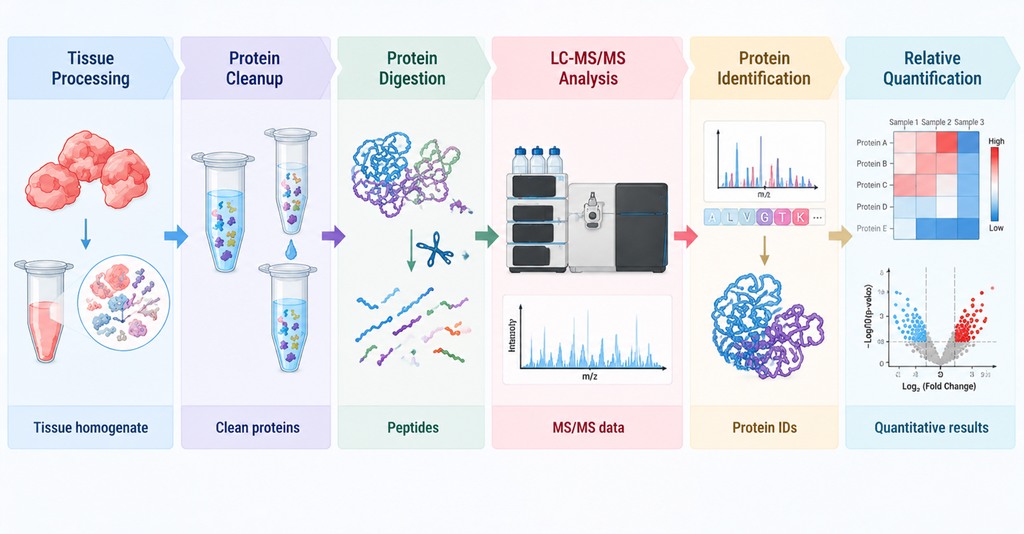
Figure 2. Workflow of Tissue Proteomics from Protein Extraction to LC-MS/MS.
Protein Identification and Relative Quantification
Data processing converts raw MS/MS spectra into peptide identifications, protein-level assignments, and relative abundance values. The search context depends on suitable protein-sequence reference information and project-appropriate analytical settings. Peptide-level evidence is evaluated first and then combined to support protein-level reporting.
1. MS/MS Spectral Matching and Peptide Identification
MS/MS spectra are evaluated against suitable protein-sequence reference information to assign candidate peptide sequences. The resulting peptide identifications provide the evidence for protein-level inference.
2. Protein-Level Inference
Identified peptides are mapped to protein sequences. Some peptide sequences are shared by related proteins or isoforms, while others provide more specific evidence. Protein-level reporting integrates the available peptide evidence into individual protein assignments or protein groups.
3. Relative Quantification
Quantitative signals associated with identified peptides can be summarized into protein-level relative abundance values according to the project’s analytical strategy. The resulting matrix supports comparisons of the same protein across samples or conditions; relative abundance is not equivalent to absolute protein concentration.
The resulting protein list and relative-abundance matrix provide foundational outputs for subsequent comparisons. More detailed interpretation of biological variation and changes in protein abundance belongs in Why Tissue Proteomics Results Vary and How to Interpret Protein Changes.
Frequently Asked Questions
1. Does a higher total protein yield always lead to more protein identifications?
No. Total yield is only one factor. Protein composition, digestion, peptide recovery, interfering components, chromatographic behavior, and the quality of MS/MS evidence also influence the number of proteins identified.
2. Should the reference database match the tissue species?
Species information helps select suitable protein-sequence reference data for peptide matching. Provide the tissue species and, where relevant, strain or other available reference information so the search context can be assessed appropriately.
3. What happens if protein or peptide recovery is insufficient?
The preparation stage associated with the loss can be reviewed alongside the remaining material and the intended analysis. Whether the project can proceed depends on the material available after review and the analytical goal.
4. Why should the lysis-buffer composition be reported for a tissue lysate?
Buffer components can affect digestion and downstream peptide preparation. Provide reagent names and concentrations when available; if the full composition is unknown, state which details are known so compatibility can be assessed.
5. How should samples be labeled in a multi-sample project?
Use a unique identifier for each sample and keep the identifier consistent across tubes, the sample sheet, and group information. Record the tissue source or region and any relevant pairing information so the resulting data can be linked to the correct material.
Conclusion
Tissue proteomics links sample assessment, protein preparation, peptide generation, LC-MS/MS acquisition, and data processing. The submitted material determines where the workflow begins and which preparation steps are needed.
How to order?