






Phosphoproteomics: How to Profile and Compare Protein Phosphorylation
Protein phosphorylation is a dynamic post-translational modification involved in cell signaling, enzyme regulation, protein interactions, and cellular responses to external stimuli. Changes in phosphorylation can occur rapidly following growth factor stimulation, drug exposure, environmental stress, or genetic perturbation.
Phosphoproteomics uses mass spectrometry to investigate protein phosphorylation across a large number of proteins and modification sites. By identifying phosphorylated proteins, localizing phosphorylation sites, and comparing their relative abundance across experimental conditions, researchers can investigate changes in cellular signaling and protein regulation. For projects requiring global phosphorylation identification and profiling, MtoZ Biolabs provides Phosphoproteomics Analysis Service. For cross-class PTM planning, see Post-Translational Modifications (PTMs) in Proteomics Research.
What Is Protein Phosphorylation?
Protein phosphorylation is a post-translational modification in which a phosphate group is covalently attached to specific amino acid residues in a protein. This process is primarily regulated by protein kinases, which transfer phosphate groups from ATP to substrate proteins, and protein phosphatases, which remove them.
Phosphorylation can alter protein charge, conformation, interactions, localization, and enzymatic activity. Through these effects, it plays important roles in cell signaling, cell cycle regulation, metabolism, and cellular responses to external stimuli. The balance between kinase and phosphatase activities dynamically regulates protein phosphorylation patterns. Changes in this balance can influence signaling pathways and other cellular processes.
Major Types and Sites of Protein Phosphorylation
Protein phosphorylation can be classified according to the chemical linkage between the phosphate group and the modified amino acid residue. Depending on the linkage, phosphorylation may involve O-phosphorylation, N-phosphorylation, or sulfur S-phosphorylation. Acyl phosphate formation represents another distinct type of phosphorylation chemistry involving carboxyl groups.
In eukaryotic proteins, the most extensively studied phosphorylation events involve three amino acid residues:
- Serine phosphorylation (pSer): Phosphate groups are attached to the hydroxyl group of serine residues. This is the most prevalent type of protein phosphorylation in many eukaryotic systems.
- Threonine phosphorylation (pThr): Phosphate groups are attached to the hydroxyl group of threonine residues. It frequently occurs alongside serine phosphorylation in kinase-mediated signaling.
- Tyrosine phosphorylation (pTyr): Phosphate groups are attached to the hydroxyl group of tyrosine residues. It plays important roles in receptor-mediated signaling, growth factor responses, and protein interaction networks.
These three modifications are examples of O-phosphorylation. Other phosphorylation events include histidine phosphorylation, which involves an N-phosphorylation linkage and is particularly relevant to prokaryotic two-component signaling systems. Phosphorylation can also occur on cysteine residues through S-phosphorylation, although these events are less commonly investigated in conventional eukaryotic phosphoproteomics.
The chemical properties and stability of different phosphorylation types can influence their detection and analysis by mass spectrometry. Conventional phosphoproteomics primarily focuses on Ser, Thr, and Tyr phosphorylation, while other phosphorylation types may require specialized analytical considerations.
What Can Phosphoproteomics Reveal?
Phosphoproteomics provides insights into protein phosphorylation across biological samples, helping researchers investigate:
- Global Phosphorylation Profiles: Identify phosphorylated proteins and sites across the proteome.
- Differential Phosphorylation: Compare phosphorylation changes between experimental conditions, treatments, or time points. (Olsen et al., 2006)
- Potential Signaling Regulation: Explore kinase-associated motifs and signaling pathways related to observed phosphorylation changes.
These findings help identify candidate phosphorylation sites and signaling processes for further biological investigation.
How Are Phosphorylation Sites and Phosphopeptides Identified?
Protein Digestion and Phosphopeptide Enrichment
Proteins are first extracted from biological samples and digested into peptides, commonly using proteolytic enzymes such as trypsin. Because phosphorylated peptides are often present at relatively low abundance compared with unmodified peptides, enrichment is commonly performed before LC-MS/MS analysis to increase their representation in the analytical sample. Different phosphorylation targets may require different enrichment approaches:
- Serine and threonine phosphorylation: Titanium dioxide (TiO₂) is commonly used to enrich phosphopeptides through interactions between phosphate groups and the titanium dioxide surface. (Larsen et al., 2005)
- Tyrosine phosphorylation: Antibody-based enrichment can selectively capture tyrosine-phosphorylated peptides, helping investigate relatively low-abundance pTyr events.
Broader enrichment selection across PTM classes is covered in How to Choose a PTM Enrichment Strategy for LC-MS/MS Analysis.
LC-MS/MS Identification of Phosphopeptides
Following enrichment, liquid chromatography-tandem mass spectrometry (LC-MS/MS) is used to separate peptides and acquire precursor and fragment ion information. High-resolution mass spectrometry provides accurate mass measurements and fragmentation data that support phosphopeptide identification. During database searching, experimental spectra are matched against theoretical peptide sequences with phosphorylation included as a variable modification. This process helps identify phosphorylated peptides and associate them with their corresponding proteins.
Phosphorylation Site Localization
Identifying a phosphorylated peptide does not necessarily establish which amino acid residue carries the phosphate group. For peptides containing multiple potential phosphorylation sites, site localization requires informative fragment ions that distinguish between alternative phosphorylation positions. For example, a peptide containing multiple serine or threonine residues may have several possible phosphorylation configurations. Site-determining fragment ions help establish which residue is supported by the MS/MS evidence.
When the available fragmentation evidence cannot distinguish between candidate residues, the localization may remain ambiguous. Site localization confidence therefore depends on peptide identification, fragment ion coverage, and the availability of informative site-specific evidence. For a broader explanation of modification site identification and localization principles, see How Are Post-Translational Modification Sites Identified and Localized?
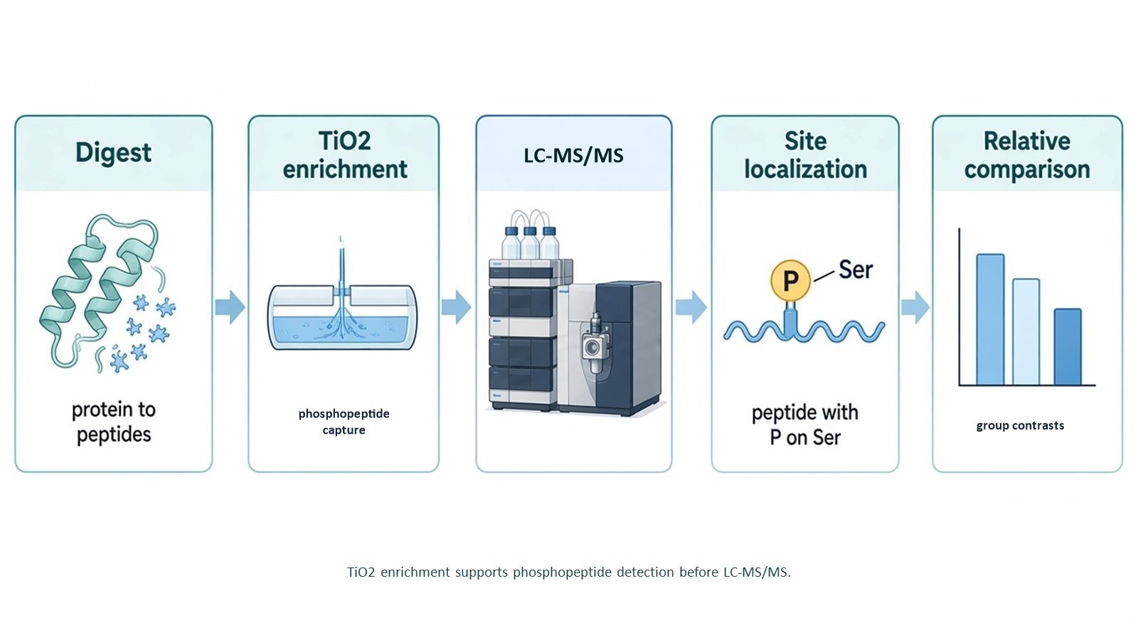
Figure 1. Phosphoproteomics moves from digestion and TiO2 enrichment to LC-MS/MS identification, localization, and optional relative comparison.
How Are Phosphorylation Changes Quantified and Compared?
Label-Free Quantification
Label-Free quantification compares phosphopeptide signals across separately analyzed samples without introducing isotopic or chemical labels. After appropriate data alignment and normalization, relative phosphopeptide abundance can be compared between experimental groups.
This approach is suitable for studies investigating phosphorylation responses across control and treatment conditions, disease models, or different time points. Consistent sample preparation, analytical performance, and biological replication are important for obtaining comparable results.
TMT-Based Quantification
Tandem Mass Tag (TMT) is an isobaric labeling strategy that allows multiple samples to be combined and analyzed within a multiplexed experiment. In phosphoproteomics, TMT supports comparative analysis across several experimental conditions, making it useful for multi-group and time-course studies. However, co-isolation interference during MS/MS analysis can cause ratio compression, potentially reducing the apparent differences between samples.
Both Label-Free and TMT approaches provide relative quantitative information about phosphopeptide abundance rather than direct measurements of absolute phosphorylation site occupancy. MtoZ Biolabs provides Quantitative Phosphoproteomics Service using Label-Free and TMT-based relative quantification. Shared quantification logic is expanded in How Is PTM Quantification Used to Compare Modification Changes Across Conditions?
PRM Analysis of Selected Phosphopeptides
When global phosphoproteomics identifies candidate phosphorylation sites of interest, researchers may wish to examine selected phosphopeptides in greater detail. Parallel Reaction Monitoring (PRM) is a targeted mass spectrometry approach that monitors predefined peptide targets and their characteristic fragment ions.
For suitable phosphopeptides, PRM can support targeted detection and relative quantitative comparisons across samples. It is particularly useful for following up on selected phosphorylation changes identified during discovery experiments.
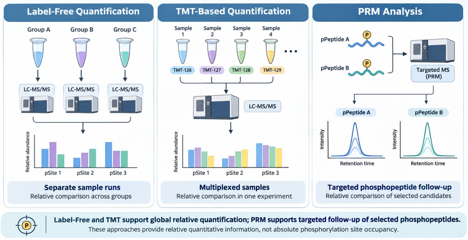
Figure 2. Quantitative Strategies in Phosphoproteomics: Label-Free, TMT, and PRM Analysis.
How Are Phosphorylation Changes Interpreted Biologically?
Identifying and comparing phosphorylation sites provides a foundation for investigating the biological processes associated with phosphorylation changes. Kinase motif analysis and pathway enrichment can help connect individual phosphosites with broader signaling networks.
Kinase Motif Analysis
Protein kinases often recognize characteristic amino acid sequence patterns surrounding their phosphorylation sites. These sequence preferences can provide clues about potential kinase-substrate relationships.
Kinase motif analysis examines amino acid residues surrounding identified phosphosites and compares their sequence patterns with known kinase recognition motifs. For example, enrichment of a particular phosphorylation motif among treatment-responsive sites may suggest the involvement of a related kinase family.
The results can help prioritize candidate kinases and guide subsequent experiments. However, motif matching does not prove that a specific kinase catalyzed a particular phosphorylation event or that its activity increased under the tested conditions.
Signaling Pathway and Functional Enrichment
Identified phosphoproteins and differentially regulated phosphorylation sites can be mapped to biological processes and signaling pathways. Gene Ontology (GO) and Kyoto Encyclopedia of Genes and Genomes (KEGG) enrichment analyses can help identify functional categories and pathways represented among the phosphoproteins. For example, phosphoproteins associated with MAPK signaling, PI3K-AKT signaling, or cell cycle regulation may be enriched in samples exposed to particular treatments.
These analyses help organize large phosphoproteomic datasets and identify biological processes that may warrant further investigation. However, enrichment of proteins associated with a pathway does not independently demonstrate pathway activation. Similarly, a change in phosphorylation does not establish a causal relationship with a biological phenotype. Interpretation should consider the experimental context, corresponding protein abundance, and supporting biological evidence.
Frequently Asked Questions
Is TiO2 enrichment interchangeable with IMAC in MtoZ phosphorylation projects?
In MtoZ phosphorylation projects, TiO2 is the dedicated enrichment route; IMAC is not offered as a standard substitute for it.
Can phosphoproteomics replace kinase activity assays?
No. Phosphosite maps generate substrate and pathway hypotheses; confirming kinase activity requires direct activity measurements or carefully controlled signaling assays.
When is tyrosine-focused enrichment considered?
Tyr-focused enrichment is considered when the biological question centers on tyrosine phosphorylation and Ser/Thr-dominated TiO2 profiles are unlikely to provide sufficient Tyr coverage. Antibody-based enrichment can then be added as a separate step.
Conclusion
After TiO2 enrichment and LC-MS/MS, phosphoproteomics establishes which phosphopeptides and sites are present and how they compare across defined groups. Motif and pathway readouts are best kept as ranked hypotheses, and protein abundance should be considered alongside phosphosite changes when modification-specific effects are claimed.
For projects involving global phosphorylation profiling, site identification, or comparative phosphorylation analysis, explore the MtoZ Biolabs Phosphoproteomics Analysis Service. Researchers are also welcome to contact our team for a free consultation about their sample types, research objectives, and analytical requirements.
References
- J.V. Olsen, B. Blagoev, F. Gnad, B. Macek, C. Kumar, P. Mortensen, M. Mann (2006). Global, in vivo, and site-specific phosphorylation dynamics in signaling networks. Cell, 127, 635-648. https://doi.org/10.1016/j.cell.2006.09.026
- M.R. Larsen, T.E. Thingholm, O.N. Jensen, P. Roepstorff, T.J.D. Jorgensen (2005). Highly selective enrichment of phosphorylated peptides from peptide mixtures using titanium dioxide microcolumns. Mol. Cell. Proteomics, 4, 873-886. https://doi.org/10.1074/mcp.T500007-MCP200
How to order?