


Main Types of Protein Glycosylation: N-Linked, O-Linked, GPI Anchors, and Glycoproteomics
-
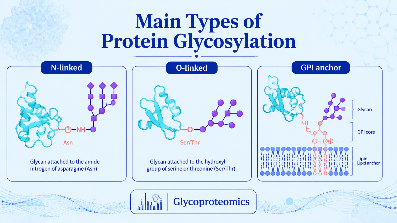
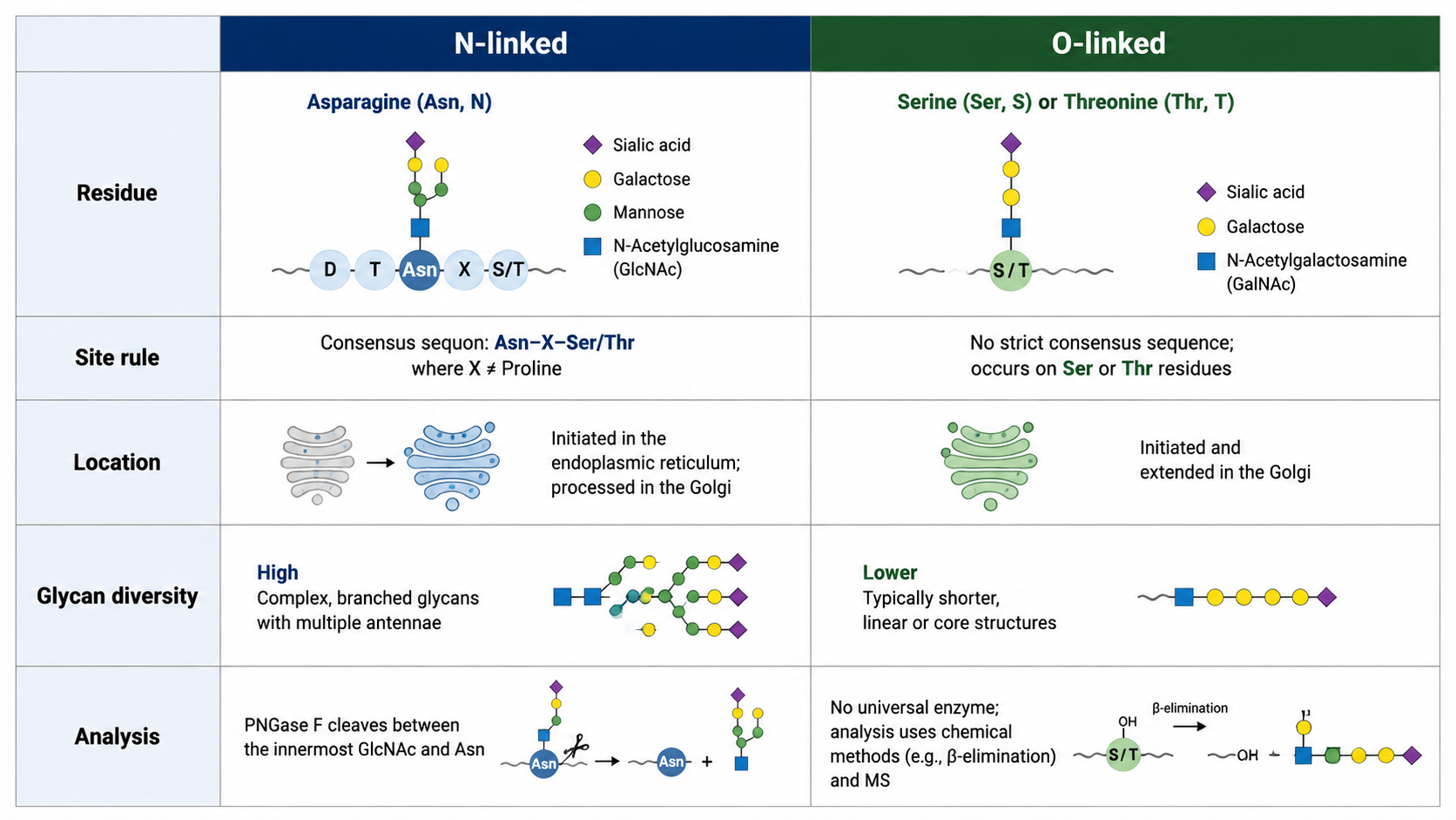
N-linked glycosylation usually occurs on asparagine within an N-X-S/T motif and is processed through the ER and Golgi.
-
O-linked glycosylation usually occurs on serine or threonine and is highly diverse, especially in mucin-like regions.
-
GPI anchors tether proteins to membranes through a glycolipid structure attached near the C-terminus.
-
Glycosylation can affect folding, stability, trafficking, receptor binding, immune recognition, and therapeutic protein function.
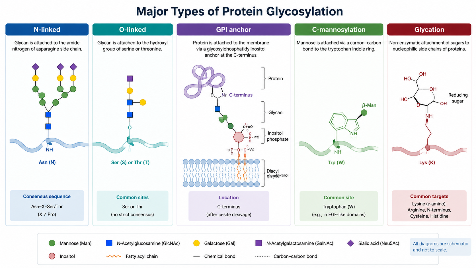
The main types of protein glycosylation are N-linked glycosylation, O-linked glycosylation, and glycosylphosphatidylinositol (GPI) anchoring. Other forms, such as C-mannosylation and non-enzymatic glycation, may also be relevant depending on the protein, organism, and research question. Each type differs in attachment site, biosynthetic pathway, structural diversity, and analytical strategy.
Key Takeaways
What Does Protein Glycosylation Mean?
Protein glycosylation is the covalent attachment of sugar structures to proteins. It is one of the most common post-translational modifications and can alter protein folding, localization, half-life, signaling, and interaction with other molecules. In biopharmaceuticals, glycosylation can also affect efficacy, safety, and batch consistency.
Related Services
Comprehensive Glycosylation Analysis Service
Glycosylation Site Analysis Service | LC-MS/MS
Glycoproteomics Analysis Service
N-Linked Glycosylation
N-linked glycosylation attaches glycans to the nitrogen atom of asparagine, most often in the consensus sequence N-X-S/T, where X is not proline. The initial glycan is transferred in the endoplasmic reticulum and later trimmed or remodeled in the ER and Golgi. N-glycans are often grouped into high-mannose, hybrid, and complex types.
O-Linked Glycosylation
O-linked glycosylation attaches sugars to oxygen atoms on serine or threonine residues. It does not depend on one simple consensus motif, which makes site prediction harder. Mucin-type O-glycosylation is common, but several O-glycan subclasses exist, including O-GlcNAc, O-mannose, O-fucose, and O-glucose.
GPI Anchors
GPI anchors are glycolipid structures that attach proteins to the outer leaflet of the cell membrane. The protein is linked near its C-terminus to a glycan-phosphatidylinositol anchor. GPI-anchored proteins can participate in signaling, adhesion, immune recognition, and regulated release from the membrane.
Other Glycosylation-Related Modifications
C-mannosylation attaches mannose to the carbon of tryptophan residues in specific contexts. Glycation is different from enzymatic glycosylation: it is a non-enzymatic reaction between reducing sugars and amino groups, often relevant in aging, diabetes, and protein stability studies.
How Is Glycosylation Analyzed?
Glycosylation can be analyzed at several levels: released glycan profiling, glycosylation site mapping, intact glycopeptide analysis, glycoproteomics, and targeted validation. Released glycan analysis gives detailed glycan composition, while intact glycopeptide LC-MS/MS links a glycan to a specific peptide site.
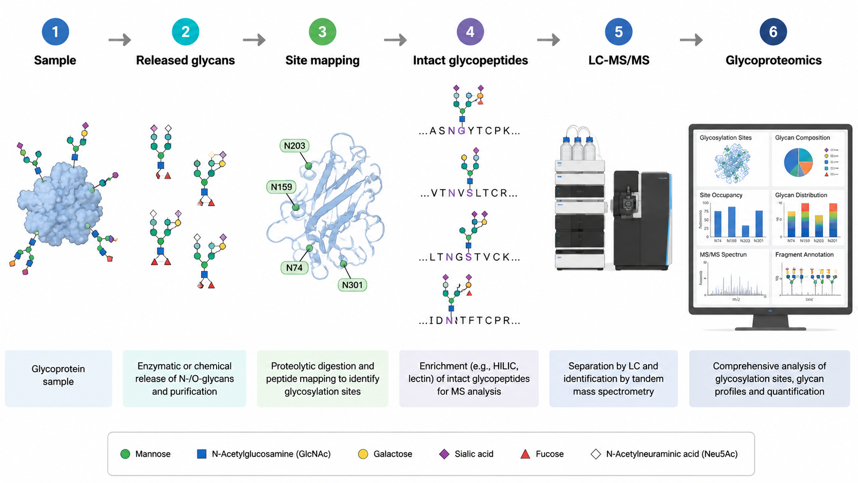
Method Selection
| Question | Useful Analysis | What It Answers? | Main Caution |
|---|---|---|---|
| What glycans are present? | Released glycan profiling | Glycan composition and abundance | Loses protein site context |
| Which sites are glycosylated? | Glycosylation site analysis | Modified residue evidence | May not fully define glycan structure |
| Which glycan is on which peptide? | Intact glycopeptide LC-MS/MS | Site and glycan together | Spectra are complex |
| How does glycosylation change across samples? | Quantitative glycoproteomics | Differential glycosylation | Requires careful normalization |
FAQ
1. What are the main types of protein glycosylation?
The main types are N-linked glycosylation, O-linked glycosylation, and GPI anchoring. Other forms include C-mannosylation and non-enzymatic glycation.
2. What is the difference between N-linked and O-linked glycosylation?
N-linked glycosylation attaches glycans to asparagine, usually in an N-X-S/T motif. O-linked glycosylation attaches sugars to serine or threonine and has more diverse site rules.
3. Why is glycosylation important?
Glycosylation can affect protein folding, stability, secretion, receptor binding, immune recognition, and biological activity.
4. How is glycosylation site analysis performed?
Site analysis is often performed by LC-MS/MS after enzymatic digestion, enrichment, or glycan-specific preparation. The goal is to identify the modified peptide and localize the glycosylation site.
Conclusion
Protein glycosylation is not one modification but a family of related modifications. N-linked glycosylation, O-linked glycosylation, and GPI anchoring differ in chemistry and biology, so the analytical strategy should match the question: glycan structure, site occupancy, intact glycopeptide identity, or quantitative glycoproteomic change.
How to order?

