


Hybridoma Monoclonal Antibody Sequencing: How to Recover VH and VL Sequences from Unstable or Lost-Producing Clones
- clear heavy chain / light chain assignment
- meaningful sequence coverage in both variable domains
- direct peptide support within or adjacent to critical CDRs
- explicit marking of unresolved or low-confidence residues
- a downstream plan for orthogonal validation
- a reconstructed set of VH and VL sequences
- peptide-level evidence linked to specific regions
- annotated ambiguity sites
- chain assignment logic supported by constant region anchoring
- an interpretation summary that separates high-confidence calls from provisional ones
Residual antibody protein can still support hybridoma monoclonal antibody sequencing after a clone becomes unstable or largely stops secreting. The practical question is whether the material left on hand still contains enough interpretable peptides for de novo sequencing, peptide mapping, and LC-MS/MS-based reconstruction of VH and VL sequences before the original antibody asset is lost.
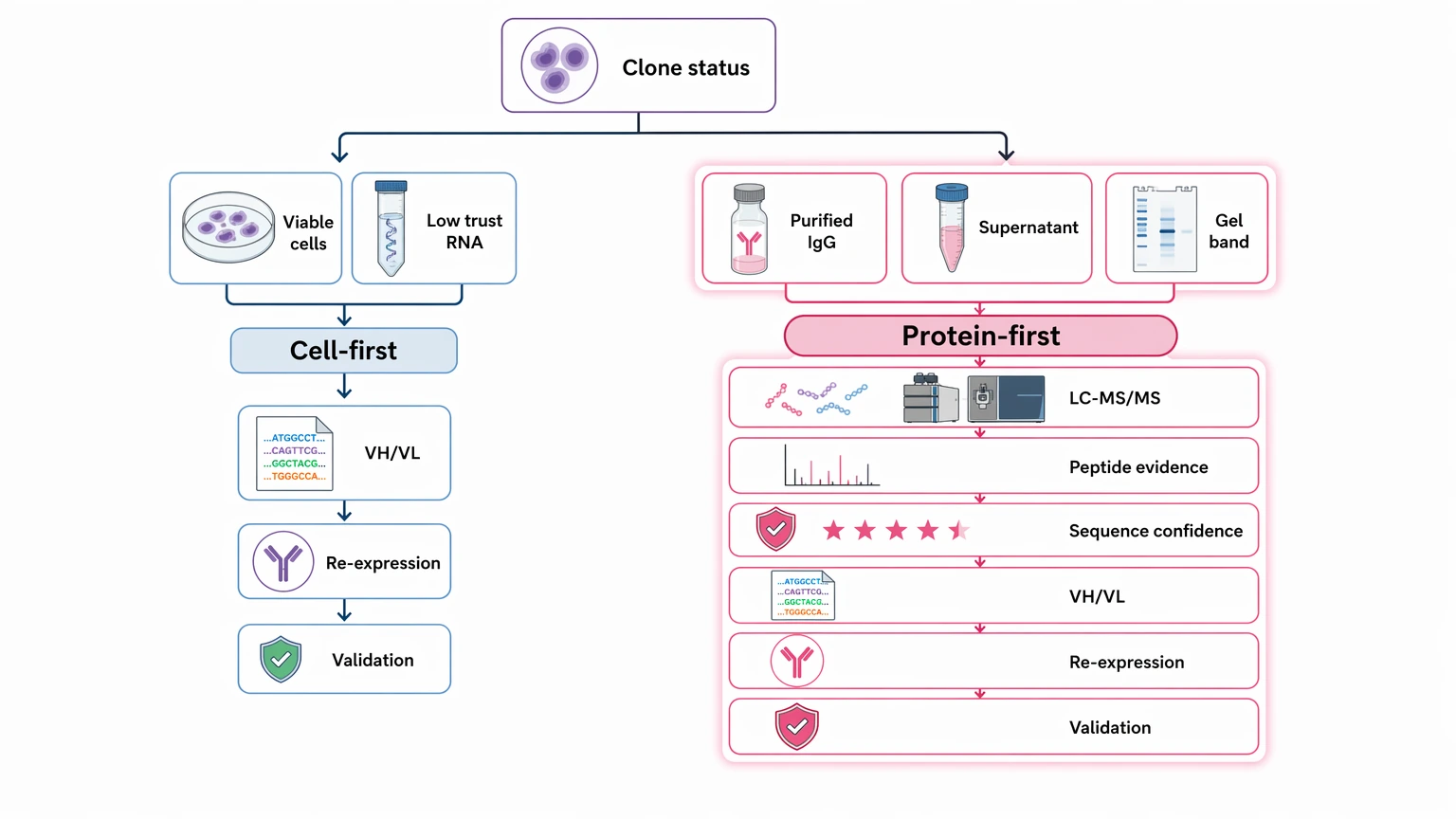
Once cell recovery is no longer reliable, the rescue path usually shifts from transcript capture to protein-derived evidence. That means sorting the material you still have, choosing preparation steps that preserve useful variable-region peptides, and deciding whether the resulting sequence confidence is strong enough to support recombinant re-expression plus follow-up orthogonal validation.
Quick Decision Guide
Use protein-based recovery first when: viable cells are scarce, transcript quality is questionable, clone purity is uncertain, or only purified antibody, supernatant, gel bands, or archived protein remains.
Prefer cell-based recovery first when: a viable, productive, and demonstrably clonal hybridoma is still available in enough quantity for reliable RT-PCR, 5' RACE, or transcript sequencing.
Best rescue candidates for LC-MS/MS: purified IgG, chain-separated bands, or antibody-enriched supernatant with detectable intact material.
Main limitation: MS/MS fragmentation can support high-confidence local sequence calls, but some positions may still remain unresolved because of leucine/isoleucine ambiguity, PTMs, incomplete sequence coverage, or weak peptide evidence across some CDRs.
Why This Problem Becomes Urgent
Sequence rescue usually turns urgent late in the life of a valuable hybridoma. A clone that once produced useful antibody can start showing poor post-thaw viability, declining secretion, mixed-population drift, or a contamination history that makes fresh cellular material harder to trust. In some cases, the cells are still around, but not with enough confidence that they still reflect the original productive state.
At that point, the central question is no longer whether the culture can be restored. It is whether the remaining antibody-derived material can still provide enough evidence to recover the variable heavy chain and variable light chain before the reagent is effectively gone.
That matters because sequence-defined recovery changes what a team can do next. Without credible VH and VL sequences, rebuilding the antibody as a recombinant reagent, comparing later preparations, reformatting the molecule, or moving the asset into a more stable expression system becomes much harder.
Why Conventional Cell-Based Rescue Can Fail Here
Cell-based routes still make sense when the hybridoma is healthy and clearly clonal. In the failure scenario discussed here, three common issues make them less dependable.
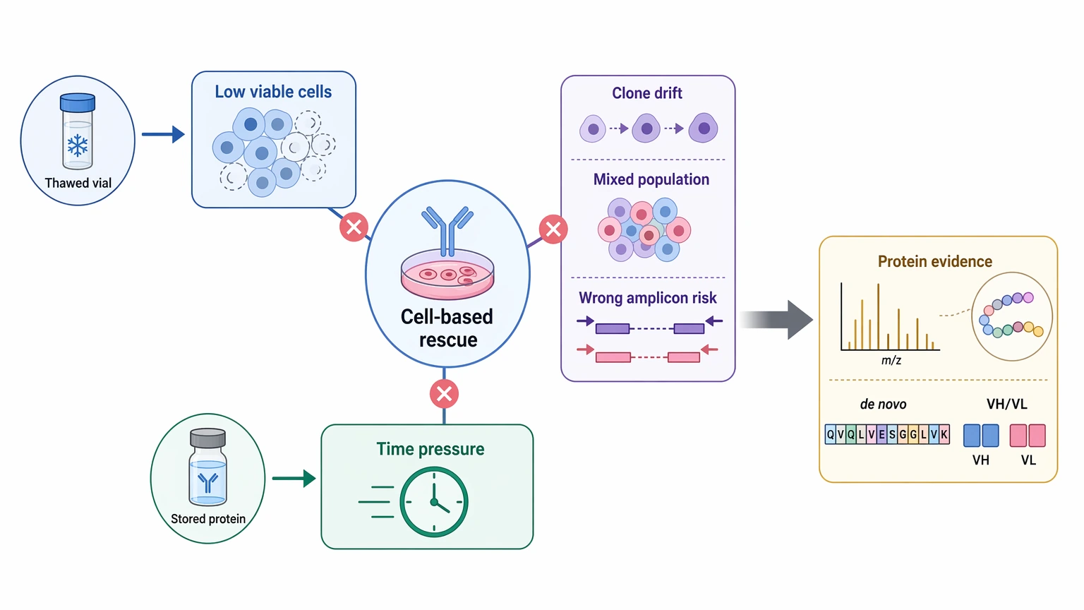
First, the number of viable cells may be too low for a clean recovery workflow. Even if a thawed vial gives some signal, that does not mean the surviving population still represents the original antibody-producing clone.
Second, transcript-based recovery can become misleading when clone drift or mixed populations are involved. A readable amplicon is not automatically the correct one.
Third, time pressure often favors the material already available. If purified antibody, stored supernatant, or excised chain bands are already in hand, delaying rescue while attempting uncertain cellular recovery may use up the last workable protein source.
That is where protein-level recovery becomes useful: the mature antibody itself may still retain enough sequence evidence for de novo sequencing, even when cells and RNA no longer offer a dependable path.
The Main Blockers That Affect Sequence Rescue
This troubleshooting problem is usually driven by a few high-impact blockers, not by every possible failure point in the lab.
1. Secretion instability leaves too little fresh antibody
A clone may still be marginally viable but produce too little intact IgG for a straightforward recovery effort. That limits peptide abundance and often pushes the project toward older, partially degraded, or low-volume protein stocks.
2. Cell viability and transcripts no longer reflect the original clone
Repeated passage, freeze-thaw stress, contamination history, or mixed populations can make cellular recovery difficult to trust. Even a technically successful transcript workflow may capture the wrong productive state.
3. Remaining protein material may be degraded or contaminated
Stored samples often carry aggregation, fragmentation, excipients, serum proteins, or other background components. These increase spectral complexity and can weaken peptide-spectrum interpretation in the variable domains.
4. Database matching alone is usually not enough for unknown variable regions
For hybridoma-derived antibodies, the closest germline or homologous reference can help with constant-region context, but it does not define the true sequence of the CDRs and nearby framework regions. A nearest-sequence match can therefore create false confidence if it is treated as a final answer rather than supporting context.
Step-by-Step Troubleshooting Workflow for VH and VL Recovery
Step 1: Start with the material you actually have
The rescue plan should begin with current sample reality, not the original experimental plan. Useful inputs can include purified IgG, old culture supernatant, reduced heavy and light chain bands, archived formulated antibody, or small residual aliquots.
The table below helps frame the first go/no-go decision.
| Sample type | Best fit | Constraint | Next step |
|---|---|---|---|
| Purified IgG | Strongest starting point for broad peptide evidence | Storage damage or PTMs may complicate interpretation | Check intactness and digestion suitability |
| Antibody-containing supernatant | Useful when secretion is low but still detectable | Background proteins may suppress antibody peptides | Enrich or purify before LC-MS/MS |
| Heavy/light chain gel bands | Helps with heavy chain / light chain assignment | In-gel recovery can reduce yield | Optimize in-gel digestion and targeted review |
| Archived formulated antibody | Often workable for rescue projects | Excipients and age-related degradation may interfere | Desalt, assess integrity, then digest |
| Trace degraded protein | May still provide partial sequence tags | Greater ambiguity and coverage gaps | Use only when no better source remains |
Takeaway: purified or chain-separated material gives the clearest route to constant region anchoring and variable-region assembly.
Service Routes to Consider
For this project scenario, readers usually compare these service routes before requesting a quote or submitting samples.
Step 2: Screen for interpretability before full de novo sequencing
Before starting a full project, confirm that the sample still looks structurally informative. The early check is simple: is intact or partly intact antibody present, and can it generate informative peptides after digestion?
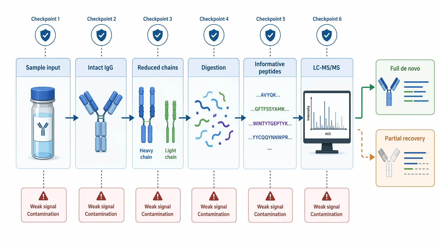
Quick intact-mass screening, reduced-chain assessment, or limited coverage-oriented LC-MS/MS can help answer that. This is not the final confirmation step. It is a triage step meant to estimate whether the sample can support useful MS/MS fragmentation, peptide mapping, and later sequence assembly.
If this screen shows dominant contamination, extensive fragmentation, or very weak antibody signal, the project may still be worth attempting, but expectations should shift toward partial recovery instead of complete assembly.
Step 3: Use preparation conditions that improve variable-region evidence
Once the sample looks usable, preparation should focus on peptide diversity and interpretability rather than raw peptide count alone. A single digest can miss key variable-region segments, especially in loop-rich CDRs or PTM-affected heavy-chain regions.
Helpful tactics often include chain separation when assignment is unclear, complementary digestion strategies to improve overlap, and selective handling of glycan-related complexity. The goal is to generate overlapping peptides that support both local residue calls and broader contig assembly across the framework regions and CDR-containing segments.
This is also the point where a feasibility discussion often matters more than a fixed template. If your team needs to judge whether a limited supernatant archive, old IgG prep, or gel-derived material still justifies sequencing work, submit your requirements to MtoZ Biolabs to evaluate your project around sample fit, LC-MS/MS workflow choice, and likely sequence-confidence limits before the remaining material is consumed.
Step 4: Assemble VH and VL sequences from peptide evidence
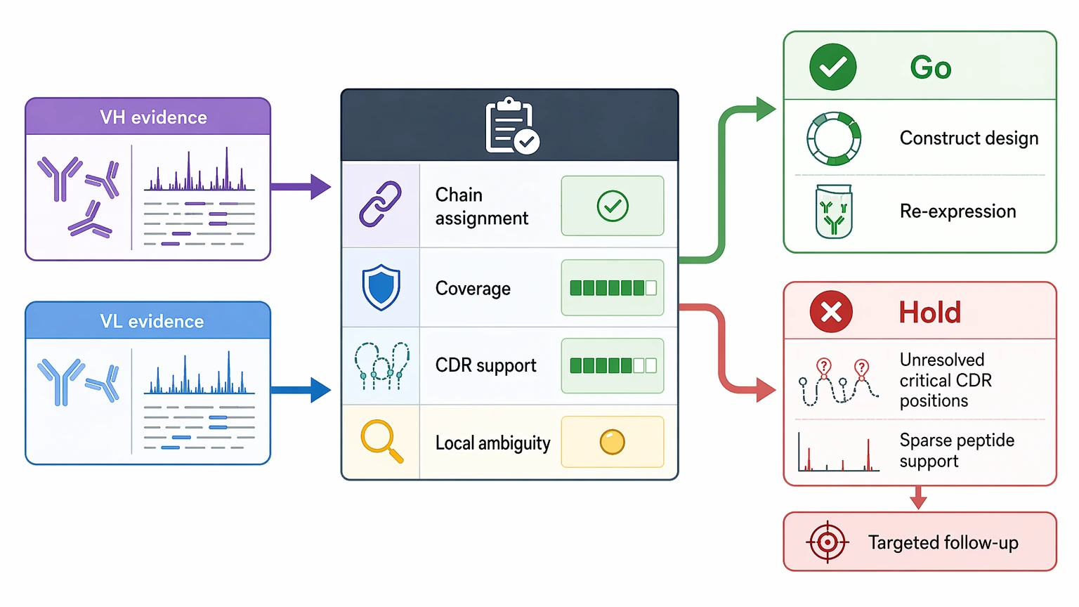
Protein-derived rescue does not come from one direct readout. It comes from combining peptide evidence into a confidence-ranked reconstruction.
| Evidence type | What it supports | Main limitation | Follow-up use |
|---|---|---|---|
| Constant-region peptides | Subclass context and chain identity | Does not define variable regions | Supports heavy chain / light chain assignment |
| Overlapping variable-region peptides | Local sequence continuity | Coverage may break across difficult regions | Builds contigs across frameworks and CDR-adjacent regions |
| CDR-containing peptides | High-value specificity evidence | Often harder to recover and interpret cleanly | Prioritize for manual review or targeted confirmation |
| De novo sequence tags | Residue inference without an exact reference | Sensitive to spectrum quality and isobaric residues | Cross-check against homologous antibody frameworks |
| Intact mass consistency | Global support for assembled chain logic | Does not resolve position-specific ambiguity | Use as orthogonal structural support |
A strong assembly usually combines constant region anchoring, overlapping peptide evidence, and internally consistent heavy/light partitioning. It should also make uncertainty visible. Common unresolved areas include isobaric residues, especially leucine/isoleucine ambiguity, along with sites affected by oxidation, deamidation, glycation, or other PTMs.
A clean-looking sequence is not enough on its own. If some positions are supported only by nearest-database inference rather than direct peptide evidence, those positions should stay flagged.
Step 5: Decide whether the output is sufficient for recombinant rescue
The key decision is not whether every residue is resolved with absolute certainty. The real question is whether the recovered variable heavy chain and variable light chain have enough direct support to justify construct design and controlled recombinant re-expression.
In practice, a usable rescue package usually includes:
If uncertainty is limited and well localized, recombinant rescue may be reasonable. If uncertainty falls on functionally important CDR positions, targeted follow-up is usually the safer next step before committing to broader downstream work.
Expected Results and Validation Strategy
The immediate deliverable from this workflow is not simply “the final sequence.” It is a structured evidence package showing what has been recovered and how confidently.
Typical immediate deliverables include:
Follow-up confirmation is a different stage. Once candidate sequences are assembled, the next question is whether they behave like the original antibody after recombinant re-expression. That later stage often includes expression testing, binding comparison, targeted peptide confirmation, or another orthogonal check focused on uncertain positions.
So a high-confidence result means more than a high peptide count. It means the variable domains are supported by interpretable overlaps, coherent chain assignment, and transparent handling of uncertainty. A partial result can still be useful if it narrows the important unknowns enough to guide the next experiment.
Key Cautions and Practical Limits
Protein-based rescue can be very useful in this setting, but its limits need to stay explicit.
Sample quality and amount set the ceiling
Trace material may still yield informative peptides, but low input reduces options for replicate preparation, chain separation, and confirmatory runs. Severe degradation also raises the chance of patchy variable-region recovery.
Controls and repeat logic still matter
A rescue workflow benefits from chain-specific checks, digestion repeats, or targeted confirmation runs when sample is available. Without those checks, it becomes harder to tell a true low-frequency signal from a preparation artifact.
Batch effects and contamination can distort interpretation
Supernatant background, keratin, excipient carryover, or residual mixed-antibody material can complicate peptide-spectrum interpretation. If clone drift is suspected, the recovered sequence should be treated as the best representation of the antibody material currently available, not automatic proof of the original single-cell state.
Sequence interpretation has built-in boundaries
Mature antibody protein does not retain signal peptide information. In addition, MS/MS fragmentation and database-assisted assembly may still leave uncertainty in PTM-affected regions, low-coverage segments, or positions involving leucine/isoleucine ambiguity. Those limits should be reported directly.
Another method may be the better next step
If variable-region evidence remains sparse after reasonable preparation, repeating the same broad workflow may only consume the last usable sample. In that case, chain-specific targeted confirmation, a complementary N-terminal method, or renewed cell-based recovery from a verified archived source may be the better next move.
When to Move Forward
Protein-based recovery becomes most convincing when the hybridoma is no longer a dependable biological source, but antibody-derived material still exists in a form that can produce interpretable peptide evidence. In that situation, the decision should rest on sample type, antibody integrity, variable-region coverage, ambiguity location, and the downstream need for recombinant rescue.
For aging hybridoma archives, low-producing rescue projects, or partially lost clones, that framework is often more practical than waiting on an uncertain cellular recovery attempt. If you need to compare the material on hand with a feasible rescue workflow, contact MtoZ Biolabs to evaluate your project and discuss sample history, remaining amount, intended recombinant re-expression, and the orthogonal validation steps that should follow sequence reconstruction.
FAQ
Can a non-secreting hybridoma still be rescued if only a historic purified antibody vial remains?
Sometimes, yes. A historic purified vial may still support recovery if intact antibody or chain-derived peptides are detectable after cleanup. The main risk is storage-related degradation, which can reduce coverage in the variable domains.
Do you need the signal peptide sequence to rebuild the antibody recombinantly?
Not necessarily for the mature antibody variable regions recovered from protein. Expression constructs can use appropriate secretion leaders, but the mature protein itself does not preserve the native signal peptide sequence.
How do you tell whether a residue call came from direct evidence or database inference?
A strong report should separate peptide-supported positions from positions inferred through homology, contig assembly, or reference alignment. If that distinction is missing, the stated sequence confidence may be overstated.
Is one digestion enzyme enough for hybridoma monoclonal antibody sequencing?
Sometimes, but not always. A single digest can leave blind spots, especially in CDR-rich or modification-prone regions. Complementary digestion strategies often improve overlap and reduce unresolved positions.
What is the main trigger for stopping and switching strategy?
The most common trigger is weak or patchy variable-region evidence after initial triage and a reasonable first-pass workflow. At that point, targeted confirmation or an alternative recovery route is usually more informative than repeating the same broad acquisition.
How to order?

