


Cell Migration Assay Service | Recombinant Collagen Evaluation
Cell migration is a fundamental biological process that underpins tissue repair, wound healing, and regenerative responses. As a critical component of the extracellular matrix (ECM), collagen not only provides structural support but also modulates the local microenvironment that governs cell motility. To evaluate the biofunctionality of recombinant collagen, standardized in vitro migration assays are essential for assessing its impact on cellular movement.
MtoZ Biolabs offers comprehensive Cell Migration Assay Service that integrates both scratch assay and Transwell migration assay. This platform provides quantitative insights into the migration-promoting effects of recombinant collagen under various treatment conditions, supporting applications in biomaterials research, tissue engineering, and functional verification of ECM products.
Services at MtoZ Biolabs
To ensure robust and reproducible data, MtoZ Biolabs’ Cell Migration Assay Service utilizes two classical in vitro assay systems to evaluate cell migration responses.
· Scratch Assay
Cells are seeded in 6-well plates and grown to confluence. A mechanical wound is introduced using a pipette tip, and cells are then treated with recombinant collagen extracts at varying concentrations. Images are captured at 0 h and 24 h to evaluate wound closure rate, serving as a direct measure of cell motility.
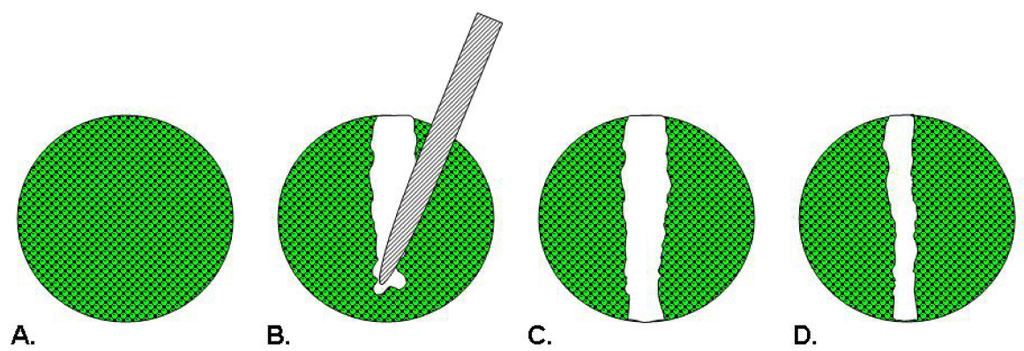
Hulkower, K. I. et al. Pharmaceutics. 2011.
Figure 1. Schematic illustration of the Scratch Assay
· Transwell Migration Assay
Cells are seeded in the upper chamber of Transwell inserts. The lower chamber contains medium with or without collagen extract to create a chemoattractant gradient. After 24 hours of incubation, migrated cells are fixed, stained, and quantified under a microscope. This approach is highly sensitive and reflects directed migration behavior.
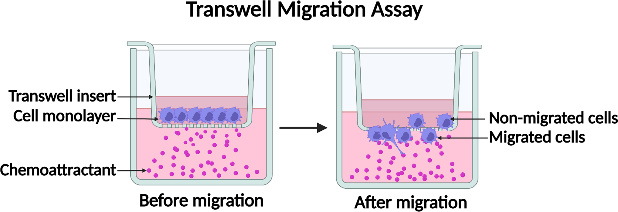
Justus, C. R. et al. Methods Mol Biol. 2023.
Figure 2. Schematic illustration of the Transwell Migration Assay
Both cell migration assays are adaptable for various mammalian cell lines, with protocols tailored to material characteristics and research objectives.
Analysis Workflow
1. Sample Preparation
Recombinant collagen samples are sterile-filtered and extracted in culture medium. Negative (blank medium) and positive (collagen reference standard) controls are included to ensure data comparability.
2. Cell Treatment & Incubation
Scratch Assay: Confluent cells are scratched and treated with test solutions. Imaging is performed at 0 h and 24 h.
Transwell Assay: Cells are seeded into the upper chamber and incubated for 24 h with test solutions. After incubation, filters are fixed and stained.
3. Image Acquisition & Data Analysis
Microscopic images are captured to evaluate wound closure rates or cell migration counts. Data are quantified and statistically compared across groups.
Why Choose MtoZ Biolabs?
✔ Robust Imaging and Quantification Platform: Equipped with high-resolution microscopy and automated image analysis tools.
✔ Standardized Experimental Workflow: Including positive controls and multi-condition testing for reliable cross-sample comparisons.
✔ Customizable Protocol Design: Cell type, treatment concentration, and medium components can be tailored to project-specific needs.
✔ Experienced Interdisciplinary Team: Scientists with dual expertise in cell biology and biomaterials ensure rigorous assay execution and interpretation.
✔ Integrated Functional Evaluation Services: Compatible with adhesion, proliferation, cytotoxicity, and other assays to support multi-angle functional assessment.
Applications
· Regenerative Medicine & Tissue Engineering: Assess ECM-mediated cell migration in scaffold-based tissue repair.
· Medical Devices & Wound Healing Materials: Evaluate bioactivity of collagen in hydrogel, membrane, or coating forms.
· Personal Care & Cosmetic Research: Verify collagen-based ingredient efficacy in promoting skin cell mobility.
· Fundamental Research: Investigate how ECM components modulate migration-related signaling pathways.
Sample Results
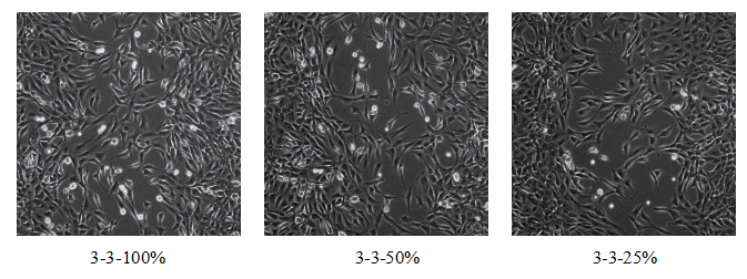
Figure 3. Microscopy Images of Scratch Areas at 0h and 24h
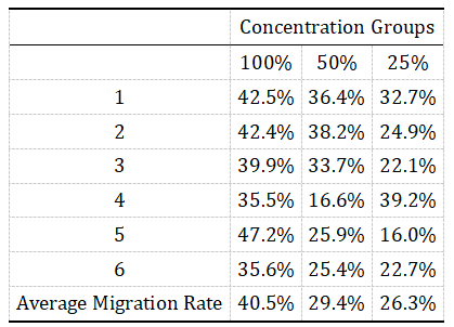
Figure 4. Quantification of Cell Migration Rates for Each Concentration
FAQ
Q1: How is a migration assay different from adhesion or proliferation assays?
Migration assays assess the ability of cells to move across a surface or through a membrane, while adhesion assays evaluate initial substrate attachment and proliferation assays measure cell growth. Together, these assays provide a complete view of material-induced cellular responses and are integral components of our Cell Migration Assay Service.
Q2: Can I submit lyophilized collagen powder for analysis?
Yes. We accept lyophilized powders, solutions, membranes, and hydrogels. For powders, ≥50 mg is recommended. We will perform standardized sterile solubilization prior to testing.
Q3: Should I choose a scratch assay or Transwell migration assay?
Scratch assays are simple and ideal for high-throughput screening of monolayer cell migration. Transwell assays better simulate chemotactic behavior and are more sensitive for detecting directional movement. Our team will help you choose the best approach based on your material and study goals.
What Could be Included in the Report?
1. Raw image files (pre- and post-treatment)
2. Quantitative migration metrics (e.g., wound closure %, migrated cell counts)
3. Experimental parameters and protocol summary
4. Optional: Statistical analysis and interpretive report
If you're interested in evaluating the migration-promoting function of recombinant collagen using our Cell Migration Assay Service, contact MtoZ Biolabs for a customized service proposal and expert consultation. We are committed to supporting your biomaterial R&D with precision and scientific excellence.
Related Services
Recombinant Collagen Biological Function Evaluation Service
Cell Proliferation Assay Service | Recombinant Collagen Evaluation
Cytotoxicity Assay Service | Recombinant Collagen Evaluation
Cell Adhesion Assay Service | Recombinant Collagen Evaluation
How to order?

