


Cell Adhesion Assay Service | Recombinant Collagen Evaluation
- Microscopic images showing pre- and post-wash adhesion morphology
- Fluorescence intensity data, adhesion percentage, and relative adhesion ratios
- A comprehensive report including methodology, statistical results, graphs, and key findings
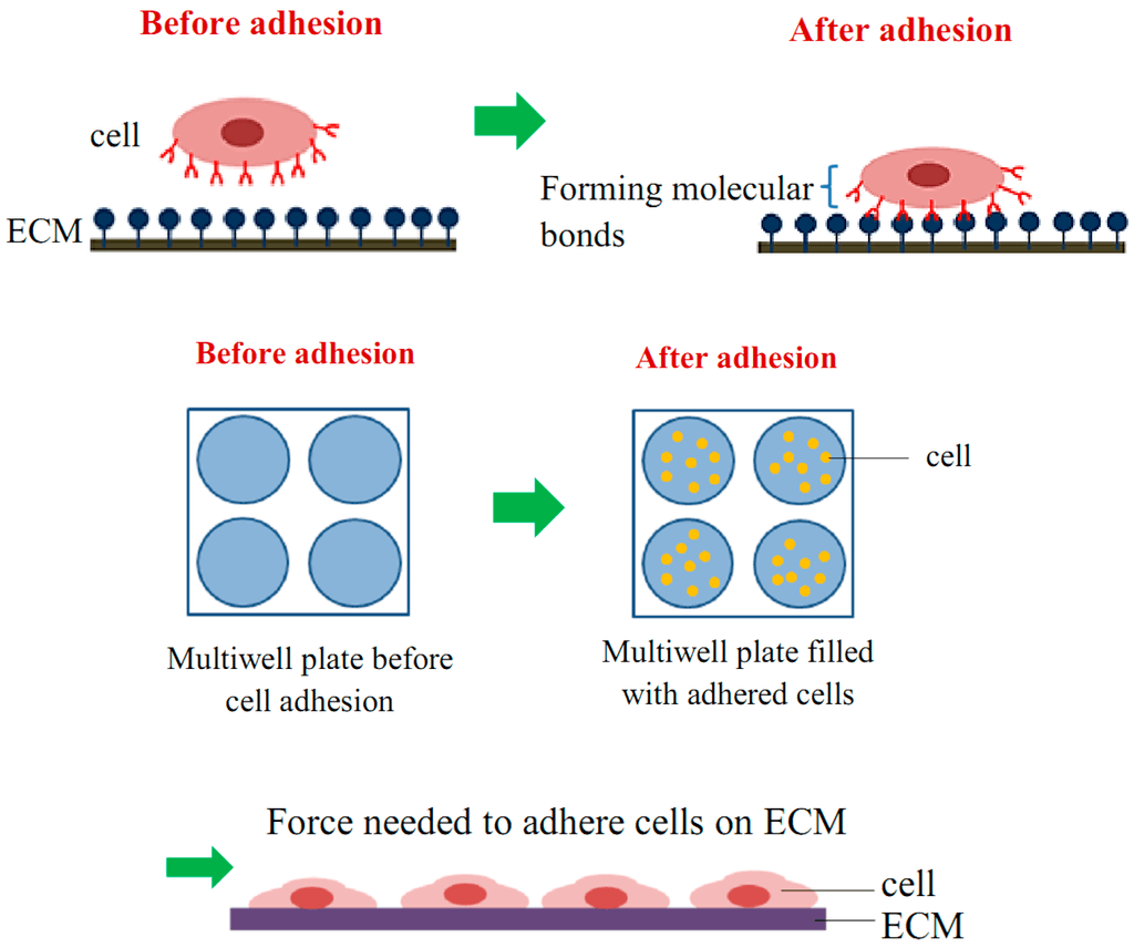
Cell adhesion is a critical early event in the interaction between cells and the extracellular matrix (ECM), playing a fundamental role in tissue architecture, signal transduction, and cellular fate determination. For ECM-derived biomaterials like recombinant collagen, the ability to promote cell adhesion is a key functional indicator.
At MtoZ Biolabs, we offer a standardized Cell Adhesion Assay Service tailored to recombinant collagen characterization. Leveraging fluorescence labeling and high-throughput microplate analysis, we provide accurate, reproducible, and quantitative insights into cell adhesion dynamics under various experimental conditions—supporting your biomaterial development, optimization, and mechanistic exploration.
Khalili, A. A. et al. Int. J. Mol. Sci. 2015.
Figure 1. Illustration of Cell Adhesion Behavior
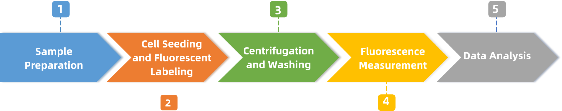
Analysis Workflow
The analysis workflow for our Cell Adhesion Assay Service is designed to ensure high precision and compatibility with a wide range of recombinant collagen formats.
1. Sample Preparation
Test materials are extracted using sterile culture media and filtered through 0.22 μm membranes to remove particulates. Negative (water) and positive (standard collagen) controls are included for benchmarking.
2. Cell Seeding and Fluorescent Labeling
NIH/3T3 fibroblasts are labeled using cell-compatible fluorescent dyes (e.g., Calcein-AM) and seeded into coated 96-well plates. Cells are incubated under standard conditions (37 °C, 5% CO₂) to allow adhesion.
3. Centrifugation and Washing
Unattached cells are removed by centrifugation and buffer washing, ensuring that only firmly adhered cells remain for evaluation.
4. Fluorescence Measurement
Fluorescence intensity is measured before and after washing using a microplate reader to quantify the number of retained cells.
5. Data Analysis
Adhesion percentage and relative adhesion ratio are calculated based on fluorescence intensity changes, providing a robust and quantitative assessment of the material’s cell adhesion–promoting properties.
Why Choose MtoZ Biolabs?
✅ Standardized Assay Workflow
A validated Cell Adhesion Assay specifically optimized for recombinant collagen evaluation, ensuring consistency and scientific rigor.
✅ Quantitative and Traceable Results
High-sensitivity fluorescent labeling combined with a standardized data analysis pipeline delivers reproducible and reliable results.
✅ Diverse Cell Type Options
Support for multiple cell lines beyond NIH/3T3, including HDF, HaCaT, MSCs, and others, tailored to different experimental models.
✅ Customized Technical Support
Flexible project customization and one-on-one technical consultation to meet specific research and development needs.
Sample Submission Suggestions
To ensure optimal experimental quality, we recommend the following:
|
Sample Type |
Requirements |
|
Solution Form |
Concentration ≥ 1 mg/mL; minimum volume ≥ 200 μL; provide buffer composition and pH; sample must be sterile and endotoxin-free |
|
Powder/Lyophilized Form |
Minimum amount ≥ 1 mg; provide batch number, dissolution conditions, and storage instructions; sample must be sterile and endotoxin-free |
Please contact our technical team in advance for projects with special requirements.
Applications
Cell Adhesion Assay Service is ideal for clients in the following sectors:
· Biomedical Research: Evaluate biocompatibility of drug delivery vectors, scaffolds, and regenerative materials.
· Cosmetic Ingredient Testing: Assess cell-adhesive properties of collagen and ECM-based actives.
· Stem Cell and Cell Therapy Research: Analyze stem cell adhesion efficiency on substrate materials.
· Basic Research: Study how material surface properties regulate cellular behavior.
Deliverables
You will receive:
Sample Results
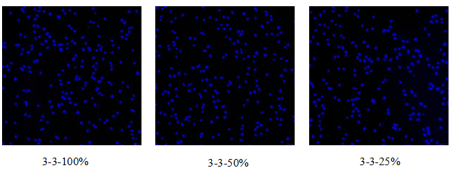
Figure 2. Microscopic Images Before and After Centrifugation
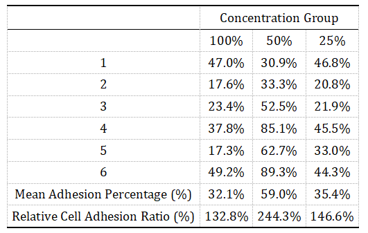
Figure 3. Adhesion Percentage and Relative Adhesion Ratio Per Group
FAQ
Q1: Is NIH/3T3 the only supported cell line?
NIH/3T3 is a common fibroblast model, but MtoZ Biolabs supports additional cell types including HDF, HaCaT, and hMSC. Custom cell line use is fully supported.
Q2: Does fluorescence labeling interfere with cell adhesion?
No. We use biocompatible dyes (e.g., Calcein-AM) validated not to affect membrane integrity or adhesion behavior under assay conditions.
Ready to evaluate the biofunctionality of your collagen-based materials?
Contact MtoZ Biolabs today for consultation or to request a tailored cell adhesion assay proposal. We are committed to delivering data-driven insights that accelerate your biomaterial development.
Related Services
Recombinant Collagen Biological Function Evaluation Service
Cell Proliferation Assay Service | Recombinant Collagen Evaluation
Cytotoxicity Assay Service | Recombinant Collagen Evaluation
Cell Migration Assay Service | Recombinant Collagen Evaluation
How to order?

