






Quantitative Histone PTM Analysis: How to Compare Modification Changes Across Conditions?
Histone post-translational modifications (PTMs) are dynamic and can change in response to cellular signals, environmental conditions, and disease-associated processes. Quantitative histone PTM analysis uses mass spectrometry to compare modification abundance across experimental groups, helping identify changes in specific histone proteins, modification sites, and modification states. Unlike the analysis of a single protein modification, histone PTM quantification may involve multiple histone proteins and numerous modification sites. Understanding these changes requires appropriate sample preparation, peptide coverage, and quantitative strategies, followed by careful interpretation of the resulting modification patterns.
Researchers planning to compare histone modification changes across experimental conditions can explore the Histone PTMs Quantitative Analysis Service offered by MtoZ Biolabs.
Quantitative Targets in Histone PTM Analysis
Quantitative histone PTM analysis can examine modification changes at different levels, from individual histone proteins and modification sites to combinations of histone marks.
Different Histone Proteins
The four core histones, H2A, H2B, H3, and H4, contain multiple modification sites and can exhibit different modification patterns across experimental conditions. Histone variants, such as H2A.Z and macroH2A, also contribute to histone diversity. Quantitative analysis can compare the relative abundance of modified peptides derived from different histone proteins, helping identify which histones exhibit changes under specific biological conditions.
Different Histone Modification Sites
A single histone protein can carry multiple PTMs at different amino acid residues. For example, histone H3 contains several frequently studied lysine modification sites, including K4, K9, K27, and K36. Comparing modification abundance at individual sites helps identify whether specific regions of a histone undergo different responses to treatment or changes in cellular conditions. For example, quantitative analysis may reveal different changes in H3K4 methylation, H3K9 methylation, and H3K27 acetylation between control and treatment groups.
Different Modification States at the Same Site
The same lysine residue can carry different methylation states, including monomethylation (me1), dimethylation (me2), and trimethylation (me3). For example, H3K27 can occur as H3K27me1, H3K27me2, or H3K27me3. These forms have distinct biological associations and can be quantified as separate modified peptide features when adequately identified and localized. Comparing different methylation states helps investigate how histone modification patterns change across experimental conditions.
Combinatorial Histone Modifications
Histones can carry multiple PTMs, and different modifications may occur on the same histone protein or within the same peptide region. These combinations are often referred to as combinatorial histone modifications. For example, H3K9ac and H3K14ac can occur together on the same histone H3 tail. Mass spectrometry can identify and quantify peptides carrying multiple modifications when the peptide sequence and fragmentation evidence support their assignments.
Comparing these modified peptides across experimental groups can help investigate changes in histone modification patterns. However, bottom-up peptide analysis generally provides information about modifications within individual peptides rather than establishing the complete modification pattern across an intact histone molecule or nucleosome
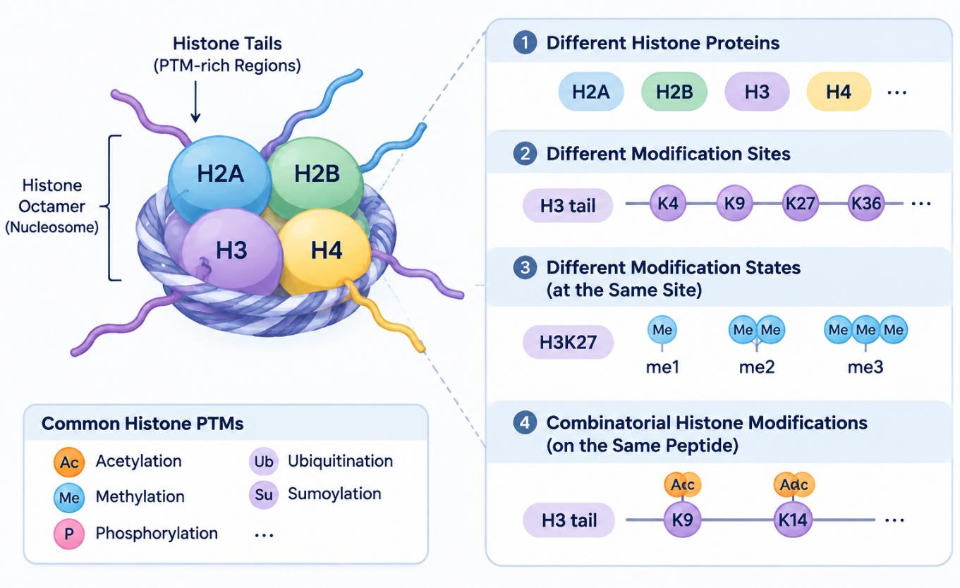
Figure 1. Quantitative Targets in Histone PTM Analysis
Sample Preparation and Peptide Coverage for Histone PTM Quantification
Reliable histone PTM quantification depends on obtaining suitable histone material and sufficient peptide coverage. Histone extraction, purification, and enzymatic digestion are particularly important because histone sequences contain numerous basic amino acid residues and modification-rich regions.
Histone Extraction and Purification
Histones are commonly extracted from isolated nuclei or chromatin-containing cellular fractions. Acid extraction and salt-based extraction are established approaches for recovering histones, followed by additional purification when required. Consistent extraction and purification conditions help reduce differences in sample composition between experimental groups. Contaminating proteins and other sample components may affect subsequent digestion, peptide recovery, and mass spectrometry analysis. Shechter et al. (2007) describe established methods for histone extraction, purification, and analysis, providing methodological background for histone sample preparation.
Histone Digestion and Protease Selection
Histones are rich in lysine and arginine residues, particularly within their N-terminal tails. Conventional trypsin digestion can therefore produce very short peptides in some histone regions, limiting sequence coverage and the detection of certain modification sites. Using multiple proteases with different cleavage specificities can generate peptides of different lengths and improve coverage of modification-rich regions. The selection of proteases should consider the target histone regions, expected PTMs, and suitability of the resulting peptides for LC-MS/MS analysis.
Peptide Length and Modification Site Coverage
Peptide length influences the identification and quantification of histone modifications. Very short peptides may provide insufficient sequence information, while longer peptides can contain multiple candidate modification sites that require adequate fragmentation evidence for localization. Appropriate digestion strategies help generate peptides that retain relevant modification information and provide sufficient sequence coverage for comparative analysis.
Reproducibility Across Experimental Groups
Sample preparation should be consistent across control, treatment, and other experimental groups. Differences in histone extraction, purification, digestion, or peptide recovery can introduce technical variation that complicates quantitative comparisons. Consistent processing and appropriate biological replication help distinguish condition-associated modification changes from differences introduced during sample preparation.
Quantitative Strategies for Comparing Histone PTM Changes
Label-Free and Tandem Mass Tag (TMT)-based approaches can support relative quantification of histone PTMs across experimental conditions. The choice depends on sample numbers, experimental design, and the analytical requirements of the study. Huang et al. (2015) provide a comprehensive review of mass spectrometry-based quantitative histone modification analysis, including the analytical strategies used to investigate changes in histone PTM abundance.
Label-Free Quantification in Histone PTM Studies
Label-Free quantification compares the relative abundance of modified peptide signals across separately analyzed samples. This approach can be applied to studies comparing histone modification changes between control and treatment groups, different disease models, or multiple time points. It also allows flexible sample numbers without requiring chemical labeling. For histone PTM studies, consistent sample preparation, digestion, and LC-MS/MS acquisition are important for comparing modified peptide abundance across samples.
TMT-Based Quantification in Histone PTM Studies
TMT is an isobaric labeling strategy that allows multiple samples to be labeled separately, combined, and analyzed within a multiplexed experiment. In histone PTM studies, TMT can support comparisons across multiple experimental conditions within the same labeling set. It is particularly useful when several treatment groups or time points need to be evaluated together.
However, co-isolation interference during MS/MS analysis can cause reporter-ion ratio compression, potentially reducing the apparent differences between samples. MS3-based acquisition can reduce this interference, as demonstrated by Ting et al. (2011), although the suitability of specific acquisition strategies depends on the analytical platform and project design.
Considerations for Comparing Multiple Histone Modifications
When multiple histone PTMs are investigated within the same study, several factors influence the comparability of quantitative results:
-
Modification compatibility: Different PTMs may require different sample preparation, enrichment, or analytical conditions.
-
Peptide coverage: Different proteases can produce different peptide populations, affecting which modification sites can be quantified.
-
Quantitative consistency: Modified peptides should be evaluated using consistent identification and quantification criteria across experimental groups.
-
Multiplexing design: For TMT experiments, samples should be assigned to labeling sets with appropriate consideration of biological groups and batch structure.
The general principles of Label-Free and TMT-based PTM quantification, including their applications and technical considerations, are discussed in How Is PTM Quantification Used to Compare Modification Changes Across Conditions?.
Interpreting Histone Modification Changes Across Conditions
Quantitative histone PTM analysis generates information about changes in modified peptide abundance across experimental groups. Interpreting these results requires considering the specific histone protein, modification site, and modification state represented by each quantified peptide.
Changes at Individual Histone Modification Sites
Comparing individual modification sites helps identify which histone marks exhibit increased or decreased abundance under different experimental conditions. For example, an increase in H3K27ac may indicate higher relative abundance of the H3K27-acetylated peptide, while a decrease in H3K9me3 may indicate lower relative abundance of the corresponding trimethylated peptide. These changes can be evaluated separately to identify modification sites that respond differently to the same treatment or biological condition.
Changes in Different Modification States
Different modification states at the same residue should be evaluated separately. For example, quantitative comparisons of H3K27me1, H3K27me2, and H3K27me3 may reveal different abundance patterns across experimental groups. An increase in one methylation state alongside a decrease in another indicates changes in the relative abundance of the corresponding modified peptides. However, these observations alone do not demonstrate direct conversion between methylation states or establish changes in site occupancy.
Combinatorial Changes Across Histone Marks
Multiple histone modifications may change simultaneously in response to a biological condition. For example, a study may observe increased H3K27ac alongside decreased H3K27me3. Such results provide evidence of changes in two distinct histone modification marks and may suggest altered chromatin-associated regulatory patterns. When multiple modifications are identified on the same peptide, their combined abundance can also be compared across experimental groups. This provides information about changes in specific multi-modified peptide forms. However, simultaneous changes in separate modification sites do not necessarily establish that the modifications occur together on the same histone molecule or nucleosome.
Relative Abundance and Histone Protein Levels
An increase in a modified peptide signal does not necessarily indicate an increase in the proportion of histone molecules carrying that modification. For example, an increase in the abundance of an H3K27ac peptide may reflect changes in the modified peptide signal, but it does not independently establish increased H3K27 acetylation occupancy. Changes in total histone abundance, histone variants, peptide recovery, and analytical response can also influence quantitative measurements. Therefore, interpreting histone PTM changes requires considering the corresponding histone protein abundance and the quantitative design. Site occupancy measurements require appropriate quantification of both modified and unmodified forms, together with suitable analytical calibration and correction strategies.
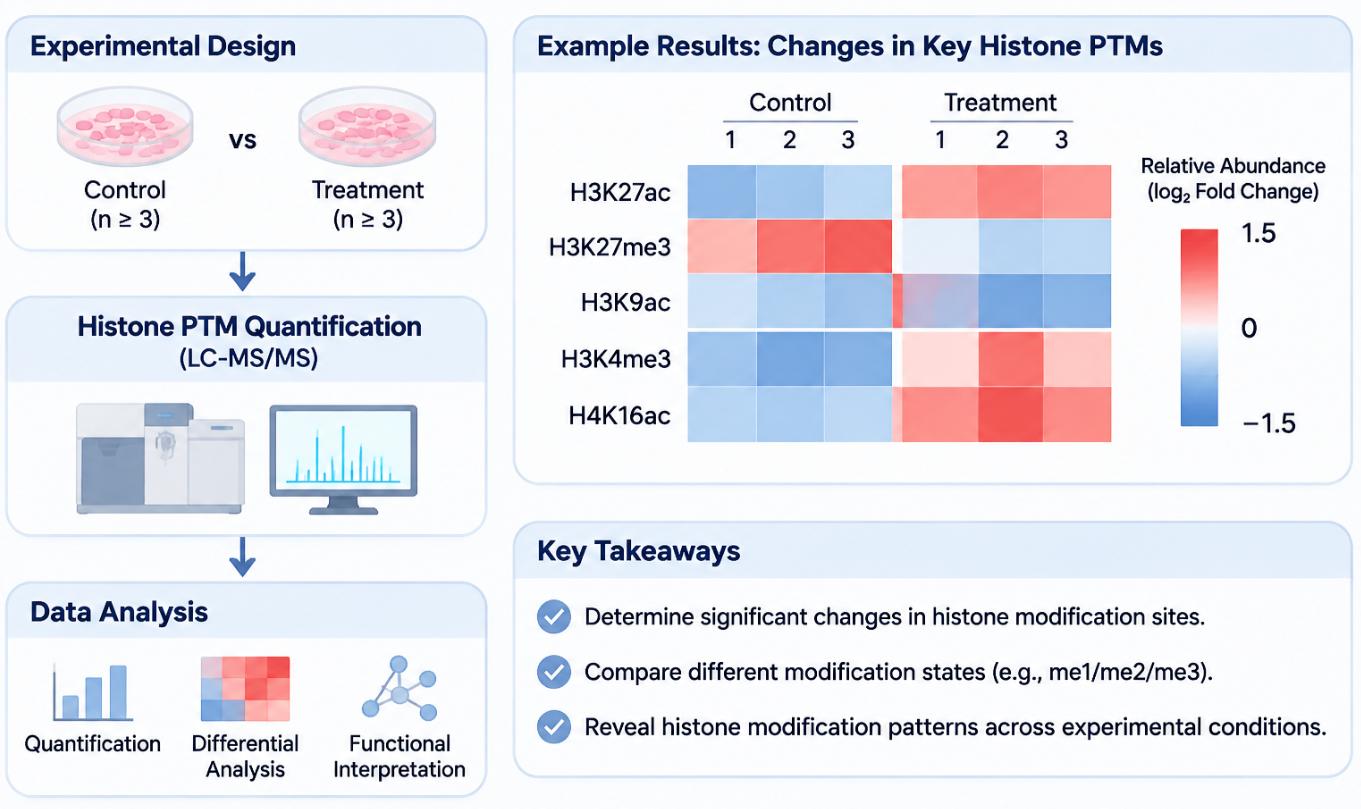
Figure 2. Workflow and Quantitative Profiling of Histone PTM Changes Between Experimental Groups.
Biological Significance of Histone Modification Patterns
Quantitative changes in histone PTMs can provide molecular evidence for investigating chromatin-associated regulation and its relationship with cellular processes. Histone modifications encompass diverse PTM types with distinct biological roles in chromatin regulation. An overview of their major types, functions, and research applications is provided in Exploring Histone Post-Translational Modifications.
Histone Marks and Chromatin Regulation
Different histone modifications are associated with distinct chromatin regulatory processes. For example, H3K27ac is commonly associated with active promoters and enhancers, whereas H3K27me3 is associated with Polycomb-mediated transcriptional repression. Changes in these marks can provide information about chromatin-associated regulatory states under different experimental conditions.
Combinatorial Histone Modifications
Histone modifications can function in combination rather than acting independently. Different marks may influence the recruitment of chromatin-associated proteins and contribute to distinct regulatory outcomes. Quantitative analysis of multiple histone marks can help investigate whether modification patterns change together or show different responses across experimental conditions. Such observations can provide candidates for further studies of interactions between histone modifications and chromatin regulation.
Histone PTM Changes and Gene Expression
Histone modification patterns are closely associated with transcriptional regulation. Quantitative changes in specific histone marks can therefore help identify potential relationships between chromatin-associated modifications and gene expression. For example, changes in histone acetylation or methylation may be investigated alongside transcriptional changes to explore potential regulatory associations. Combining quantitative histone PTM data with transcriptomic analysis or chromatin-binding experiments can provide additional evidence for understanding how histone modifications relate to gene regulation. These integrated approaches help connect site-specific modification changes with broader chromatin and transcriptional patterns.
Frequently Asked Questions
How should biological replicates be set for a histone PTM comparison?
At least three independent biological replicates per group are generally recommended for quantitative histone PTM analysis. Additional replicates may be needed when samples show high biological variability, multiple experimental conditions are compared, or smaller modification changes need to be detected. Repeated injections of the same sample are technical replicates and do not replace independent biological replicates.
Should total histone abundance be measured in the same LC-MS/MS analysis?
Measuring total histone abundance alongside modified peptides is useful for interpreting histone PTM changes. When suitable unmodified histone peptides can be reliably detected and quantified in the same LC-MS/MS analysis, they can provide a reference for evaluating changes in modified peptide abundance relative to total histone levels. If the sample preparation or analytical method limits the detection of unmodified peptides, a separate measurement using matched sample material may be needed.
Should I choose Label-Free or TMT for quantitative histone PTM analysis?
Label-Free is suitable for flexible sample numbers and studies involving large sample sets. TMT is useful when multiple experimental groups can be combined into a multiplexed analysis. The choice also depends on sample preparation and labeling compatibility with the target modifications.
Conclusion
Quantitative histone PTM analysis enables researchers to compare modification changes across different histone proteins, amino acid residues, and modification states. By examining individual sites alongside broader modification patterns, researchers can identify condition-associated changes and investigate how different histone marks relate to chromatin regulation. Reliable comparisons depend on appropriate histone preparation, sufficient peptide coverage, and suitable quantitative strategies. Combining histone PTM measurements with complementary molecular analyses can further help establish the biological significance of observed modification changes.
MtoZ Biolabs provides Histone Post-Translational Modification Analysis Service to support the identification and analysis of multiple histone modification types and sites. Researchers interested in comparing histone PTM changes across experimental conditions can discuss their sample types, target modifications, and research objectives with our team to explore a suitable analytical approach. Contact MtoZ Biolabs for a free consultation. All services are intended for research use only.
References
- D. Shechter, H.L. Dormann, C.D. Allis, S.B. Hake (2007). Extraction, purification and analysis of histones. Nat. Protoc., 2, 1445-1457. https://doi.org/10.1038/nprot.2007.202
- H. Huang, S. Lin, B.A. Garcia, Y. Zhao (2015). Quantitative proteomic analysis of histone modifications. Chem. Rev., 115, 2376-2418. https://doi.org/10.1021/cr500491u
- L. Ting, R. Rad, S.P. Gygi, W. Haas (2011). MS3 eliminates ratio distortion in isobaric multiplexed quantitative proteomics. Nat. Methods, 8, 937-940. https://doi.org/10.1038/nmeth.1714
How to order?