






Phage Display Antibody Analysis vs Mass Spectrometry: Method Selection and Research Use Cases
Phage Display Antibody Analysis and Mass Spectrometry Answer Different Research Questions
If your immediate goal is to discover or enrich new binders from an antibody library, start with phage display antibody analysis. If you already have antibody material and need molecular characterization, such as antibody sequence confirmation, peptide mapping, intact mass analysis, LC-MS/MS profiling, or post-translational modification assessment, start with mass spectrometry. If you need both new candidates and analytical evidence to explain binding behavior, sample heterogeneity, or epitope-related questions, a staged workflow often uses phage display upstream and mass spectrometry downstream.
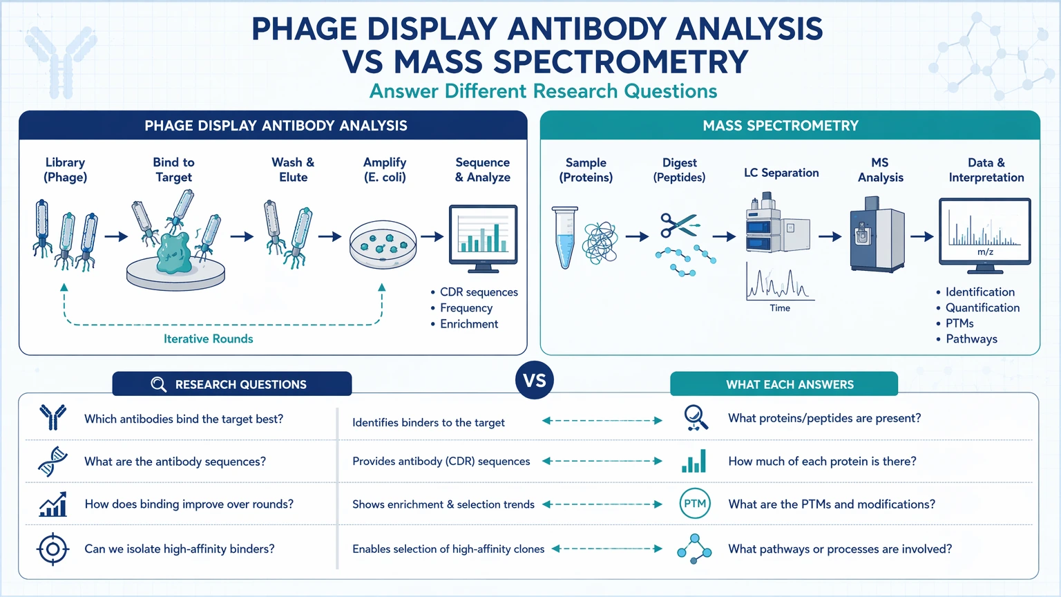
That is the real decision behind phage display antibody analysis vs mass spectrometry. These methods are often compared as if they solve the same problem, but they operate at different points in an antibody research program. Phage display is a selection platform built for binder selection and clone enrichment. Mass spectrometry is an analytical measurement platform built for molecular characterization and verification.
For immunology, antibody engineering, and translational assay teams, that distinction affects project timing and data quality. A discovery-stage program can lose momentum if it begins with detailed analytical characterization before suitable monoclonal antibody candidates exist. A characterization-stage program can stall when teams continue screening for binders even though the real issue is sample integrity, sequence liability, or an unexpected post-translational modification.
What Phage Display Antibody Analysis Is Best Suited For
Phage display is generally the better choice when the team needs to find binders rather than explain a molecule already in hand. In practice, that means starting with an antibody library, performing biopanning against a target, and recovering enriched clones for downstream ranking and validation.
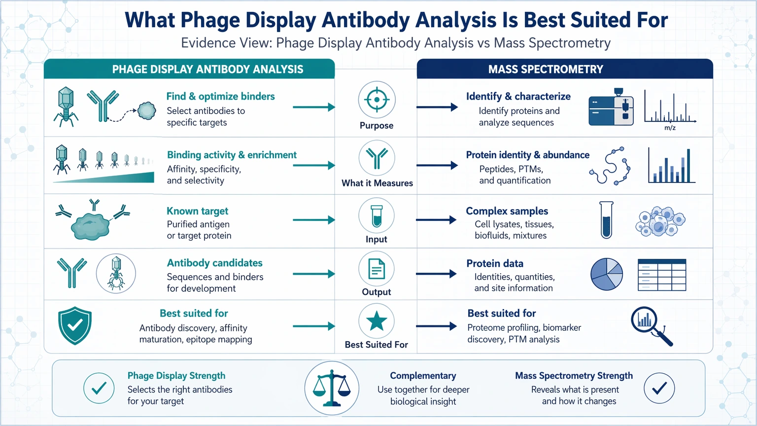
Phage display is often selected for:
- discovery of binders against a new or difficult antigen
- binder selection from large antibody library formats
- clone enrichment across iterative biopanning rounds
- antigen specificity screening
- early separation of candidate populations
- affinity maturation after an initial binder is identified
- exploratory work centered on the question, “Can we generate a candidate that binds this target?”
Its output is not a full molecular characterization package. The usual outputs are enriched clones, screening results, and candidate sequences that still require confirmation through follow-up assays such as ELISA, SPR, or BLI, along with sequencing and other orthogonal characterization steps.
This distinction matters because phage display can show which binders are worth advancing, but it does not replace LC-MS/MS, peptide mapping, or intact mass analysis when the program later needs to verify antibody sequence, assess glycosylation or oxidation, or investigate why two clones with similar screening performance behave differently after purification.
Phage display also becomes more attractive when target presentation shapes the screening strategy. Teams may need to account for whether the antigen is a recombinant protein, peptide, membrane-associated construct, or another format that changes biopanning behavior and apparent antigen specificity. In those cases, the value of phage display comes from controlled selection pressure and iterative enrichment, not from direct molecular readout.
What Mass Spectrometry Is Best Suited For
Mass spectrometry should be prioritized when the main question is no longer “Which binder can we find?” but “What exactly is present in this antibody sample?” or “What molecular feature explains the observed behavior?”
For antibody programs, MS-based workflows are commonly used for:
- antibody sequence confirmation
- peptide mapping
- intact mass analysis
- LC-MS/MS characterization of digested antibody samples
- sequence coverage assessment
- detection of post-translational modification categories such as glycosylation, deamidation, oxidation, or truncation
- compositional profiling and impurity review
- developability assessment support
- orthogonal characterization after screening or functional assays
- epitope-related workflows such as HDX-MS or cross-linking mass spectrometry
The key difference from phage display is that mass spectrometry usually starts with material already present in the sample. It does not generate new binders from an antibody library. Instead, it identifies, confirms, or profiles molecular features in purified antibodies, antibody digests, antigen-antibody complexes, or related preparations.
This makes MS especially useful when teams already have several monoclonal antibody candidate leads and need to understand why one is more suitable for downstream work. A clone may show inconsistent binding, altered stability, unusual chromatographic behavior, or disagreement between functional readouts. In that setting, LC-MS/MS and related workflows can help determine whether the cause is sequence mismatch, incomplete processing, glycosylation differences, deamidation, oxidation, or another sample-level feature.
MS also supports some forms of epitope mapping, but the readout differs from binder selection. HDX-MS can indicate protected regions and binding-associated conformational changes. Cross-linking mass spectrometry can support interface analysis in suitable systems. These are characterization approaches, not substitutes for library-based antibody discovery.
Side-by-Side Comparison: Phage Display Antibody Analysis vs Mass Spectrometry
The table below summarizes the main planning implications for the method choice.
| Dimension | Phage Display Antibody Analysis | Mass Spectrometry |
|---|---|---|
| Primary purpose | Discovery and enrichment of binders | Molecular characterization of existing material |
| Typical input | Antibody library, phage-displayed variants, target antigen | Purified antibody, peptide digest, complex mixture, antigen-antibody complex |
| Core workflow | Biopanning, clone enrichment, screening, recovery | Sample preparation, digestion or intact analysis, LC-MS/MS or related acquisition, data interpretation |
| Main output | Candidate binders, enrichment trends, screening hits | Antibody sequence information, sequence coverage, intact mass, PTM profile, compositional data |
| Best stage | Early discovery and hit generation | Lead triage, characterization, developability assessment, translational assay preparation |
| Strength for antigen specificity | Strong in screening and selection context | Indirect unless paired with an epitope-related MS workflow |
| Strength for antibody sequence confirmation | Usually requires downstream sequencing support | Strong analytical role when material is available |
| Strength for PTM analysis | Limited | Better suited for glycosylation, deamidation, oxidation, and truncation review |
| Strength for epitope mapping | Often limited to selection-based clues | Can support HDX-MS or cross-linking mass spectrometry |
| Main limitation | Does not fully characterize molecular heterogeneity | Does not generate new binders from a library |
Use these differences to align the analytical method with the biological question and validation plan.
The practical takeaway is straightforward: if the next decision is about *finding* candidates, phage display is often the better first step. If the next decision is about *understanding* candidates at the molecular level, mass spectrometry is usually the better first step.
Which Method Fits Antibody Discovery, Characterization, and Translational Research Scenarios
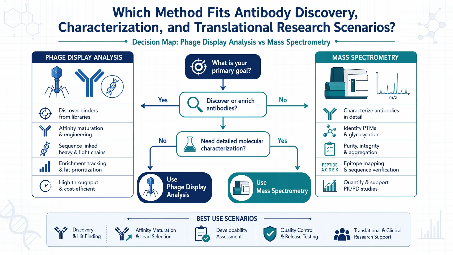
Scenario 1: You need new binders against a difficult target
Choose phage display first.
This is the clearest use case for phage display antibody screening. When the project starts with a target but no suitable antibodies, the immediate challenge is antibody library diversity, biopanning design, and clone enrichment. The first question is whether a binder can be recovered with acceptable antigen specificity and whether follow-up screening can narrow the pool to a manageable candidate set.
Mass spectrometry may become useful later, after clone recovery and expression, but it is not the primary tool for discovering those binders.
Scenario 2: You already have antibody leads but need molecular confirmation
Choose mass spectrometry first.
If the team has purified antibodies and needs to confirm antibody sequence, examine sequence coverage, compare lots, or profile post-translational modification patterns, MS is the more direct route. Peptide mapping and intact mass analysis can support clone triage when one candidate behaves differently from another despite similar screening results.
This is the clearest form of the antibody discovery vs molecular characterization divide. The project no longer needs binder generation. It needs molecular evidence.
Scenario 3: Functional data look inconsistent
Prioritize mass spectrometry if the concern is sample integrity or molecular heterogeneity. Return to phage display only if the evidence suggests that the wrong binders were advanced earlier.
For example, if a clone loses apparent performance after purification, the issue may involve oxidation, deamidation, truncation, glycosylation variation, or another compositional feature. MS can test those possibilities directly. If no meaningful molecular issue is found, the team may then revisit target format, screening bias, or clone selection strategy upstream.
Scenario 4: You need epitope-related information before assay development
Usually start with mass spectrometry when the question is analytical or structural, especially for HDX-MS or cross-linking mass spectrometry. Start with phage display when the need is still candidate generation or exploration of binding populations.
This distinction prevents a common planning error: treating all epitope questions as the same type of problem. Epitope mapping by MS is not the same as selecting binders against an antigen through phage display.
When to Use Both Methods in the Same Antibody Research Program
Many teams do not need a winner in the phage display vs mass spectrometry for antibodies discussion. They need a staged workflow that reduces uncertainty at each step.
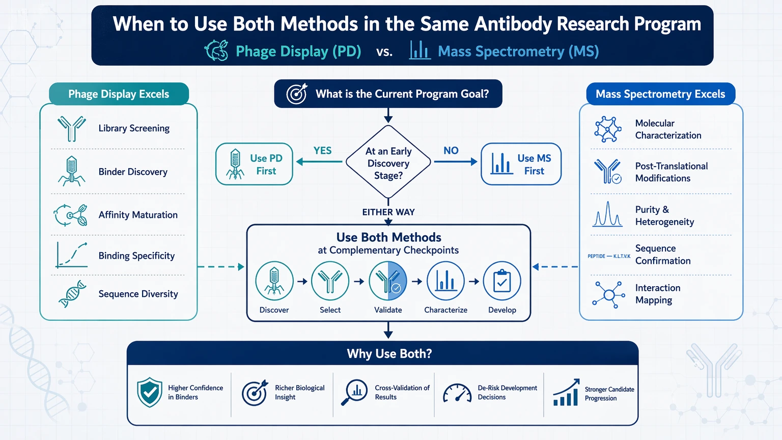
A combined strategy is often useful when the program moves through the following sequence:
1. Phage display for discovery Use an antibody library to perform biopanning and recover enriched binders.
2. Clone-level screening and sequencing Rank candidate clones for antigen specificity and early performance.
3. Mass spectrometry for characterization Apply peptide mapping, LC-MS/MS, or intact mass analysis to confirm molecular identity and check for liabilities or heterogeneity.
4. Epitope-related or developability follow-up Use HDX-MS, cross-linking mass spectrometry, or targeted PTM review when downstream assay behavior raises specific questions.
This combined workflow is especially useful when teams want to reduce decision risk before investing in expanded expression, engineering, or translational assay setup. It connects candidate finding with orthogonal characterization instead of assuming that screening data alone is enough.
A simple checkpoint is this: if your next decision would change based on molecular detail, add MS. If your program cannot move forward because no suitable binders exist yet, start with phage display.
If you are planning that transition, submit your requirements to MtoZ Biolabs for a workflow review based on antibody stage, sample type, and analytical objective.
How to Evaluate the Right Analytical Route for Your Project
A practical selection framework is to match the method to the next irreversible project decision.
Choose phage display when:
- you need new binders
- the starting material is an antibody library rather than purified antibody
- the near-term decision is clone selection
- the project requires biopanning and enrichment before deep molecular analysis is useful
- affinity maturation is part of the plan
Choose mass spectrometry when:
- you already have antibody material in hand
- the near-term decision is analytical confirmation or root-cause investigation
- you need peptide mapping, sequence coverage, or intact mass analysis
- you need to assess glycosylation, deamidation, oxidation, or related post-translational modification patterns
- you need orthogonal characterization before developability assessment or assay transfer
Combine both when:
- phage display output needs downstream confirmation
- clone ranking alone does not explain performance differences
- molecular heterogeneity could affect lead selection
- epitope-related analytical work is needed after discovery
- translational assay planning would benefit from discovery data plus molecular verification
The most useful request to send a service partner is not “Which platform is better?” A short project brief is more actionable if it states your target type, available material, current assay evidence, and the next decision your team must make.
Conclusion: Method Selection Should Follow the Research Decision, Not a Technology Label
Phage display antibody analysis and mass spectrometry are not interchangeable methods. Use phage display when your program needs binder selection, clone enrichment, or affinity maturation from an antibody library. Use mass spectrometry when your program needs molecular characterization of existing antibody material, including antibody sequence support, peptide mapping, intact mass analysis, PTM review, or epitope-related analytical work. Use both when discovery outputs must be verified, explained, or advanced through orthogonal characterization.
Phage display does not fully resolve molecular heterogeneity. Mass spectrometry does not create new binders. For many antibody teams, the most productive next step is not choosing one platform in isolation, but defining which question comes first and which follow-up data would reduce uncertainty fastest.
To scope that choice in a research setting, contact us at MtoZ Biolabs or evaluate your project with details on target format, sample type, and current assay findings so the workflow matches the decision your team needs to make.
FAQ
Are phage display and mass spectrometry direct alternatives in antibody research?
Usually not. Phage display addresses discovery and binder selection from an antibody library, while mass spectrometry addresses characterization of molecules already present in the sample. They may appear to be alternatives only when the project goal has not been separated into discovery, confirmation, and root-cause analysis.
When should a team move from phage display into mass spectrometry?
Move into MS when candidate material exists and the next question requires molecular evidence. Common triggers include disagreement between screening and purified-sample behavior, uncertainty about antibody sequence, concern about glycosylation or oxidation, or the need for peptide mapping and intact mass analysis before lead progression.
Can mass spectrometry replace sequencing of recovered phage-display clones?
Not in the same way. Recovered clone identity is usually established through sequencing workflows tied to clone recovery and expression. MS becomes more informative after antibody material is available for sequence-related characterization, PTM analysis, or composition review.
Is mass spectrometry always needed after phage display?
Not always, and not always at the same depth. Early binder triage may proceed with screening plus basic orthogonal confirmation. Deeper LC-MS/MS characterization becomes more valuable when the program must compare candidates, investigate liabilities, or explain unexpected assay behavior before additional investment.
What sample features can limit mass spectrometry in antibody programs?
Common constraints include low sample amount, poor purity, incomplete digestion, matrix complexity, and mismatch between the sample state and the analytical question. A purified monoclonal antibody candidate is generally easier to interpret than a mixed or poorly defined preparation. HDX-MS and cross-linking mass spectrometry may also require tighter control of sample handling and complex formation.
How should translational assay teams frame a method-selection request?
Include the target format, available sample type, current assay observations, whether the immediate need is discovery or characterization, and the next project decision. That information makes it easier to determine whether phage display, LC-MS/MS, or a combined antibody analysis workflow is the better starting point.
Service Routes for Study Planning
For teams moving from method selection into execution, these service paths connect assay design, validation, and interpretation needs.
How to order?




