


Glycoproteomics in Neuroscience: Mapping Neural Glycosylation, Biomarkers, and Drug Targets
-
Neural glycoproteins regulate cell recognition, axon guidance, synapse formation, receptor trafficking, and extracellular signaling.
-
Glycoproteomics can identify disease-associated changes in glycosylation before total protein abundance changes are obvious.
-
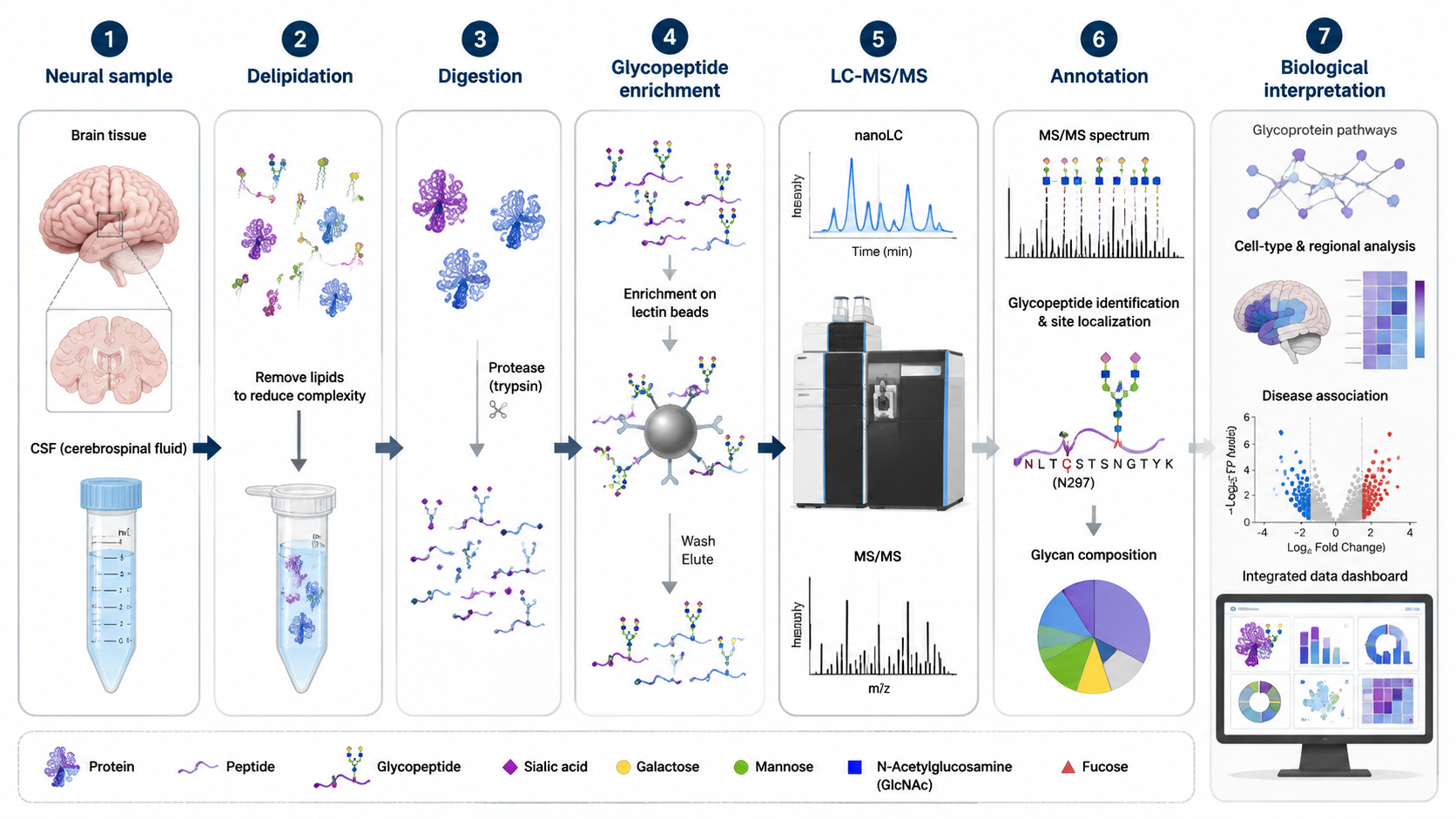
Brain tissue and cerebrospinal fluid require careful sample cleanup because lipids, salts, and abundant proteins interfere with glycopeptide analysis.
-
Enrichment, DDA/DIA acquisition, multi-stage fragmentation, and specialized bioinformatics improve glycopeptide identification.
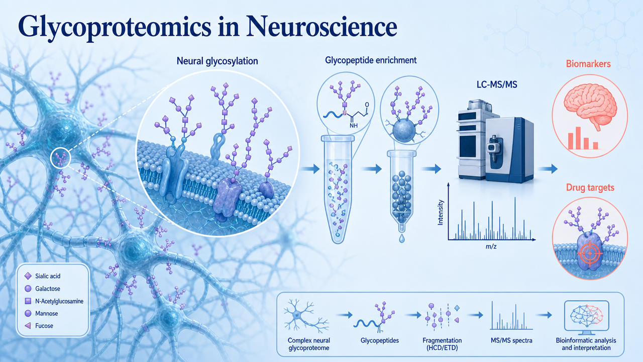
Glycoproteomics in neuroscience studies how protein glycosylation changes in the brain, cerebrospinal fluid, neurons, glia, synapses, and neural disease models. It connects glycan structures and glycosylation sites with neuronal development, synaptic signaling, neuroinflammation, neurodegeneration, and drug target biology.
Key Takeaways
What Does Glycoproteomics Measure in Neuroscience?
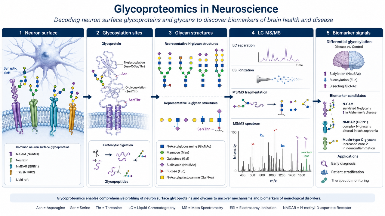
Glycoproteomics measures glycosylated proteins, glycosylation sites, attached glycan compositions, and abundance changes across biological conditions. In neuroscience, these measurements are especially relevant for membrane proteins, secreted proteins, extracellular matrix proteins, receptors, adhesion molecules, and synapse-associated proteins.
Related Services
DIA-Based Glycoproteomics Services
Quantitative Glycoproteomics Service
Glycoproteomics Analysis Service
Glycosylation Site Analysis Service | LC-MS/MS
Glycoproteomics Data Analysis Service
Why Glycosylation Matters in the Nervous System?
The nervous system is rich in glycoproteins. Glycans help define how cells recognize each other, how neurons migrate, how axons find targets, and how synapses stabilize. Surface glycoproteins also influence receptor localization, neurotransmission, immune signaling, and blood-brain barrier interactions.
In disease, glycosylation can change through altered enzyme activity, inflammation, cellular stress, aging, or protein trafficking defects. These changes may affect protein folding, ligand binding, receptor activation, and clearance.
Technical Workflow
A neural glycoproteomics workflow usually starts with sample preparation from brain tissue, cerebrospinal fluid, neuronal cultures, organoids, or sorted cell populations. The workflow then removes interfering lipids and salts, digests proteins, enriches glycopeptides or glycoproteins, analyzes them by LC-MS/MS, and uses bioinformatics to assign peptide sequence, glycosylation site, glycan composition, and abundance.
Main Technical Challenges
Neural samples are difficult because glycoproteins may be low abundance, membrane-associated, highly heterogeneous, and mixed with lipids or abundant structural proteins. Glycopeptide spectra are also complex because peptide fragments and glycan fragments appear together.
Useful solutions include optimized delipidation, lectin or hydrophilic enrichment, multi-protease digestion, DDA plus DIA acquisition, stepped collision energy, glycan oxonium ion extraction, tissue-specific databases, and manual review of high-priority glycopeptides.
Applications in Neuroscience
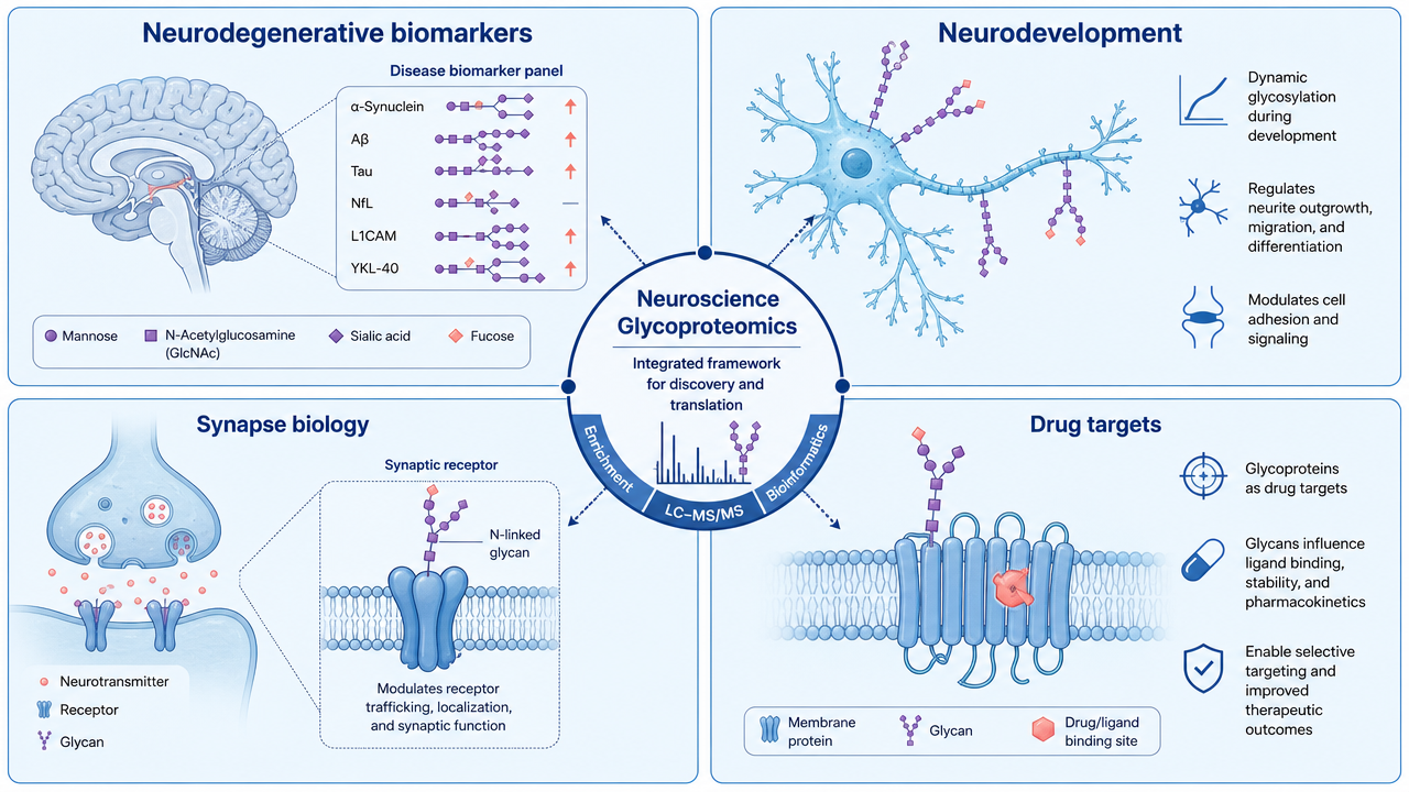
1. Neurodegenerative Disease Biomarkers
In Alzheimer disease, Parkinson disease, amyotrophic lateral sclerosis, and other neurodegenerative disorders, glycosylation changes may reflect inflammation, synaptic dysfunction, protein aggregation, or altered clearance. Glycoproteomics can help prioritize glycoprotein biomarkers in tissue, cerebrospinal fluid, or plasma.
2. Neurodevelopmental Mechanisms
During development, glycosylation affects axon guidance, synapse formation, migration, and receptor signaling. Profiling glycosylation across developmental stages can identify regulatory nodes that are missed by total protein abundance alone.
3. Drug Target Discovery
Many neural drug targets are membrane glycoproteins. Glycan structures can alter receptor conformation, ligand binding, trafficking, and antibody recognition. Glycoproteomics can therefore support target nomination and mechanism-of-action studies.
Method Selection Considerations
| Study Goal | Useful Approach | What Can It Reveal | Main Caution |
|---|---|---|---|
| Discover neural glycoproteins | Enrichment plus DDA LC-MS/MS | Broad glycopeptide inventory | Low-abundance sites may be missed |
| Compare disease and control groups | Quantitative glycoproteomics | Differential glycosylation | Batch design is critical |
| Improve coverage in complex samples | DIA-based glycoproteomics | More consistent quantitation | Requires strong spectral libraries or software |
| Localize glycosylation sites | Site-focused LC-MS/MS analysis | Site-specific modification evidence | Fragmentation may be incomplete |
FAQ
1. What is glycoproteomics in neuroscience?
Glycoproteomics in neuroscience is the mass spectrometry-based study of glycosylated proteins, glycosylation sites, and glycan structures in neural cells, brain tissue, cerebrospinal fluid, or neurological disease models.
2. Why are glycoproteins important in the brain?
Glycoproteins help regulate cell recognition, axon guidance, synapse formation, receptor localization, neurotransmission, and immune signaling in the nervous system.
3. What samples can be used for neural glycoproteomics?
Common samples include brain tissue, cerebrospinal fluid, neuronal cultures, organoids, glial cells, plasma, and disease model tissues.
4. What makes neural glycoproteomics technically difficult?
Neural samples contain many lipids and abundant structural proteins, while glycopeptides are often low abundance and structurally heterogeneous.
Conclusion
Glycoproteomics gives neuroscience studies a layer of information that standard proteomics may miss: where glycosylation occurs, which glycans are attached, and how those changes relate to neural function or disease. For biomarker discovery and target biology, that site-level context often matters as much as total protein abundance.
How to order?

