


Sparse VH/VL Coverage in Antibody Protein Sequencing: Digest, Purity, and LC-MS/MS Recovery Fixes
- VH or VL is only partially assembled, with gaps in framework or CDR regions
- CDR3 shows weak or missing MS/MS support despite acceptable Fc peptide coverage
- heavy-chain and light-chain evidence appears inconsistent across digestion runs
- host protein background dominates while antibody variable peptides remain sparse
- database search returns only low-confidence matches for variable-region peptides
- the project timeline requires both chains, but only one is confidently recovered
- confidently assembled VH and VL sequences or clearly flagged ambiguous segments
- CDR annotation where spectral support is adequate
- coverage maps showing peptide support across variable regions
- notes explaining any unresolved CDR3 or L/I ambiguity
- recommendations for expression validation if the sequence will enter production
- Repeat LC-MS/MS with revised digestion when sample quality remains acceptable
- Intact mass or peptide mapping when confirmation against a reference is sufficient
- Hybridoma PCR recovery when viable cells remain and speed is the priority
Introduction
A purified IgG sample can look acceptable on a Coomassie gel and still produce disappointing sequencing results. LC-MS/MS may run cleanly, yet variable-region coverage remains thin, CDR3 evidence is weak, or heavy-chain and light-chain assembly stops short of a usable VH/VL pair. For teams preparing recombinant expression, legacy clone rescue, or documentation submissions, sparse coverage creates immediate schedule risk.
Sparse VH/VL coverage in antibody protein sequencing usually reflects sample preparation or acquisition limits rather than fundamental unsuitability of the method. Low IgG purity, insufficient material, single-protease digestion, glycosylation interference, or prior sample mishandling can all reduce peptide evidence before assembly begins. Submitting the same material again without changing preparation or scope often yields the same incomplete outcome.
If your team is facing thin variable-region coverage from a legacy IgG lot or a first-time recombinant submission, MtoZ Biolabs can Assess coverage gaps and recommend the most efficient recovery path before material is resubmitted.
Common Signs That Coverage Is Too Thin
Researchers often contact a sequencing provider after seeing one or more of the following patterns:
These outcomes are common with legacy hybridoma-derived IgG, crude purification pools, polyclonal enrichments, and samples stored for long periods under suboptimal conditions. The key question is whether the current IgG material can support deeper variable-region coverage with a revised workflow.
Why Variable-Region Coverage Falls Short
Before resubmitting antibody or switching methods, review the most frequent barriers.
1. Insufficient IgG Purity
Host proteins, albumin, transferrin, or free light chain can suppress relevant peptide signals and complicate assembly.
2. Limited Antibody Amount
Low load reduces the number of high-quality MS/MS spectra available for CDR3 interpretation.
3. Single-Protease Digestion
One enzyme may miss critical overlapping peptides in variable regions, especially around CDR boundaries.
4. Glycosylation and Heterogeneity
Heavy Fc glycosylation can dominate the LC-MS/MS profile while VH/VL peptides remain underrepresented.
5. Aggregation or Chemical Modification
Oxidation, deamidation, or aggregation can reduce digest efficiency and fragment ion quality.
6. Scope Mismatch
A project optimized for quick VH/VL confirmation may not include the repeat acquisition or additional proteases needed for difficult legacy IgG.
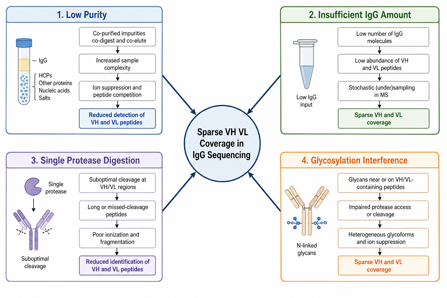
Figure 1. Sparse variable-region coverage usually reflects purity, digestion depth, or LC-MS/MS depth rather than method failure alone.
Related Services
| Customer Need | Recommended Service Direction |
|---|---|
| Need protein-level antibody sequence from purified IgG | De Novo Antibody Sequencing Service |
| Need full antibody sequencing support | Antibody Sequencing Service |
| Need MS-based antibody sequence recovery | Mass Spectrometry Based Antibody Sequencing Service |
| Need IgG-specific sequencing workflow | IgG Antibody Sequencing Service |
| Need heavy and light chain sequence recovery | Antibody Light and Heavy Chain Sequencing Service |
| Need sequence recovery from hybridoma cells instead | Hybridoma Antibody Sequencing Service |
Recovery Fixes That Most Often Improve Coverage
Use a structured review rather than repeating the same submission.
Fix 1: Reassess IgG Purity Before Anything Else
Review SDS-PAGE, SEC-HPLC, or prior QC traces if available. Additional affinity cleanup or buffer exchange may be required before variable-region peptides become detectable above background.
Fix 2: Increase Usable Antibody Input When Material Allows
If the first run used minimal IgG, a higher load may improve CDR3 spectral depth. Discuss safe upper limits with the provider so repeat extraction remains possible if needed.
Fix 3: Expand Digestion Strategy
Multi-enzyme digestion often improves overlap across VH and VL, especially when a single protease leaves CDR-adjacent gaps. Repeat analysis with an alternative enzyme set is a common next step for legacy IgG.
Fix 4: Repeat LC-MS/MS With Targeted Acquisition Settings
Weak but present variable-region peptides may benefit from repeat injection, longer acquisition, or optimized fragmentation settings rather than immediate method switching.
Fix 5: Revisit Project Scope and Fallback Options
If IgG quality cannot be improved and coverage remains insufficient, evaluate whether viable hybridoma cells remain for or whether orthogonal support a narrower documentation goal.
Figure 2. Purity review, multi-enzyme digestion, and repeat LC-MS/MS are the highest-leverage fixes for thin variable-region coverage.
IgG Submission Checklist Before Resending Material
Sample quality remains the strongest predictor of coverage depth.
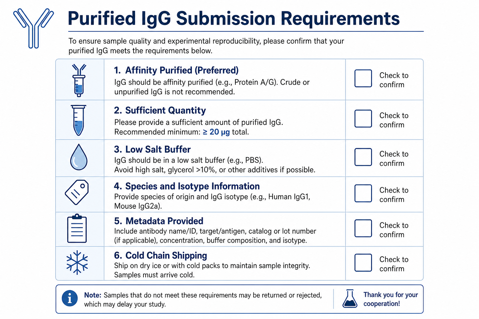
Figure 3. Feasibility review before shipment reduces repeat submissions and shortens time to usable VH/VL evidence.
Include species, isotype, purification method, expected subclass, storage history, and any prior QC data when available. If multiple aliquots exist, submit the sample with the best documented purity and the most recent integrity check.
When resubmitting after a failed run, also share the prior coverage map or partial sequence report. That information helps the analysis team avoid repeating the same digestion or acquisition strategy.
What Improved Coverage Should Look Like
A successful recovery should deliver more than a partial contig. Expected outputs may include:
Validation options depend on project goal:
Key Cautions
Do not treat strong Fc peptide coverage as proof of complete VH/VL recovery. Constant-region depth can look excellent while CDR3 remains unresolved.
Do not assume binding activity guarantees sequencing success. Functional IgG can still fail coverage targets when purity or amount is borderline.
Do not skip metadata on isotype and purification history. These details guide digestion design and review strategy.
Do not commit to expression design until both chains meet the project's confidence standard.
Frequently Asked Questions
1. Should I resubmit the same IgG without changes?
Only after reviewing purity, amount, and prior LC-MS/MS depth. Repeating the same workflow on unchanged material rarely improves CDR3 coverage.
2. Can crude supernatant IgG be sequenced?
Sometimes, after enrichment or additional purification. Coverage is usually better from affinity-purified material.
3. What if only one chain is recovered confidently?
Partial recovery may still inform next steps, but expression design typically requires both VH and VL. Targeted follow-up is usually needed.
4. Can glycosylated IgG still reach usable VH/VL coverage?
Yes, when variable-region peptides are detected clearly enough for assembly. Fc glycosylation alone does not always block VH/VL recovery.
5. How can I reduce resubmission delays?
Submit the cleanest available IgG, provide complete metadata, and request feasibility review before shipping.
Conclusion
Sparse VH/VL coverage in antibody protein sequencing is often a solvable preparation or acquisition problem rather than a dead end. By reviewing IgG purity, digestion depth, LC-MS/MS quality, and project scope before resubmitting material, teams can often recover the variable-region evidence needed for expression design or documentation.
When coverage remains insufficient, MtoZ Biolabs can Plan a revised IgG workflow , , or hybridoma-based alternatives when cells remain available. Contact the technical team to review sample status and the fastest path to usable sequence data.
How to order?

