


SDS-PAGE Protein Detection Methods: Western Blot, Mass Spectrometry, and 2D-PAGE Beyond Gel Electrophoresis
-
SDS-PAGE is a separation method; protein detection usually requires Western blot, staining plus MS, or higher-resolution 2D-PAGE.
-
Western blot is best when a specific target protein must be confirmed with antibody-level specificity.
-
Mass spectrometry is best when band identity, sequence evidence, or deeper proteomic characterization is required.
-
2D-PAGE adds isoelectric-point separation and can improve resolution for complex mixtures, isoforms, and some modified proteins.
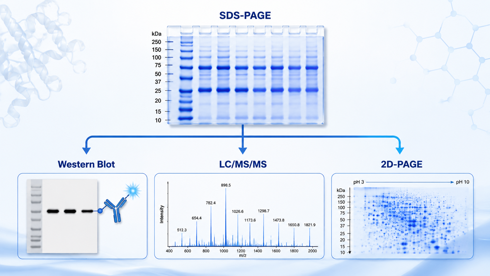
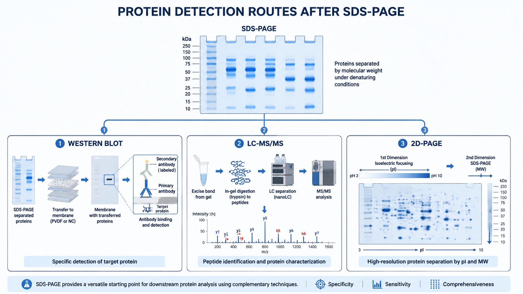
SDS-PAGE separates proteins mainly by molecular weight, but separation alone is rarely the final answer. Researchers usually need a second detection step to confirm identity, estimate abundance, check purity, or move from a visible band to a protein name. Western blot, mass spectrometry, and two-dimensional gel electrophoresis are the three most common follow-up routes after SDS-PAGE, and each answers a different experimental question.
Key Takeaways
What Does SDS-PAGE Detect by Itself?
SDS-PAGE can show whether proteins are present in a sample, whether major bands match expected molecular weights, and whether a preparation looks clean or heterogeneous. Coomassie, silver, or fluorescent staining makes protein bands visible, while pre-stained ladders help estimate size.
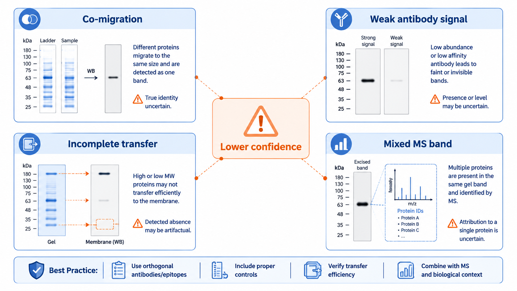
That visual readout is useful, but it does not prove protein identity. Two different proteins can migrate near the same molecular weight, and post-translational modifications can shift apparent size. That is why most serious workflows add a downstream detection method.
Related Services
SDS-PAGE Based Protein Purity Analysis Service
Western Blot after SDS-PAGE
Western blot transfers separated proteins from the gel to a membrane, then uses antibodies to detect a target. It remains one of the most practical ways to answer questions such as whether a protein is present, whether expression changed between conditions, or whether a band likely corresponds to the expected antigen.
The strength of Western blot is specificity. A good antibody can distinguish one protein from a crowded lane. The limitation is coverage: each antibody usually interrogates one target or a small related family, so it is not a discovery method for unknown bands.
Mass Spectrometry after SDS-PAGE
Mass spectrometry is the main route when researchers need to identify what a band actually contains. A common workflow excises the gel band, digests proteins into peptides, and analyzes the peptides by LC-MS/MS. Database search then links spectra to protein identities.
MS-based band analysis is especially useful for unknown bands, purity checks, pull-down validation, and moving from a visible gel pattern to sequence-level evidence.
Two-Dimensional Gel Electrophoresis (2D-PAGE)
2D-PAGE separates proteins first by isoelectric point and then by molecular weight in a second SDS-PAGE dimension. That extra separation can resolve protein isoforms, charge variants, and some post-translationally modified forms that overlap in one-dimensional gels.
The tradeoff is workflow complexity: sample preparation, focusing, staining, spot picking, and downstream MS or image analysis all require more hands-on control than a standard 1D gel.
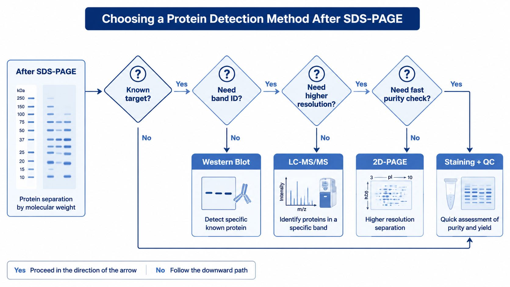
How to Choose the Right Detection Method?
| Goal | Best Starting Method | Why It Fits | Main Limitation |
|---|---|---|---|
| Confirm one known protein | Western blot | High target specificity | Limited to available antibodies |
| Identify unknown bands | LC-MS/MS after band excision | Sequence-level evidence | More time and analysis effort |
| Compare proteoforms or complex mixtures | 2D-PAGE | Better separation than 1D SDS-PAGE alone | Lower throughput and more technical variability |
| Check sample purity quickly | SDS-PAGE staining plus purity analysis | Fast visual readout | Does not prove identity by itself |
Typical Workflow Combinations
1. Targeted Expression or Pathway Studies
Run SDS-PAGE, transfer to membrane, and use Western blot when antibodies are reliable and the question is limited to one or a few proteins.
2. Band Identification or Contaminant Analysis
Run SDS-PAGE, excise bands of interest, and use in-gel digestion plus LC-MS/MS when identity must be established.
3. Complex Proteome Comparison
Use 2D-PAGE when isoform separation matters, then pick spots for MS identification or use image analysis to compare spot patterns across samples.
Common Limitations Across Gel-Based Detection
All gel-based workflows share practical constraints. Sample preparation, loading amount, gel percentage, and run conditions affect migration and band appearance. Detection limits also differ across staining, Western blot, and MS.
FAQ
1. What methods can detect proteins after SDS-PAGE besides gel electrophoresis itself?
The most common options are Western blot for targeted detection, mass spectrometry for band identification, and 2D-PAGE for higher-resolution separation before further analysis.
2. When should I use Western blot instead of mass spectrometry?
Use Western blot when you already know the target protein and have a suitable antibody. Use mass spectrometry when you need to identify unknown bands or confirm what proteins are present in a gel slice.
3. Can SDS-PAGE alone prove protein purity?
No. SDS-PAGE can suggest purity based on band pattern, but identity confirmation usually requires Western blot, MS, or another orthogonal method.
4. Why use 2D-PAGE if SDS-PAGE already separates proteins?
2D-PAGE adds separation by isoelectric point, which can resolve isoforms and modified proteins that overlap in a standard one-dimensional SDS gel.
Conclusion
SDS-PAGE is an excellent first step for protein separation, but the detection method defines what you can claim afterward. Western blot is the practical choice for known targets, mass spectrometry is the strongest route for band identity, and 2D-PAGE helps when higher resolution is worth the extra workflow complexity.
How to order?

