






Released N-Glycan Profiling vs. Glycopeptide Mapping for Antibody Glycosylation Analysis
Introduction
Antibody glycosylation analysis often brings two related approaches into the same project discussion: released N-glycan profiling and glycopeptide mapping. Both use mass spectrometry and both report glycan information on monoclonal antibodies, yet they answer different structural questions. A biologics team may request glycopeptide mapping when what it actually needs is a fast Fc glycan distribution for lot comparison. A CMC group may rely on released glycan profiling alone when site occupancy on a bispecific antibody also needs to be documented. The confusion is understandable because both workflows target N-linked glycans on therapeutic antibodies.
Released N-glycan profiling cleaves N-linked glycans from the protein and analyzes the released glycan pool to quantify major glycan structures and relative abundances. Glycopeptide mapping digests the antibody into peptides that retain glycans at glycosylation sites, then uses LC-MS/MS to assign glycan structures to specific peptide sequences and residues. Choosing the wrong route can waste sample, delay reporting, and produce data that do not support the intended comparability, process control, or site-specific review decision.
Understanding the key differences helps teams define the right antibody glycosylation analysis scope before sample preparation and reporting begin.
When Researchers Compare Released N-Glycan Profiling and Glycopeptide Mapping
This comparison usually appears when a project needs glycan evidence on a monoclonal antibody but the exact analytical route has not been defined.
Common scenarios include Fc glycan lot comparison during mAb development, where the goal is glycoform distribution rather than residue-level site assignment; comparability review after a process change, where glycan composition shifts must be quantified across reference and test lots; biosimilar glycan similarity assessment, where major N-glycan populations must be compared within an analytical package; bispecific or multi-site antibody review, where glycan structures must be linked to specific glycosylation positions; and follow-up after released glycan profiling, when a composition difference requires site-specific confirmation on the affected glycopeptide.
In each case, the decisive factor is whether the project needs glycan composition at the population level or site-specific glycan assignment on defined peptide sequences.
Four Comparison Dimensions That Matter Most
A useful comparison should focus on the glycosylation question rather than instrument platform alone.
Structural information level.
Released N-glycan profiling reports glycan structures after cleavage from the protein. Glycopeptide mapping reports glycan structures linked to specific peptide sequences and glycosylation sites.
Sample preparation route.
Released N-glycan profiling uses enzymatic or chemical release followed by glycan cleanup and profiling. Glycopeptide mapping uses proteolytic digestion, glycopeptide enrichment, and LC-MS/MS of intact glycopeptides.
Primary analytical question.
Released profiling asks which glycan structures are present and in what relative abundance. Glycopeptide mapping asks which glycan structures occur at which sites on the antibody.
Expected deliverable.
Released profiling usually produces glycan composition tables and relative abundance profiles. Glycopeptide mapping usually produces site-specific glycopeptide assignments, occupancy notes, and annotated spectra for key glycopeptides.
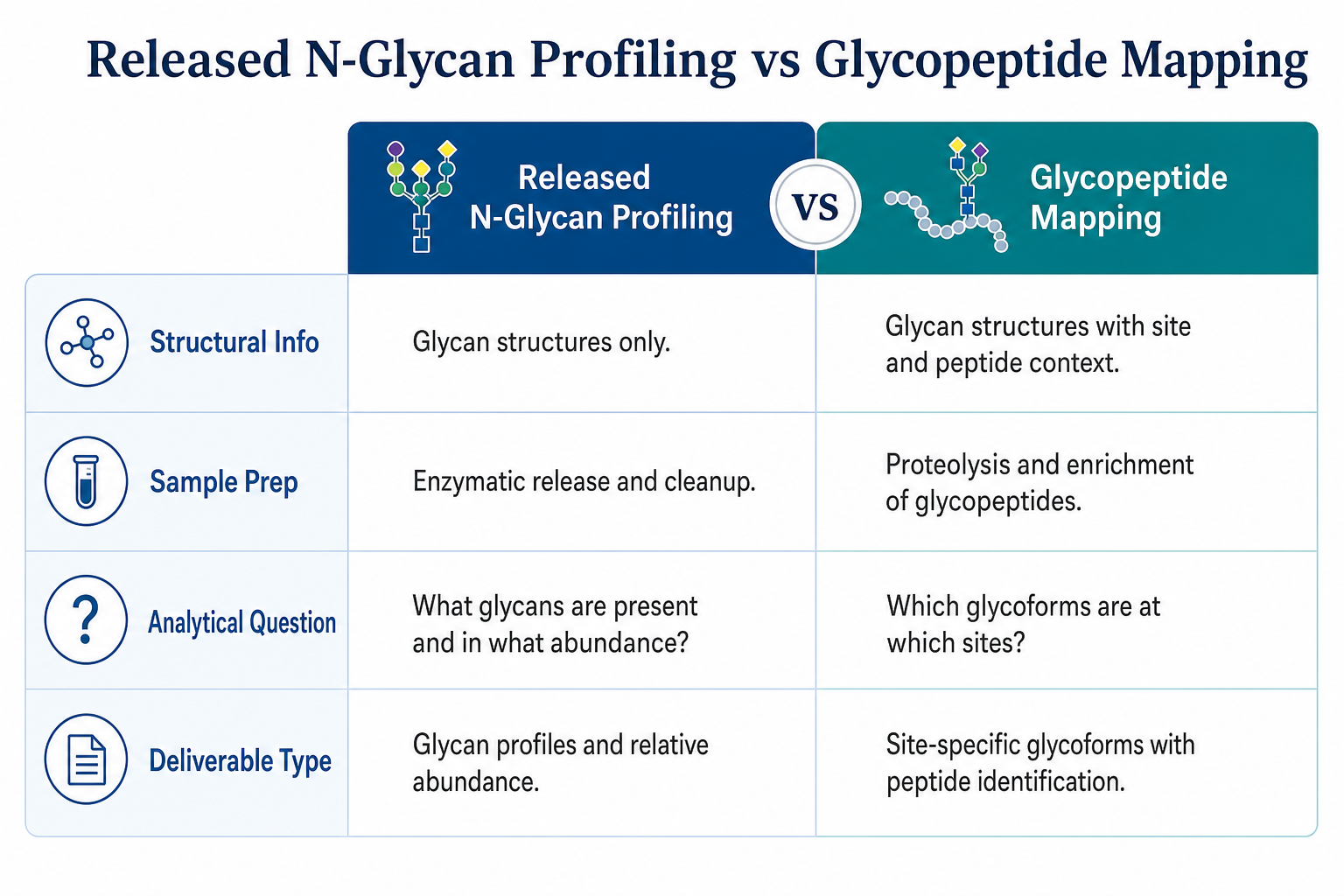
Figure 1. Released N-glycan profiling and glycopeptide mapping differ in structural resolution, preparation route, analytical goal, and reporting format.
How Released N-Glycan Profiling Works for Antibodies
Released N-glycan profiling begins with cleavage of N-linked glycans from the antibody using PNGase F or suitable release chemistry. Released glycans are cleaned, labeled when required for separation, and analyzed by LC-MS, UPLC with fluorescence detection, or MALDI-MS depending on the workflow.
For monoclonal antibodies, this approach is widely used to profile Fc-associated glycan populations, including core fucosylation, galactosylation, sialylation, and high-mannose content. The strength of released profiling is efficient glycan composition data and strong support for lot comparison, process monitoring, and development-stage profiling when site linkage is not the immediate reporting need.
Its main limitation is loss of direct site context. Released glycans represent the pooled N-glycan population and do not by themselves show which glycan structures occur at Asn297 versus any additional glycosylation sites on complex antibody formats.
How Glycopeptide Mapping Works for Antibodies
Glycopeptide mapping applies a bottom-up proteomics strategy to glycosylated antibodies. The sample is digested with an enzyme such as trypsin or another protease suited to the antibody format. Glycopeptides are enriched using HILIC, lectin affinity, or graphitized carbon workflows, then analyzed by LC-MS/MS with fragmentation modes that preserve glycan and peptide information.
For monoclonal antibodies, glycopeptide mapping is commonly used to assign glycan structures to the Fc Asn297-containing peptide and to any additional N-linked sites on engineered or multi-domain formats. The strength of glycopeptide mapping is site-specific glycan assignment, occupancy review, and direct linkage between peptide sequence and glycoform identity.
Its main limitation is workflow complexity. Glycopeptide enrichment, fragmentation optimization, and data interpretation require more method development than released glycan profiling, especially for low-abundance sites or highly heterogeneous glycoform mixtures.
Related Services
Teams comparing released N-glycan profiling and glycopeptide mapping often evaluate both service types before finalizing antibody glycosylation analysis scope. Relevant options include:
N-Glycan Profiling Service | N-Glycan Analysis
N-Glycan Profiling Service | HILIC-UHPLC MS
Glycosylated Peptides Analysis Service
Glycosylation Sites and Glycoform Analysis Services
Comprehensive Glycosylation Analysis Service
Researchers unsure which workflow fits their antibody sample and reporting goal can consult MtoZ Biolabs to review glycosylation sites, sample type, and the deliverable format required.
Side-by-Side Comparison for Antibody Glycosylation Analysis
The descriptions above show why the two approaches are complementary rather than interchangeable. The table below summarizes practical differences for antibody glycosylation analysis planning.
|
Dimension |
Released N-Glycan Profiling |
Glycopeptide Mapping |
|---|---|---|
|
Core question |
What glycan structures are present and in what abundance? |
Which glycans occur at which glycosylation sites? |
|
Site linkage |
Indirect; pooled N-glycan population |
Direct; peptide-sequence and residue linked |
|
Typical antibody focus |
Fc glycan distribution on standard IgG |
Fc site and any additional N-linked sites |
|
Main workflow |
Glycan release, cleanup, profiling |
Digestion, enrichment, glycopeptide LC-MS/MS |
|
Primary deliverable |
Glycan composition and relative abundance |
Site-specific glycopeptide assignments |
|
Best fit |
Lot comparison, process monitoring, biosimilar glycan review |
Site occupancy, multi-site antibodies, site-specific comparability |
|
Throughput |
Higher for routine glycan profiling |
Lower and more method-specific |
|
Main limitation |
No direct site assignment |
More complex for low-abundance or heterogeneous sites |
Which Method Fits Different Antibody Glycosylation Goals
Choose released N-glycan profiling when
the project needs Fc glycan composition for lot comparison, the antibody has a standard IgG format with one dominant Fc N-glycan site, the reporting goal is glycoform distribution rather than site occupancy, or the team needs efficient profiling across multiple development batches.
Choose glycopeptide mapping when
site-specific glycan assignment is required, the antibody contains more than one relevant N-linked site, occupancy at a defined glycosylation position must be documented, or released glycan data show a shift that requires site-level confirmation.
Use both in sequence when
released profiling defines the glycan population shift and glycopeptide mapping then confirms which site or glycopeptide class accounts for the difference; or when a biologics program uses released profiling for routine monitoring and glycopeptide mapping for targeted site-specific review during comparability or biosimilar assessment.
Researchers should define whether the immediate decision depends on glycan composition alone or site-linked glycoform identity. That distinction usually clarifies the workflow faster than instrument choice alone.
Decision Recommendations by Project Type
|
Project Type |
More Suitable First Method |
Why |
|---|---|---|
|
mAb Fc glycan lot comparison |
Released N-glycan profiling |
Efficient glycan distribution data for routine review |
|
Process change comparability |
Released N-glycan profiling |
Major glycan population shifts are compared quickly |
|
Biosimilar glycan similarity |
Released N-glycan profiling |
Composition comparison is often the primary need |
|
Bispecific antibody glycan review |
Glycopeptide mapping |
Multiple sites require site-specific assignment |
|
Site occupancy documentation |
Glycopeptide mapping |
Direct linkage to peptide sequence and residue |
|
Glycoengineered mAb characterization |
Combined workflow |
Composition and site linkage may both be required |
|
Unexpected glycan shift investigation |
Glycopeptide mapping after profiling |
Site-level confirmation follows population-level detection |
|
Early development screening |
Released N-glycan profiling |
Fast baseline profiling before deeper site mapping |
These recommendations are starting points. Antibody format, site number, glycan heterogeneity, and reporting urgency can shift the final plan.
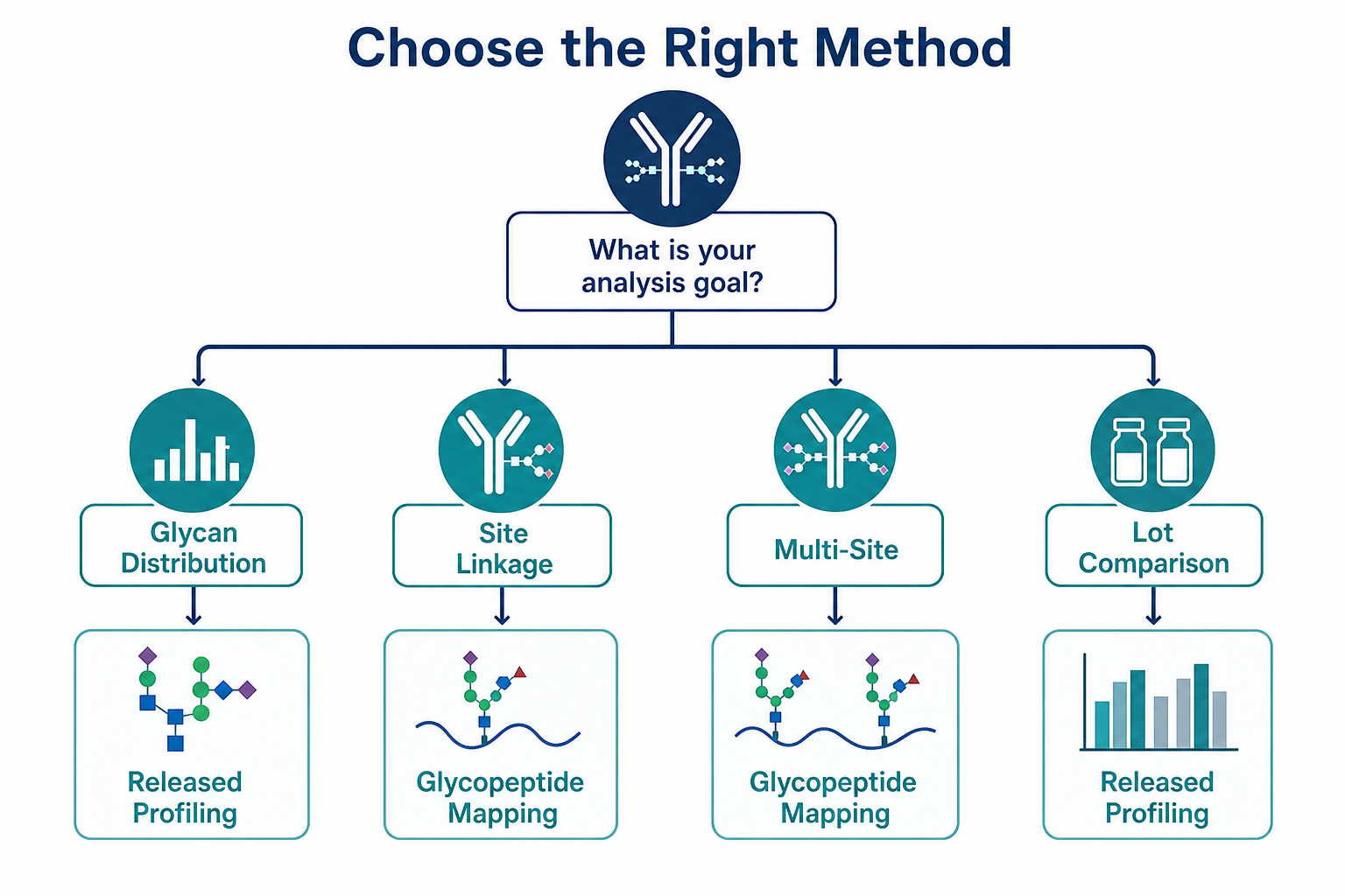
Figure 2. Site linkage requirement and reporting goal are the main factors in choosing released N-glycan profiling or glycopeptide mapping.
Combined Workflows and Practical Limits
Many antibody glycosylation programs use released N-glycan profiling as the primary monitoring route and add glycopeptide mapping for selected site-specific questions. Released profiling may define whether galactosylation, fucosylation, or high-mannose content has shifted between lots. Glycopeptide mapping may then confirm whether the shift is confined to the Fc site or distributed across additional glycosylation positions.
Released N-glycan profiling is not a substitute for site-specific glycan assignment when the decision depends on residue linkage or multi-site occupancy. Glycopeptide mapping is not the most efficient first step for routine glycan population monitoring across many development batches. The better method is the one that produces the glycan evidence type required for the next antibody characterization milestone with the least rework.
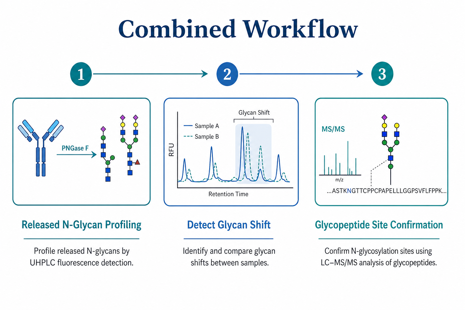
Figure 3. A combined antibody glycosylation workflow often uses released N-glycan profiling first and glycopeptide mapping for selected site-specific questions.
Frequently Asked Questions
1. What is the main difference between released N-glycan profiling and glycopeptide mapping?
Released N-glycan profiling quantifies glycan structures after cleavage from the antibody. Glycopeptide mapping assigns glycan structures to specific peptide sequences and glycosylation sites.
2. Is released N-glycan profiling enough for routine mAb glycan monitoring?
Often yes when the focus is Fc glycan distribution and lot comparison on a standard IgG format. Glycopeptide mapping may still be needed when site occupancy or multi-site assignment is part of the reporting requirement.
3. When is glycopeptide mapping the better first choice?
Glycopeptide mapping is often better when site-specific glycan assignment, occupancy review, or multi-site characterization is central to the antibody glycosylation analysis decision.
4. Can one antibody project use both methods?
Yes. Many programs use released N-glycan profiling for population-level glycan review and glycopeptide mapping for targeted site-specific confirmation.
5. What sample information is most important before choosing a method?
Antibody format, number of N-linked sites, reporting goal, comparability context, and whether site linkage or glycan composition alone is required are the most important planning inputs.
Conclusion
Released N-glycan profiling and glycopeptide mapping support antibody glycosylation analysis at different structural levels. Released profiling is usually the better first method for Fc glycan distribution, lot comparison, process monitoring, and biosimilar glycan review. Glycopeptide mapping is often the better choice when site-specific glycan assignment, occupancy documentation, or multi-site characterization must be resolved directly. Many successful antibody glycosylation programs combine both routes rather than relying on one method alone. The most suitable approach becomes clear once site linkage needs, antibody format, and reporting goal are defined. Researchers choosing between released N-glycan profiling and glycopeptide mapping for antibody glycosylation analysis can contact MtoZ Biolabs to review sample type, glycosylation sites, and the reporting format required before the next project phase.
How to order?




