






Quantitative Proteomics: Strategies, Advantages, and Challenges
A quantitative proteomics run can look complete and still fail the claim the study needs. Protein identifications may be thin, missing cells may dominate one group, coefficients of variation may exceed the expected biological effect, or ratios may pile near one. Those patterns map to study design, protein input, labeling or acquisition settings, and analysis rules. Listing label-based versus label-free strategies does not locate the break. Name the abnormal pattern, test which stage produced it, and decide whether remaining samples need more input, a different acquisition choice, a redesigned labeling set, or a targeted follow-up.
What an Unusable Quantitative Result Looks Like
Troubleshooting starts from a pattern in the protein matrix, not from a method name. Four patterns cover most stalled projects. Identification depth is far below what the matrix and load should support. Missing values concentrate in one group, one run block, or the low-intensity tail. Technical or pooled-control variation is larger than the contrast the paper must report. Fold changes collapse toward one, or an entire group moves together in a way that copies processing day.
Each pattern can appear in unlabeled data-dependent acquisition (DDA), unlabeled data-independent acquisition (DIA), or tandem mass tag (TMT) experiments. The same protein count can be adequate in depleted plasma and inadequate in a well-extracted cell lysate. A missing-value rate that is expected at the detection floor is fatal if it tracks the treatment factor. Throughput and coverage claims hold only after input, chromatography, and the quantitative signal type match the contrast.
Trace the Failure Along the Workflow
A practical check follows the peptide path. Collection and extraction decide whether comparable protein entered digestion. Digestion and cleanup decide which peptides exist. Labeling, when used, decides whether channels can be mixed. Chromatography and mass spectrometry decide which ions are sampled. Search, quantity extraction, and normalization decide which numbers enter the contrast. An early defect will survive later software.
Key parameters sit at those gates. Protein amount and concentration control digestion completeness and, for TMT, labeling occupancy. Residual detergent or salt can empty a run without changing biology. Enzyme-to-protein ratio and missed-cleavage rate change the peptide set. For unlabeled DDA, cycle time, dynamic exclusion, and peak width control precursor sampling. For unlabeled DIA, isolation-window placement and library provenance control extracted fragments. For TMT, co-isolation width and MS2 versus MS3 reporter readout control ratio accuracy (Ting et al. 2011). Run order, labeling-batch structure, and match-between-runs settings can create group differences that were never biological.
Name the abnormal pattern, then test the stage that can produce it.
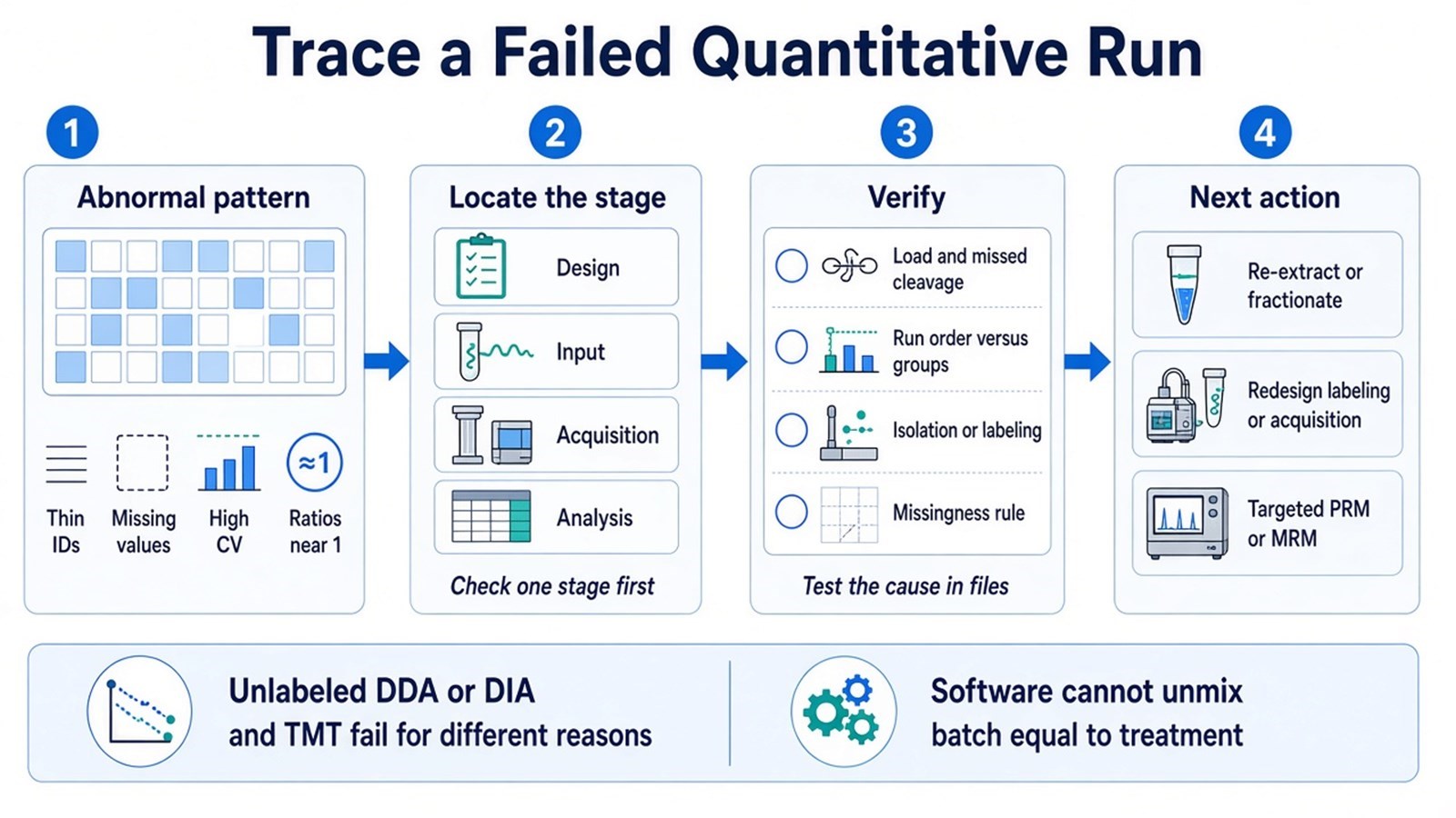
Figure 1. An unusable quantitative result is traced from the observed pattern back to design, protein input, acquisition or labeling, or analysis rules.
If groups were collected on different days, lysed in different buffers, or freeze-thawed unevenly, later peak-area software cannot restore comparability.
Researchers can refer to The Importance and Common Methods of Protein Quantification Analysis for more detailed sample-planning guidance.
If identification, missingness rules, and the statistical contrast are still open, the matrix should be rebuilt before a new digest is queued. Researchers can refer to How to Analyze Quantitative Proteomics Data for how those rules are set before a differential call is reviewed.
Failure Modes That Change the Next Experiment
Coverage collapse and missing values
Shallow identification after adequate load often points to suppression, over-complex chromatography, or DDA undersampling. Many peptide species can be present while only a fraction receive MS2 events in a data-dependent run (Michalski et al. 2011). Missing cells at low intensity are expected. Missing cells that copy one treatment arm, one injection block, or one TMT channel are not. Check chromatogram shape, peptide load, missed-cleavage rate, and whether empty samples share a processing day.
Do not impute a group-structured hole and treat the fill as a measured fold change. Match-between-runs can recover identifications and can transfer incorrect assignments if mass and time tolerances are too open (Cox et al. 2014). If new names are still required, options include more input, fractionation, or DIA. If names already exist, a targeted assay is usually more efficient than another discovery injection.
High variation and batch confounding
A coefficient of variation (CV) that exceeds the planned effect size will hide real changes and inflate noise hits. Technical injection replicates describe instrument noise. They do not replace biological replicates for an animal or patient claim. Pooled quality-control samples show whether drift is global. If principal-component separation copies labeling set, digestion day, or instrument block, the contrast is confounded.
Interleave groups in extraction, labeling, and acquisition. Normalization removes a global loading offset. It does not unmix a batch that is identical to the experimental factor. When remaining sample is limited, report the confound and redesign. A stronger batch model on the same files will not recover that contrast.
Ratios piled near one, or ratios that cannot be trusted
In TMT, reporter-ion intensities mix the target peptide with co-isolated ions in the same isolation window. Interference compresses ratios toward one, so true changes look smaller than they are (Ting et al. 2011). Check isolation interference, unused channels, and channel balance. Narrower isolation, further fractionation, or an MS3 reporter readout can reduce that distortion and will cost ions or multiplex capacity. Incomplete labeling or a failed quench can scramble channels. Re-searching the same raw files will not repair chemistry.
Unlabeled peak-area ratios can also collapse when chromatography is unstable or when the same peptide is integrated on a shoulder in one sample and on the apex in another. Overlay extracted chromatograms for a few abundant proteins and a few candidates before accepting a volcano plot.
Low-abundance proteins hidden by matrix
Plasma, serum, and some tissues are dominated by high-abundance proteins. Extra discovery time on the same digest often adds the same abundant peptides. Depletion, more starting material, or a later targeted assay may be required before a low-abundance candidate is credible (Geyer et al. 2017). Modified peptides usually need enrichment and more input than unmodified protein quantification.
Acquisition mode and labeling remain separate choices. In MtoZ Biolabs quantitative proteomics offerings, unlabeled digests are commonly processed as DDA-based label-free or as DIA, while TMT workflows are typically acquired in DDA to read reporter ions. Compare the routes on the peptides and matrix that failed.
How to Choose the Next Action
The useful test is whether the abnormal pattern is explained, whether remaining sample can support a new digest, and whether the protein list is still open.
|
Observed pattern |
Stage to check first |
Verification |
Next action that often helps |
|---|---|---|---|
|
Thin IDs despite planned load |
Input, digestion, ionization |
Load assay, missed cleavage, chromatogram |
Re-extract or clean up; consider fractionation |
|
Missingness tracks one group or block |
Design and run order |
Processing calendar versus group labels |
Rebuild the cohort order; do not impute the hole |
|
CV larger than the planned effect |
Replicates and drift |
Pooled controls, injection versus biological CV |
Add biological replicates or stabilize chromatography |
|
Fold changes piled near one in TMT |
Isolation and labeling |
Interference metrics, channel balance |
Fractionate, tighten isolation, or review MS3; relabel if chemistry failed |
|
One group moves as a processing batch |
Confounded design |
PCA or clustering versus digestion day |
Redesign; software cannot separate batch from treatment |
|
Low-abundance targets still absent |
Matrix dynamic range |
Abundant-protein dominance in the ID list |
Deplete, increase input, or move named peptides to targeted MS |
|
Names already exist and discovery still fails |
Assay type |
Whether new names are still required |
Freeze the list and design PRM or MRM |
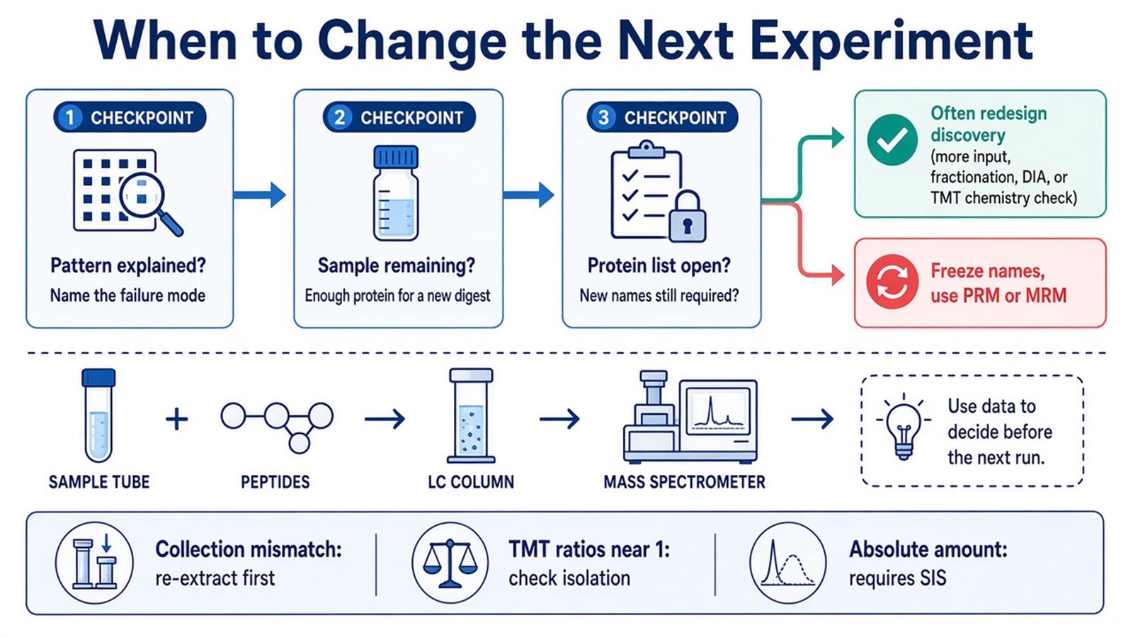
Figure 2. After a failed quantitative run, the next experiment depends on whether the pattern is explained, whether sample remains, and whether new protein names are still required.
Re-running the same unlabeled DDA method on leftover vials repeats the same sampling limits. Switching to DIA can improve fragment completeness for an open list when a suitable library or library-free workflow is available. TMT can reduce some run-to-run variation inside one labeling set and still compress ratios if isolation is dirty. None of those switches repairs a collection mismatch. Absolute amount still needs a matched stable isotope-labeled internal standard (SIS).
If the remaining task is a closed peptide panel, researchers can refer to Advantages and Disadvantages of MRM/PRM Techniques instead of another discovery strategy list.
Researchers who already have remaining sample and a defined contrast can also review the MtoZ Biolabs Quantitative Proteomics Service for project-specific feasibility and analysis planning.
Frequently Asked Questions
1. Does a low protein identification count mean the mass spectrometer failed?
A thin identification list can come from low protein input, residual detergent, digestion failure, over-complex chromatography, or DDA undersampling. Compare load, chromatogram quality, and missed-cleavage rate before attributing the result to the detector.
2. Should missing values be filled so that every protein can be tested?
Imputation can enable a statistical test and can invent a difference that was never measured. High missingness in every group is a poor discovery claim. Missingness that copies one group should be treated as a design or detection problem, not as a number to complete.
3. Is TMT always more reproducible than label-free quantification?
TMT can reduce some run-to-run variation because labeled peptides are combined before acquisition. Co-isolation can still compress ratios, and a failed labeling reaction can scramble channels. Unlabeled methods avoid reporter interference and remain sensitive to chromatography and run order.
4. When should the project leave discovery and use targeted MS?
Move to PRM or MRM when the protein list can be frozen and fragment-level confirmation is the remaining task. Keep discovery acquisition while new names are still required. Another discovery injection will not confirm a short candidate list.
5. Can software remove a batch that is identical to treatment?
Normalization and batch models can adjust global offsets and some additive structure. They cannot recover a contrast when every treated sample was processed on a different day from every control. Interleave groups in the next digest.
MtoZ Biolabs can provide quantitative proteomics analysis for unlabeled, TMT, and targeted projects, including troubleshooting of identification depth, missingness, variation, and ratio quality when project files and study design are available.
If further evaluation of remaining sample, acquisition choice, labeling design, or discovery versus targeted routing is needed, project details can be submitted for a feasibility review.
How to order?




