



A laboratory planning protein quantification often already has tissue, cells, or plasma, and still has to decide which LC-MS/MS route can turn that material into a comparable protein matrix. Bulk dye assays, antibody detection, and peptide-level mass spectrometry are all called protein quantification, yet they measure different quantities and fail for different sample reasons. For discovery or targeted MS work, extraction, digestion, buffer chemistry, and group-to-group consistency usually limit the result before the acquisition method name does. A method list becomes useful only after sample type, starting amount, storage history, and the intended comparison are specified.
Why Sample Handling Decides Whether Quantification Methods Work
Protein quantification in an MS project means turning a biological specimen into peptides whose signals can be compared across samples. Dye assays such as bicinchoninic acid (BCA) or Bradford report bulk protein for loading control. Antibody assays report an epitope under the conditions of that blot or ELISA. LC-MS/MS quantification reports peptide-derived protein values after digestion. Mixing those three readouts as interchangeable methods hides the fact that each one is only as good as the protein that survived collection, lysis, and cleanup.
The scientific question also has to match the sample. Relative discovery asks whether the same protein changes between groups in one study. Absolute targeted assays ask how much of a predefined peptide is present, usually with a matched isotope standard. Post-translational modification (PTM) work asks a third question and typically needs more starting material than unmodified protein quantification. If groups were collected on different days, lysed in different buffers, or freeze-thawed unevenly, later software cannot restore comparability. Researchers can refer to How to Analyze Quantitative Proteomics Data for how that matrix is normalized and tested after digestion.
Sample Types and Planning Amounts
Starting matrix decides lysis chemistry, contaminant load, and how much protein can be recovered before digestion. The amounts below are planning guides for general label-free, data-independent acquisition (DIA), or tandem mass tag (TMT) protein quantification. Universal acceptance cutoffs should not be inferred from the table. PTM enrichment, high-abundance protein depletion, and very low-input designs need a separate amount review.
Sample type |
Planning amount |
Minimum often reviewed |
Prep constraint |
|---|---|---|---|
Soft animal tissue |
20 mg |
10 mg |
Remove fat and extra connective tissue, snap-freeze promptly |
Tumor or adjacent tissue |
30 mg |
15 mg |
Keep lesion and adjacent pieces anatomically defined |
Hard tissue such as bone or cartilage |
100 mg |
50 mg |
Mechanical disruption is part of extraction |
Adherent or suspension cells |
1 x 10^7 cells |
2 x 10^6 cells |
PBS wash, submit a dry pellet |
Microbes |
50 mg or 50 uL |
20 mg or 20 uL |
Repeated PBS washes before freezing |
Serum or plasma, no depletion |
10 uL |
5 uL |
Hemolysis and clotting conditions affect the proteome |
Serum or plasma, with depletion |
50 uL |
25 uL |
Depletion spends sample to unmask lower-abundance proteins |
Urine |
1 mL |
0.5 mL |
Debris should be removed before freezing |
User-extracted protein solution |
20 uL |
10 uL |
Buffer composition must be disclosed before digestion |
Plant tissues need more mass than animal soft tissue because of cell wall and secondary-metabolite load; soft plant material is often planned around 2 g, with a 1 g review floor. Formalin-fixed paraffin-embedded (FFPE) sections can be used, with a common plan of 20 sections and a 10-section review floor, and they travel at room temperature. Almost all other quantitative proteomics samples should stay frozen and ship on dry ice. Protein extracts on the label-free service route are also planned around 20 ug protein when concentration, not volume, is the limiting number.
Collection conditions should be matched across groups: dissection timing, wash buffer, anticoagulant for plasma, and the interval before snap-freezing. Serum is prepared without anticoagulant; plasma is usually collected in EDTA tubes. Pink or red plasma indicates hemolysis and can shift abundant erythrocyte proteins into the quantitative matrix.
Preprocessing Workflow and Key Parameters
Quantitative proteomics preprocessing has a fixed order even when the later acquisition method changes. Collection and snap-freezing protect protein integrity. Extraction releases protein into a buffer that must be compatible with digestion or must be exchanged later. An amount check, often a BCA or similar bulk assay, sizes digestion and labeling. Reduction, alkylation, and proteolytic digestion, most often with trypsin, convert proteins into peptides. Cleanup removes detergent, salt, and debris that suppress ionization. Only then does LC-MS/MS acquisition begin.
Comparable peptide signals require matched extraction and digestion across the cohort.
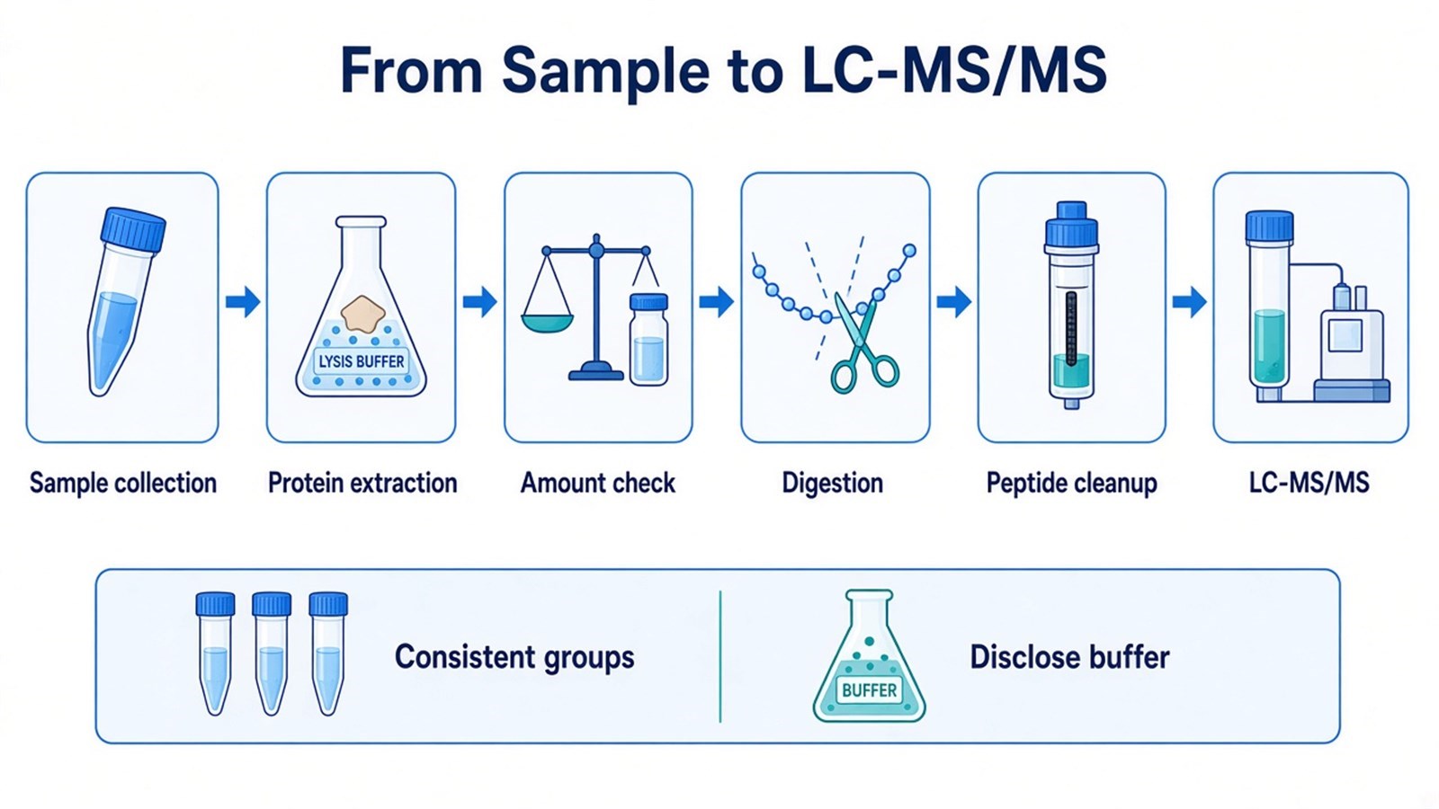
Figure 1. Protein quantification by LC-MS/MS starts with collection, extraction, amount check, digestion, and peptide cleanup before acquisition.
Key parameters sit at those steps. Lysis buffer detergent, chaotrope, and protease-inhibitor choices control yield, but residual SDS or high salt will damage chromatography and ionization if they are not removed. Filter-aided methods such as FASP were developed to move proteins out of SDS-rich lysates into digestion-compatible conditions (Wisniewski et al. 2009). Protein amount and concentration control how completely digestion and, for TMT, labeling can be performed; under-loading and over-loading both distort later ratios. Digestion time, enzyme-to-protein ratio, and missed-cleavage rate change which peptides exist to be quantified. For label-free work, each digest remains a separate injection, so chromatography and run order have to stay coherent, as formalized in intensity-based LFQ workflows (Cox and Mann 2008). For TMT, peptides from a labeling set are combined after tagging, which reduces some run-to-run variation and concentrates residual interference into co-isolated precursors.
Acquisition mode and labeling remain separate choices. DDA and DIA describe how precursors are fragmented. Label-free and TMT describe whether samples stay independent or are chemically coded into one mixture. In MtoZ Biolabs quantitative proteomics offerings, unlabeled digests are commonly acquired as DDA-based label-free or as DIA, while TMT workflows are typically acquired in DDA to read reporter ions. DIA is not a synonym of label-free, and DDA is not a synonym of TMT.
Limits That Remain After Extraction
Extraction cannot invent protein that was never in the tube, and it cannot equalize groups that were handled differently. Plasma and serum span many orders of abundance, so albumin and other high-abundance proteins occupy a large fraction of peptide signal unless depletion or a targeted assay is planned (Anderson and Anderson 2002; Geyer et al. 2017). Depletion improves access to lower-abundance proteins and also removes proteins of possible biological interest. That trade-off belongs in the study plan, not in post-hoc interpretation.
Repeated freeze-thaw cycles fragment proteins and create missed-cleavage or non-specific modification patterns that look like biological change. Detergent, glycerol, high salt, and polymeric additives in a client-prepared lysate can block digestion or suppress peptide ions; buffer composition has to be known before a protein solution is queued. Nucleic-acid viscosity, lipid load, and plant polyphenols reduce extraction efficiency and can clog filters or columns. FFPE crosslinking lowers digestion yield and changes the peptide set relative to fresh-frozen tissue, so FFPE and fresh-frozen groups should not be treated as one quantitative cohort.
Missing values in label-free or DIA matrices often start in sample prep: low protein input, incomplete digestion, or a peptide that never ionized. A missing protein value is not proof of biological absence. Batch structure created at lysis, cartridge lot, or digestion day will still appear after normalization.
Choosing a Quantification Route From the Current Sample
Method choice should start from the specimen on hand, the comparison that must be defended, and whether extra material exists for a second digest.
Current sample state |
Label-free or unlabeled DIA is often useful |
TMT is often useful |
Targeted MS is often useful |
|---|---|---|---|
Many independent samples or later add-on groups |
Independent injections scale with cohort size |
One labeling set has a fixed channel count |
Panel size is limited by scheduling |
Tight same-batch comparison of a closed set |
Run-order and chromatography must be controlled |
Combined labeled peptides reduce some run effects |
Useful after candidates are already named |
Plasma or other high-dynamic-range fluids |
Depletion or extra input may still be required |
Depletion still has to be matched across channels |
Predefined peptides can be measured without a full proteome |
Client lysate with unknown detergent |
Buffer exchange must be proven first |
Labeling fails if digestion is incomplete |
Target peptides still need a clean digest |
Low input near the planning floor |
Depth and missingness will suffer |
Channel balance needs enough peptide per sample |
Enrichment or a shorter panel may be required |
Need for amount or concentration |
Relative fold change is the native readout |
Relative reporter ratios are the native readout |
Matched isotope standards support amount claims |
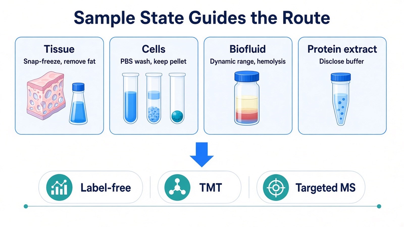
Figure 2. Tissue, cell, biofluid, and protein-extract constraints should be fixed before a label-free, TMT, or targeted MS route is locked.
Antibody methods remain appropriate when a single known protein and a validated antibody already answer the biological question. They do not generate a proteome-wide quantitative matrix and they do not remove the need for intact, consistently processed protein. If the target list is already closed, parallel reaction monitoring (PRM) or multiple reaction monitoring (MRM) should be planned as a separate assay, including peptide selection and matrix-matched validation. Researchers can refer to Advantages and Disadvantages of MRM/PRM Techniques for how those targeted routes are judged after names exist.
Researchers who already have defined samples, comparison groups, or a specific project objective can also review the MtoZ Biolabs Quantitative Proteomics Service for project-specific feasibility and analysis planning. Unlabeled discovery routes can also be reviewed through the Label-Free Quantitative Proteomics Service when sample number and group design favor that acquisition path.
Frequently Asked Questions
1. Do BCA or Bradford results replace LC-MS/MS protein quantification?
A bulk dye assay estimates total protein so that digestion or gel loading can be sized. It does not identify proteins and it does not report protein-to-protein ratios across a proteome. Use it as an input quality check. Treat LC-MS/MS peptide signals as the quantitative result of the proteomics experiment.
2. How should a limited sample be handled if it is near the planning amount?
Send the real wet weight, cell count, or volume, plus storage history and buffer details, before the sample is consumed in a non-MS assay. Low input increases missingness and can block TMT channel balance or PTM enrichment. A feasibility review can decide whether to proceed with discovery quantification, a shorter targeted panel, or additional material.
3. Can a protein solution containing SDS be submitted for quantification?
SDS lysates are common in extraction and incompatible with direct LC-MS/MS injection. The detergent has to be depleted or exchanged, and the buffer recipe has to be disclosed so that cleanup can be planned. A protein solution shipped without composition information delays method assignment and can waste limited material.
4. When is high-abundance protein depletion useful in plasma or serum?
Depletion is useful when the biological question sits below the abundant plasma proteins and enough volume exists to spend on depletion. It is less useful when albumin or other depleted proteins are themselves of interest, or when the remaining volume would fall below a workable digest. The depletion protocol must be the same for every group.
5. What project information is needed before sample shipment?
Species, sample type, group labels, biological replicates, the comparison that must be tested, available amount, storage temperature, freeze-thaw history, and any buffer or detergent already added. For protein extracts, a full buffer list is required. For plasma, the anticoagulant and hemolysis status should be stated.
MtoZ Biolabs can provide quantitative proteomics analysis for tissues, cells, biofluids, and protein extracts, including unlabeled, TMT, and targeted workflows matched to sample state and study design.
If further evaluation of sample amount, buffer compatibility, depletion needs, or method choice is required, project details can be submitted for a feasibility review.
