






Phage Display Antibody Analysis vs ELISA: Method Selection and Research Use Cases
Quick Answer
Use phage display antibody analysis when the next decision is about which binders are emerging from an antibody library, how clone enrichment changes across panning rounds, and which sequences merit further work. Use ELISA when the next decision is whether recovered candidates show antigen-binding confirmation in a defined plate-based assay and how to compare clones under the same assay conditions. In many antibody discovery programs, the methods fit best in the same screening workflow: phage display supports binder selection and sequence-informed candidate prioritization, while ELISA supports hit validation, clone ranking, and target specificity checks after clone recovery.
Why This Comparison Matters for Research Teams
Teams searching for phage display antibody analysis vs ELISA are usually not asking which method is better in the abstract. They are trying to avoid choosing the wrong tool for the next step.
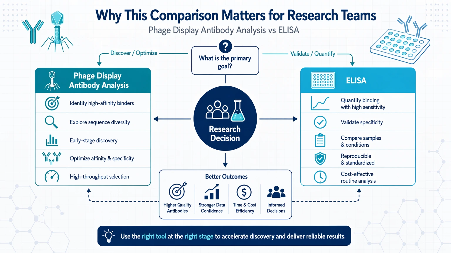
A discovery group may already have data from biopanning and need to know whether more sequence analysis is warranted before clone screening. Another team may already have monoclonal phage clone candidates or expressed antibodies and want a more direct path to antigen-binding confirmation. A translational assay group may also need to explain why ELISA alone cannot describe library behavior, display bias, or sequence diversity.
The practical question is straightforward: what decision comes next? If the next decision is about library-derived binder selection, phage display analysis is the more direct fit. If the next decision is about comparative binding signal in a standardized assay format, ELISA is usually the better fit.
What Phage Display Antibody Analysis Measures
Phage display antibody analysis addresses discovery-stage questions tied to an antibody library. It does not only ask whether binding occurred. It asks which binders were enriched, which clones recur, how diversity changes during selection, and whether the campaign is converging on a manageable candidate set.
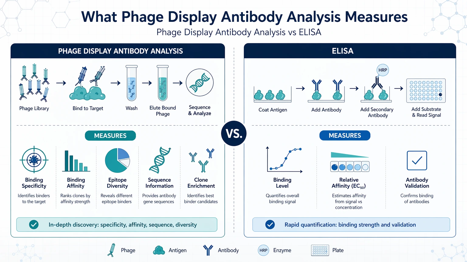
Typical questions answered by phage display analysis include:
- How did clone enrichment change across biopanning or panning rounds?
- Which monoclonal phage clone populations became dominant?
- Is the selected pool still diverse, or is it converging on a small set of related binders?
- Do repeated hits reflect genuine binder selection or display bias?
- Which sequences should advance to expression, clone screening, or affinity maturation?
This analysis often combines clone picking, sequence analysis, frequency tracking, and round-to-round comparison. The output is not a single binding signal. It is a framework for interpreting enrichment patterns, redundancy, sequence diversity, and candidate prioritization.
That distinction matters because phage display can show whether a selection campaign is producing many related clones with limited novelty or preserving multiple binder families. ELISA does not answer that library-level question on its own.
What ELISA Measures in Antibody Research
ELISA is a plate-based assay that detects and compares antigen binding under defined assay conditions. In antibody research, teams commonly use it after clone recovery or antibody expression to determine whether a candidate binds a recombinant antigen, whether target specificity can be separated from control conditions, and how clones compare by relative binding signal.
Typical questions answered by ELISA include:
- Does this candidate show antigen-binding confirmation against the coated antigen?
- How do several clones compare on the same assay plate?
- Is there clear separation between target signal and background signal?
- Does a clone behave similarly across control antigen, tagged antigen, or alternate antigen format conditions?
- Which candidates should advance to a follow-up binding assay or downstream functional assay?
ELISA is useful when the team needs standardized clone ranking across many wells, replicates, and controls. It is less informative when the team needs to understand why a clone appeared during selection, whether a sequence family is overrepresented, or whether a recovered hit reflects library bottlenecking rather than broad binder quality.
ELISA also depends strongly on assay format. Coating effects, antigen orientation, tag exposure, and plate-surface behavior can all shape the observed binding signal. A strong ELISA readout is informative, but it should not automatically be interpreted as a direct measure of affinity or developability.
Phage Display Antibody Analysis vs ELISA: Core Differences
The most useful comparison is based on the research question rather than the instrument or protocol.
The table below summarizes the main planning implications for the method choice.
| Comparison Point | Phage Display Antibody Analysis | ELISA |
|---|---|---|
| Primary purpose | Interpret binder selection from an antibody library | Confirm and compare binding in a plate-based assay |
| Typical workflow stage | Discovery and post-selection interpretation | Screening and validation after clone recovery |
| Input material | Library outputs, enriched pools, monoclonal phage clone sets, sequences | Expressed antibodies, phage-derived clones, purified or semi-purified samples |
| Main readout | Clone enrichment, sequence diversity, hit frequency, binder selection patterns | Binding signal, background signal, target specificity comparison |
| Strength | Explains selection behavior and supports candidate prioritization | Supports antigen-binding confirmation and clone ranking |
| Limitation | Does not by itself prove assay-format binding performance | Does not explain library composition or enrichment history |
| Common risk | Overinterpreting enrichment as proof of functional relevance | Overinterpreting signal intensity as proof of affinity |
Use these differences to align the analytical method with the biological question and validation plan.
In practical terms, phage display analysis explains what the selection campaign produced and how it changed over time. ELISA shows whether selected candidates bind under the assay conditions that were chosen.
When to Choose Phage Display Antibody Analysis
Choose phage display antibody analysis when the next project decision depends on understanding the output of selection rather than simply confirming that some binders exist.
This is often the better fit in the following situations:
1. You are still working at the antibody library level
If the project centers on naïve, immune, synthetic, or affinity-matured libraries, sequence-informed analysis is often more useful than immediate plate screening. At this stage, the main question is whether panning rounds are converging productively or narrowing in a misleading way.
2. Clone enrichment matters more than a single positive signal
A candidate that appears repeatedly across rounds may deserve attention, but repeated recovery can also reflect selection artifacts or display bias. Phage display analysis helps separate frequency patterns from simple yes-or-no binding calls.
3. You need sequence diversity to guide candidate prioritization
If ten apparent hits collapse into two related sequence families, lead selection may change. You may keep representatives from each family instead of ranking clones only by screening signal, which helps preserve diversity before downstream optimization.
4. The program will move into affinity maturation
When affinity maturation is planned, early sequence analysis adds context for which parent binders to retain. An ELISA-first path may miss sequence families that deserve engineering attention even if their initial binding signal is not the strongest.
5. You need to explain the selection campaign internally or to an external partner
Platform selection often has to be justified. Library history, hit frequency, and clone redundancy are easier to interpret and defend when phage display analysis has already clarified what the campaign enriched.
If your team is deciding whether to continue sequence-level interpretation before clone screening, submit your requirements to MtoZ Biolabs to evaluate a screening workflow that matches the actual decision point rather than treating phage display and ELISA as interchangeable steps.
When ELISA Is the Better Fit
ELISA is the better fit when the library-selection question has already been answered well enough and the next decision is about comparative binding performance in a defined assay.
1. You already have a manageable set of candidates
Once the project has been narrowed to recovered clones or expressed antibodies, ELISA often becomes the more direct route for hit validation. It can show which clones produce a reproducible binding signal against the target and which are dominated by background signal or nonspecific binding.
2. You need antigen-binding confirmation against a specific antigen format
For many teams, the immediate question is simple: does the candidate bind the recombinant antigen used in the next phase of work? ELISA is well suited to that question, especially when target and control conditions are included on the same plate.
3. You need clone ranking under standardized conditions
ELISA supports side-by-side comparison across many clones, concentrations, or replicate wells. That makes it useful for narrowing a long list into a smaller set for orthogonal binding assay work or downstream functional testing.
4. The translational assay team is not working from an active library campaign
Some groups inherit candidates after discovery. They may not need additional phage display-derived interpretation if binder selection history is already clear. In that case, ELISA may be the more direct option for confirming target specificity and screening behavior.
5. You need a practical bridge to the next assay
ELISA is often used as a handoff assay before more detailed characterization. It does not replace sequence analysis when sequence-level decisions are still open, but it can efficiently filter candidates before more resource-intensive follow-up.
One limitation remains: ELISA readouts are shaped by antigen format. A coated peptide, a tagged recombinant antigen, and a folded ectodomain may not produce the same clone ranking. That is why plate design and control selection matter as much as the assay itself.
When to Combine Both Methods in One Workflow
In many programs, the strongest choice is not phage display antibody analysis or ELISA alone. It is a staged workflow that reduces decision risk.
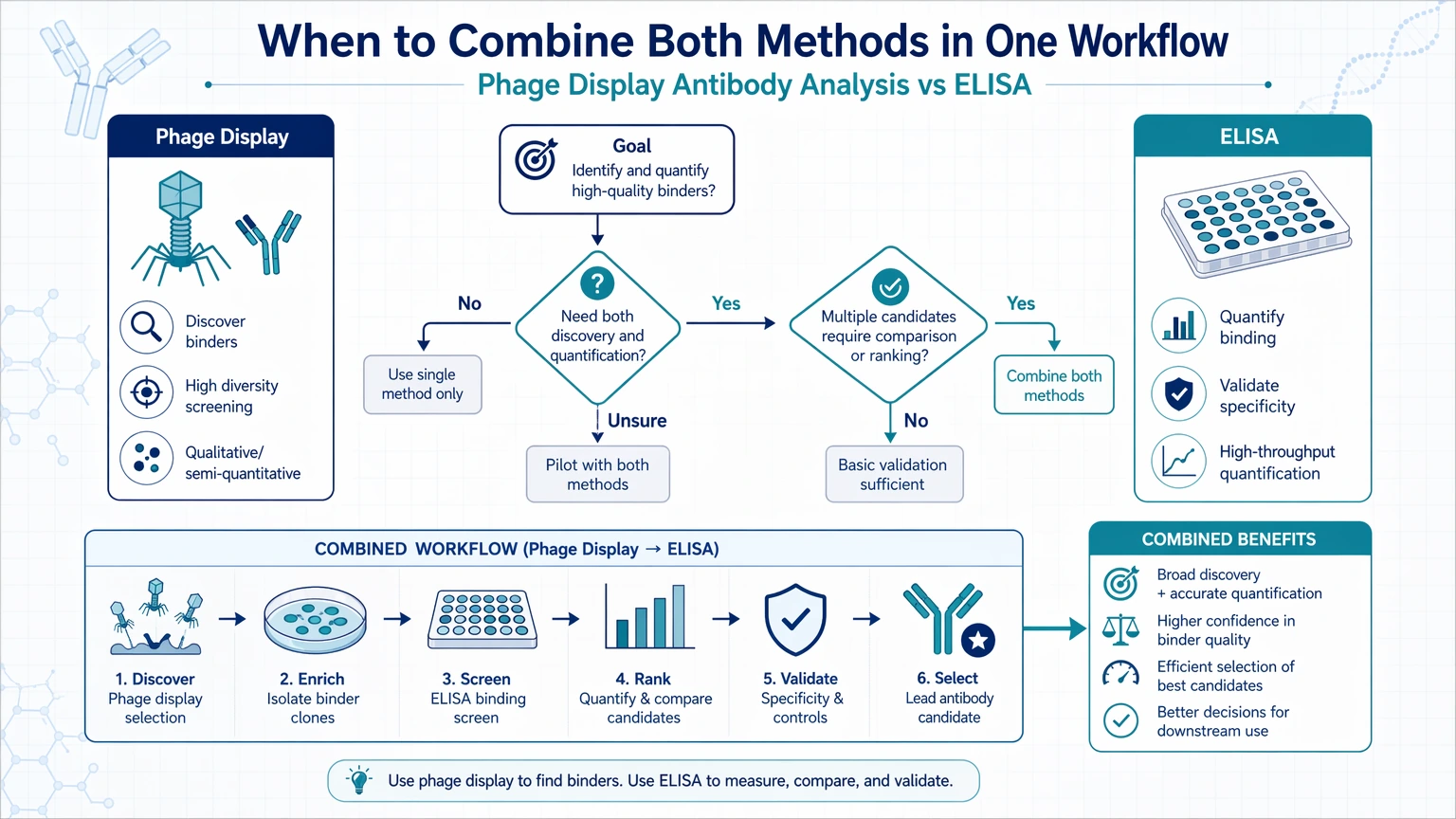
A common sequence is:
1. Run phage display selection against the target. 2. Track clone enrichment across panning rounds. 3. Recover monoclonal phage clone candidates and analyze sequence diversity. 4. Prioritize representatives from enriched and distinct sequence families. 5. Use ELISA for antigen-binding confirmation and target specificity comparison. 6. Advance selected candidates to expression studies, affinity maturation, or a downstream functional assay.
This combined approach helps avoid two common mistakes.
The first is moving to ELISA too early and screening a biased or redundant clone set without understanding library composition. That can create a shortlist dominated by near-duplicate binders.
The second is stopping after phage display analysis and assuming enrichment alone is enough for hit validation. A sequence that expands during selection still needs confirmation in a defined binding assay.
This is often the point where outside support becomes useful. Teams can contact us at MtoZ Biolabs to evaluate a project workflow when the bottleneck is in phage display screening interpretation, candidate prioritization, or ELISA follow-up design before the next handoff.
Method Selection Checklist for Antibody Discovery Teams
Use the checklist below to choose the next method based on the decision directly in front of you.
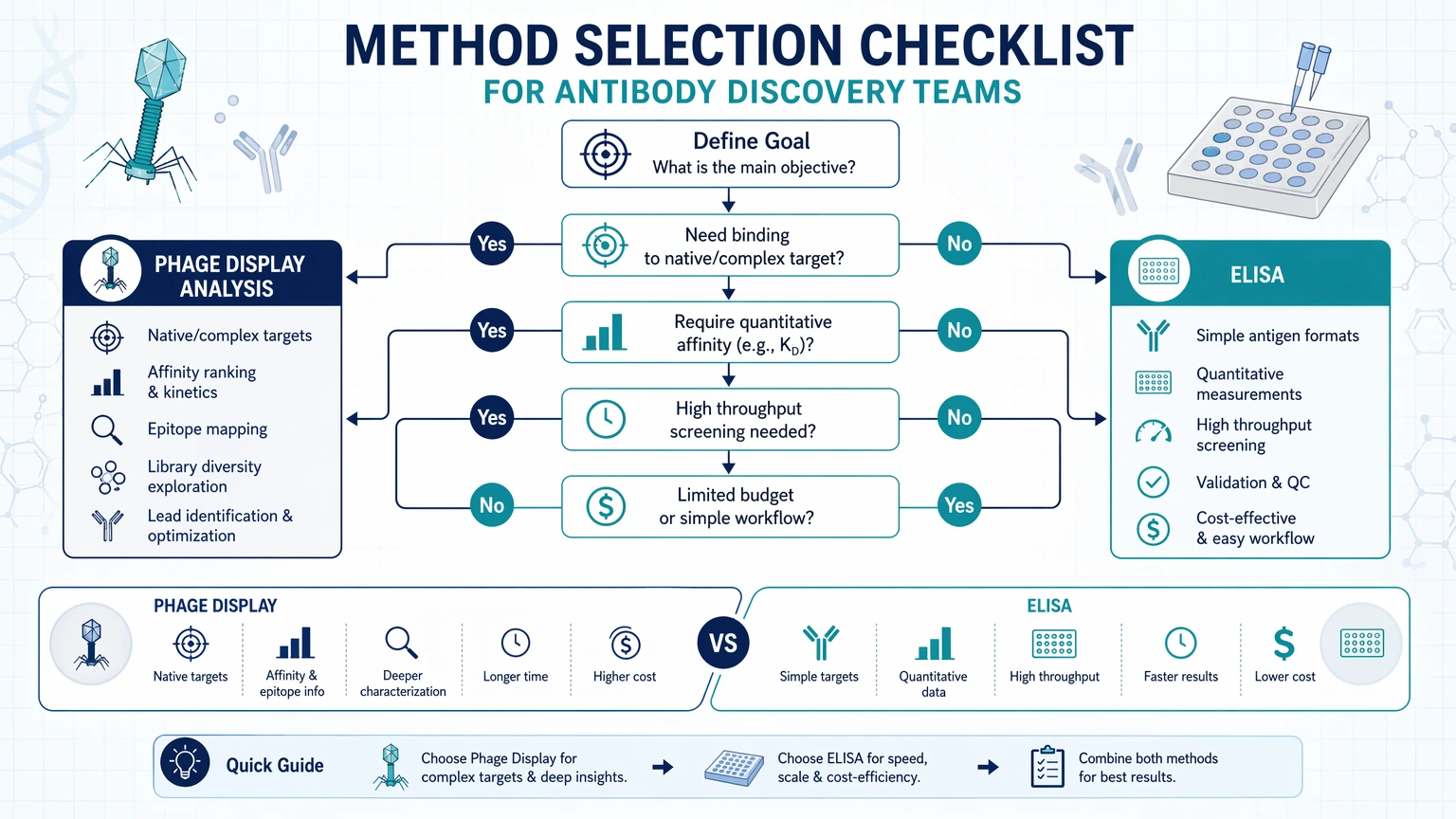
Choose phage display antibody analysis first if:
- Your main material is still an antibody library or enriched phage pool
- You need to interpret panning rounds rather than only test recovered hits
- Clone enrichment and hit frequency will influence selection strategy
- Sequence diversity is needed for lead selection
- You suspect display bias or overrepresentation of similar clones
- Candidate prioritization depends on sequence-informed grouping
Choose ELISA first if:
- You already have recovered clones or expressed antibodies
- The next question is antigen-binding confirmation
- You need comparative clone ranking on the same plate
- The team needs target specificity checks against controls
- The required output is binding signal, not library interpretation
- You are preparing for a downstream functional assay and need an initial filter
Use both in sequence if:
- You need to move from binder selection to hit validation
- You want to preserve sequence diversity before narrowing by assay signal
- The shortlist must be supported by both enrichment logic and assay data
- Your recombinant antigen is compatible with a meaningful plate-based assay
- Internal stakeholders need confidence that candidate selection was not driven by one readout alone
Service Routes for Study Planning
For teams moving from method selection into execution, these service paths connect assay design, validation, and interpretation needs.
Conclusion
Phage display antibody analysis and ELISA answer different research questions, so they should not be treated as automatic substitutes. Phage display is the stronger choice when the decision concerns binder selection from an antibody library, clone enrichment across panning rounds, sequence diversity, and candidate prioritization. ELISA is the stronger choice when the decision concerns antigen-binding confirmation, target specificity, background signal control, and clone ranking in a plate-based assay.
The methods often work best together. Phage display analysis reduces uncertainty about what the library produced. ELISA reduces uncertainty about which selected candidates perform in a defined screening format. If your workflow is stalled between those two decision points, map the research question, input material, and downstream consequence of a wrong call before choosing the assay. If you need project-planning support, submit your requirements so the workflow can be aligned with your antigen format, clone set, and selection stage.
FAQ
Is phage display antibody analysis a substitute for ELISA?
Usually not. Phage display analysis explains selection outcomes from an antibody library, while ELISA tests binding behavior in a defined assay format. A team can postpone one method in some workflows, but substituting one for the other often leaves a gap in either library interpretation or hit validation.
Can ELISA rank candidate binders without sequence-level context?
Yes, ELISA can rank clones by relative binding signal, but that ranking may be incomplete. If several wells represent sequence-redundant binders, the apparent top performers may reflect one dominant family rather than broad sequence diversity. Sequence context helps determine whether the screen captured unique options or repeated versions of the same binder class.
What does phage display analysis reveal that ELISA usually does not?
It can show clone enrichment trends, redundancy across monoclonal phage clone picks, sequence diversity, and possible display bias across panning rounds. Those outputs are directly relevant when the team is deciding which binders to carry forward from library selection.
Can a strong ELISA signal be used as a direct affinity readout?
Not on its own. ELISA signal is influenced by coating density, antigen orientation, wash conditions, detection setup, and background signal. It is useful for comparative screening and antigen-binding confirmation, but affinity claims usually require a more appropriate follow-up method.
When is recombinant antigen format most likely to distort ELISA interpretation?
Risk increases when the coated antigen differs substantially from the target’s native presentation, when purification tags alter epitope exposure, or when adsorption to the plate changes conformation. Under those conditions, ELISA can still support screening, but target specificity and clone ranking should be interpreted with the format constraint in mind.
What is a practical minimum workflow after successful panning rounds?
A common minimum path is to review clone enrichment and sequence diversity after selection, recover representative candidates, and then run ELISA-based hit validation against target and control conditions. That sequence provides both selection context and assay confirmation before more resource-intensive follow-up studies.
How to order?




