



The mass spectrometry PRM quantification method is a closed-panel targeted workflow that isolates scheduled peptide precursors, fragments them, and records high-resolution product-ion spectra for quantitative extraction on liquid chromatography-tandem mass spectrometry (LC-MS/MS) (Peterson et al. 2012). Parallel reaction monitoring (PRM) is not another name for selected reaction monitoring (SRM). SRM and multiple reaction monitoring (MRM) pass one predefined product ion per dwell on a triple quadrupole (Lange et al. 2008). PRM stores the product-ion spectrum so fragments can be reviewed after acquisition (Gallien et al. 2012). The method review below covers workflow, locked parameters, limits, and project fit. It is not an environmental or food-residue methods page, not a biomarker discovery brochure, and not a claim that PRM is automatically more precise than SRM in every matrix.
PRM quantification tables support relative change of predefined peptides across frozen groups. Amount on named peptides needs stable isotope-labeled internal standard (SIS) peptides and a calibration model in the study matrix (Gerber et al. 2003). Full-scan product-ion recording supplies richer fragment evidence. It does not remove matrix suppression or create a diagnostic cutoff by itself (Bantscheff et al. 2012).
In MtoZ Biolabs targeted offerings, the mass spectrometry PRM quantification method is scoped when the peptide panel is closed and high-resolution scheduling fits the cohort block. Service intake appears in Parallel Reaction Monitoring (PRM) Service.
What the Mass Spectrometry PRM Quantification Method Covers
Four method blocks belong in the brief and stay separated.
Sample block covers matched extraction, protein amount check when required, digestion, and cleanup to an injection-ready peptide load.
Method-development block locks precursor m/z values, retention windows, isolation width, collision energy, resolution, and fill-time rules on matrix-matched digests.
Acquisition block injects study and quality control (QC) samples through a locked liquid chromatography (LC) gradient into scheduled PRM MS/MS.
Informatics block extracts fragment chromatograms, reviews interference, applies relative normalization or SIS calibration, and tests only predefined contrasts.
The PRM quantification method remeasures a frozen shortlist with parallel product-ion spectra. It does not equal SRM naming, and it does not profile unnamed proteins from the same file.
PRM Quantification Versus SRM/MRM Quantification Methods
Method choice follows list status and fragment-review need.
SRM and MRM quantification methods lock transitions before acquisition. Throughput depends on dwell time and peak width. PRM quantification locks precursors and reviews fragments after acquisition when matrix-matched SRM ratios remain unstable. Route choice after matrix pilot appears in Parallel Reaction Monitoring vs MRM: Which Fits?.
Calling PRM "selective reaction monitoring" mixes two method names. Selected reaction monitoring is the SRM/MRM transition method. Parallel reaction monitoring is the high-resolution product-ion method.
Discovery data-dependent acquisition (DDA) or data-independent acquisition (DIA) quantifies an open or semi-open proteome. The PRM quantification method starts after candidates are named and versioned. PRM technology framing appears in PRM-Targeted Proteomics Technology.
Post-translational modification (PTM) quantification in PRM requires the modified peptide on the closed list and, for many sites, enrichment. Unenriched PRM files do not map PTM occupancy by default.
End-to-End Mass Spectrometry PRM Quantification Workflow
A practical PRM quantification method spine usually runs as follows.
- Freeze contrast, replicates, and relative versus amount claim.
- Close the peptide panel and map each peptide to its protein or site.
- Match extraction, digest, and load across all contrast arms.
- Develop precursor m/z, retention windows, and collision energies on matrix-matched digests.
- Lock isolation width, resolution, fill-time rules, and cycle time versus peak width.
- Size biological n plus QC, blanks, and SIS if used.
- Acquire interleaved study and QC injections on the locked LC-MS/MS method.
- Extract product-ion chromatograms; review interference across multiple fragments.
- Normalize or apply SIS calibration as declared; test predefined contrast only; archive method and software versions.
Steps 1 through 3 are sample and panel blocks. Steps 4 through 9 are method development, acquisition, and quantification blocks.
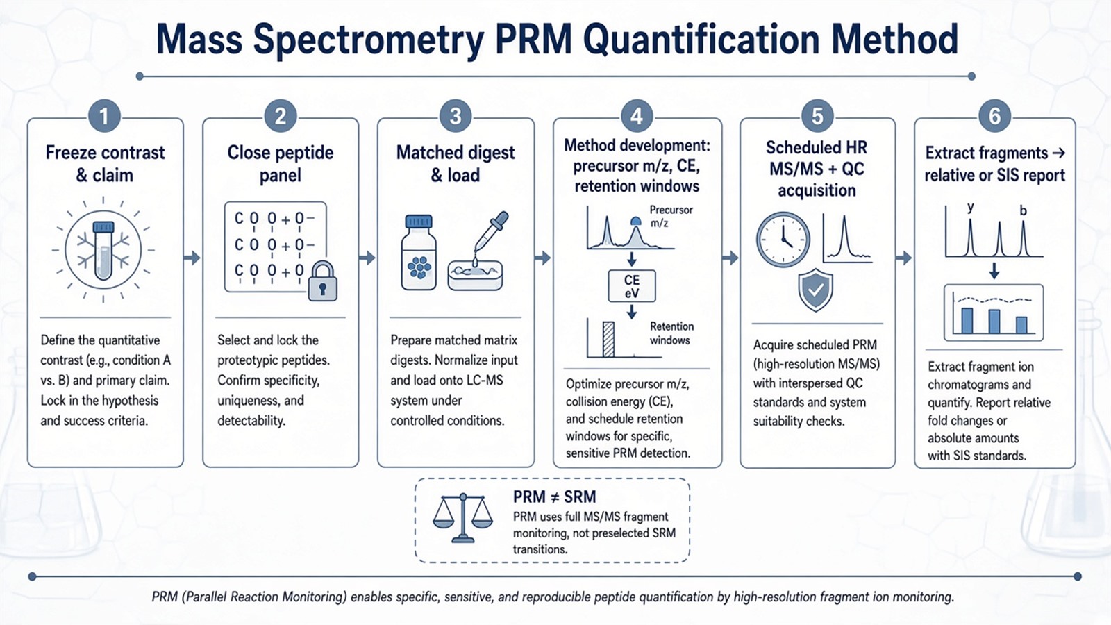
Figure 1. PRM quantification depends on matrix-matched method development and parallel product-ion extraction before group contrasts are tested.
Critical Parameters in the PRM Quantification Method
Layer |
Parameter that must be declared |
Failure mode if ignored |
|---|---|---|
Naming |
PRM versus SRM/MRM method name |
Wrong scan mode in the record |
Panel |
Peptide list version and protein mapping |
Moving targets mid cohort |
Precursor |
m/z, charge, retention window |
Missed or wrong isolation |
MS/MS |
Collision energy, resolution, fill time |
Unstable fragment chromatograms |
Schedule |
Cycle time versus peak width |
Undersampled peaks |
Prep |
Matched extraction and digest |
Peptide-pool shift as biology |
Load |
Mass on column from pilot |
Saturation or dropped precursors |
SIS |
Spike step aligned to claim |
Relative peaks read as concentration |
QC |
Pools, blanks, and bracket pattern |
Undetected batch drift |
Software |
Integration version archived |
Non-reproducible peaks |
Limits follow the same table. The PRM quantification method needs high-resolution LC-MS/MS performance and fragment-level review capacity. That is a resource cost, not a free accuracy upgrade over every SRM method. Capturing all fragment ions does not by itself raise precision when prep or chromatography drifts. Specialized software is required, but software choice does not replace locked contrast design. Biomedical, environmental, and food applications outside peptide proteomics use different analyte classes and claim units.
How to Choose the PRM Quantification Method Versus Other Routes
Use list status, method-development outcome, claim unit, and instrument context together.
Project need |
Route that often fits |
Recheck before committing |
|---|---|---|
Closed peptides, need post-acquisition fragment review |
Mass spectrometry PRM quantification |
Force SRM without interference evidence |
Closed peptides, stable SRM/MRM transitions |
SRM or MRM quantification |
Whole-panel PRM without need |
Open proteome relative profiling |
Discovery DDA or DIA first |
PRM on unnamed proteins |
Amount on named peptides |
PRM with SIS and curve |
Relative peaks as concentration |
Method transfer, same matrix |
Acquisition plus QC |
Assume free method rebuild |
PTM site amount question |
Modified-peptide PRM plus enrichment design |
Unenriched PRM as site occupancy |
Biomarker validation intent |
Closed PRM panel plus validation design |
PRM table as clinical test |
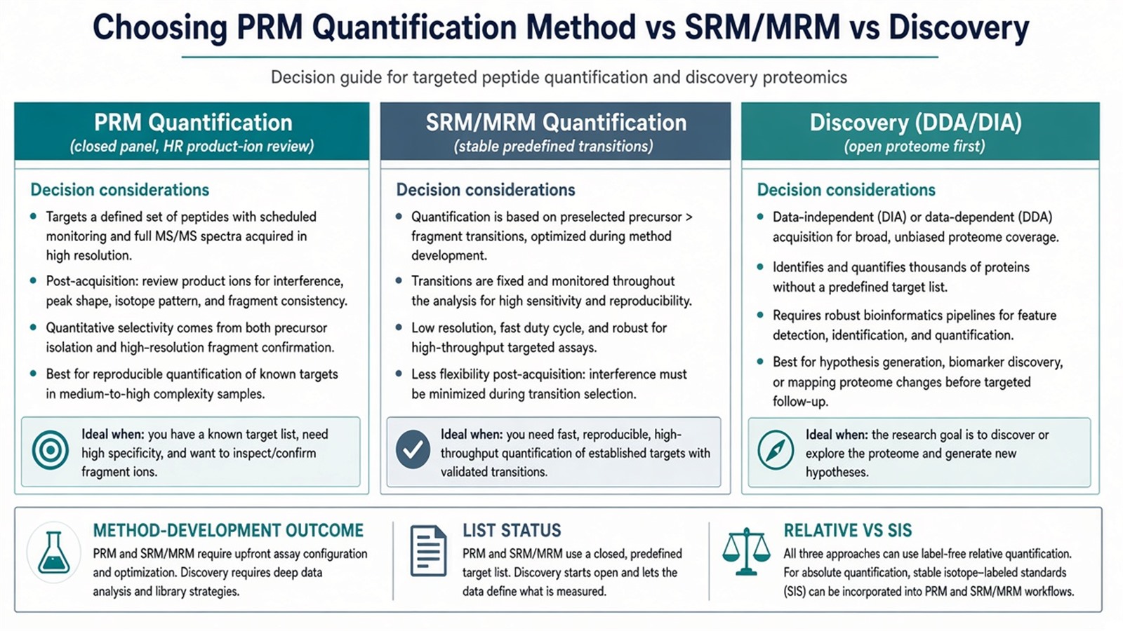
Figure 2. Choose the PRM quantification method when a closed panel needs high-resolution product-ion review; keep SRM/MRM or discovery when list status or transitions say so.
Teams with closed peptide targets, method-development notes, and contrast maps can review MtoZ Biolabs Parallel Reaction Monitoring (PRM) Service when those fields are declared in the project record.
Frequently Asked Questions
1. Is the PRM quantification method the same as SRM?
No. SRM monitors predefined transitions. PRM records high-resolution product-ion spectra for scheduled precursors. The two names should not be treated as synonyms in the methods record.
2. Does full-scan product-ion detection make PRM always more precise than SRM?
No. Fragment richness helps interference review. Precision still depends on matched prep, locked LC, scheduling, and QC. Matrix-matched pilots decide which method fits.
3. What does method development include beyond picking m/z values?
Method development locks retention windows, collision energy, isolation width, resolution, fill-time rules, and cycle time on matrix-matched digests, not only neat-standard m/z lists.
4. Must every PRM quantification project use SIS peptides?
No. Relative contrasts can run light-only when that claim is declared. SIS and a curve become necessary when amount units are required.
5. When should projects stay on SRM/MRM quantification?
When matrix-matched transitions are already stable on a triple quadrupole and post-acquisition fragment review is not required, SRM or MRM remains the matching quantification method.
MtoZ Biolabs can discuss mass spectrometry PRM quantification method scoping when peptide panel, method parameters, and contrast design are documented.
If further evaluation of PRM versus SRM/MRM, SIS calibration, or discovery-to-PRM handoff is needed, project details can be submitted for a feasibility review.
Reference
- A.C. Peterson, J.D. Russell, A.A. Klammer, P.F. Doubleday, H. Liu, J.N. Anderson, M.J. MacCoss (2012). Parallel reaction monitoring for high resolution and high mass accuracy quantitative, targeted proteomics. Mol. Cell. Proteomics, 11, 1475-1488. https://doi.org/10.1074/mcp.O112.020271
- S. Gallien, E. Duriez, A. Crone, M. Kellmann, T. Moehring, T. Domon (2012). Targeted proteomic quantification on quadrupole-orbitrap mass spectrometers. Mol. Cell. Proteomics, 11, 1709-1723. https://doi.org/10.1074/mcp.O112.019232
- V. Lange, P. Picotti, B. Domon, R. Aebersold (2008). Selected reaction monitoring for quantitative proteomics: a tutorial. Mol. Syst. Biol., 4, 222. https://doi.org/10.1038/msb.2008.61
- S.A. Gerber, J. Rush, O. Stemman, M.W. Kirschner, S.P. Gygi (2003). Absolute quantification of proteins and phosphoproteins from cell lysates by tandem MS. Proc. Natl. Acad. Sci. U.S.A., 100, 6940-6945. https://doi.org/10.1073/pnas.0832254100
- M. Bantscheff, S. Lemeer, H.U. Haab, B. Kuster (2012). Quantitative mass spectrometry in proteomics: a critical review. Anal. Bioanal. Chem., 404, 939-965. https://doi.org/10.1007/s00216-012-5892-4
