






Lysosomal Proteomics in Neurodegenerative Disease Research
Introduction
Neurodegenerative disease projects often reach a familiar design question: if lysosomal function is impaired, which proteins in lysosome-enriched fractions actually change. A team studying Alzheimer's disease models may need more than bulk brain or cell proteomes. Another team working on Parkinson's or related neurodegeneration hypotheses may need organelle-focused cargo evidence before selecting follow-up targets.
Lysosomal proteomics in neurodegenerative disease research addresses that need by measuring proteins from lysosome-enriched preparations and comparing them across disease, genotype, or treatment conditions. This article explains what the approach can support in neurodegeneration and Alzheimer's-related research designs, how studies should be planned, and which interpretation limits keep conclusions research-appropriate.
Why Neurodegeneration Studies Use Lysosomal Proteomics
Many neurodegeneration hypotheses involve lysosomal clearance, autophagy-lysosome pathway stress, or altered handling of disease-related proteins. Bulk proteomics can miss or dilute organelle-restricted changes. Lysosomal proteomics concentrates the measurement on lysosome-enriched material so disease-linked protein differences are more likely to be visible.
In practical project terms, lysosomal proteomics neurodegeneration studies are used to:
- inventory proteins detected in lysosome-enriched fractions from disease models
- compare lysosomal proteome profiles between disease and control conditions
- rank candidate proteins for later orthogonal follow-up
- connect organelle-level protein changes to autophagy or clearance hypotheses
These outputs support research design and mechanism exploration. They do not by themselves establish clinical diagnosis or disease classification.
Where Alzheimer's and Related Research Designs Fit
Lysosomal proteome Alzheimer's disease research typically asks whether lysosome-enriched protein composition changes in cellular or tissue models that reflect disease-relevant stress. Similar logic applies to other neurodegeneration models when lysosomal dysfunction is part of the hypothesis.
Useful study frames include:
- disease versus control lysosome-enriched proteomes in human, mouse, or rat models
- genotype or mutation contrasts that affect lysosomal function
- treatment or rescue conditions expected to remodel lysosomal protein cargo
- autophagy-related interventions where lysosome-associated protein changes are the readout
In each case, the claim should stay at the level of lysosome-enriched protein evidence from the chosen model system.
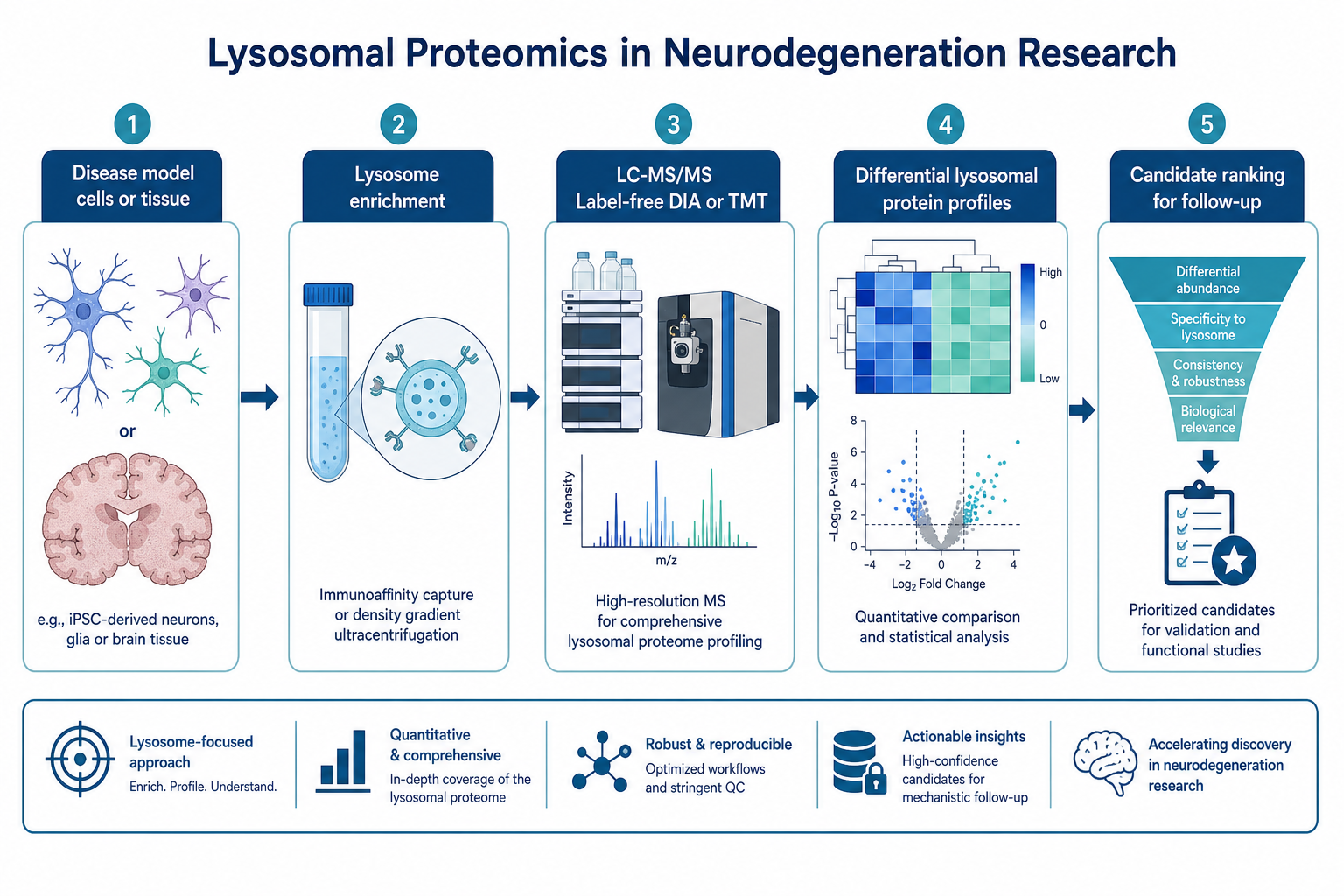
Figure 1. Neurodegeneration-focused lysosomal proteomics links disease-model enrichment to LC-MS/MS comparison and candidate ranking.
Study Design Considerations for Neurodegeneration Projects
Define an organelle-level claim first
State whether the project needs cargo inventory, differential lysosomal proteome comparison, or candidate discovery for follow-up. A clear claim prevents the study from drifting into bulk proteome interpretation.
Choose starting material that matches enrichment capacity
Cell lines, primary cells, fresh tissue, and frozen tissue can enter a lysosome enrichment and proteomics path. Human, mouse, and rat systems are routinely considered. Other species should be assessed case by case.
If the only available materials are serum, plasma, CSF, or urine, lysosome-component separation must already be completed by the client before submission. Enrichment from those fluids is outside the standard laboratory enrichment path.
Match enrichment across comparison arms
Disease and control arms should share matched lysosome enrichment chemistry and handling. Unequal enrichment efficiency can create false neurodegenerative signatures.
Select quantification after cohort structure is clear
Label-free, DIA, and TMT are all supported for lysosome-enriched proteomics. Label-free fits flexible pilots. DIA fits broader comparative cohorts. TMT fits predefined multiplexed group maps.
Plan interpretation boundaries early
Standard analysis can include differential protein screening plus GO, KEGG, and PPI views. Lysosome-specific annotation packages beyond those standard outputs are not part of the default analysis set. Membrane and luminal proteins are not reported as separately resolved classes.
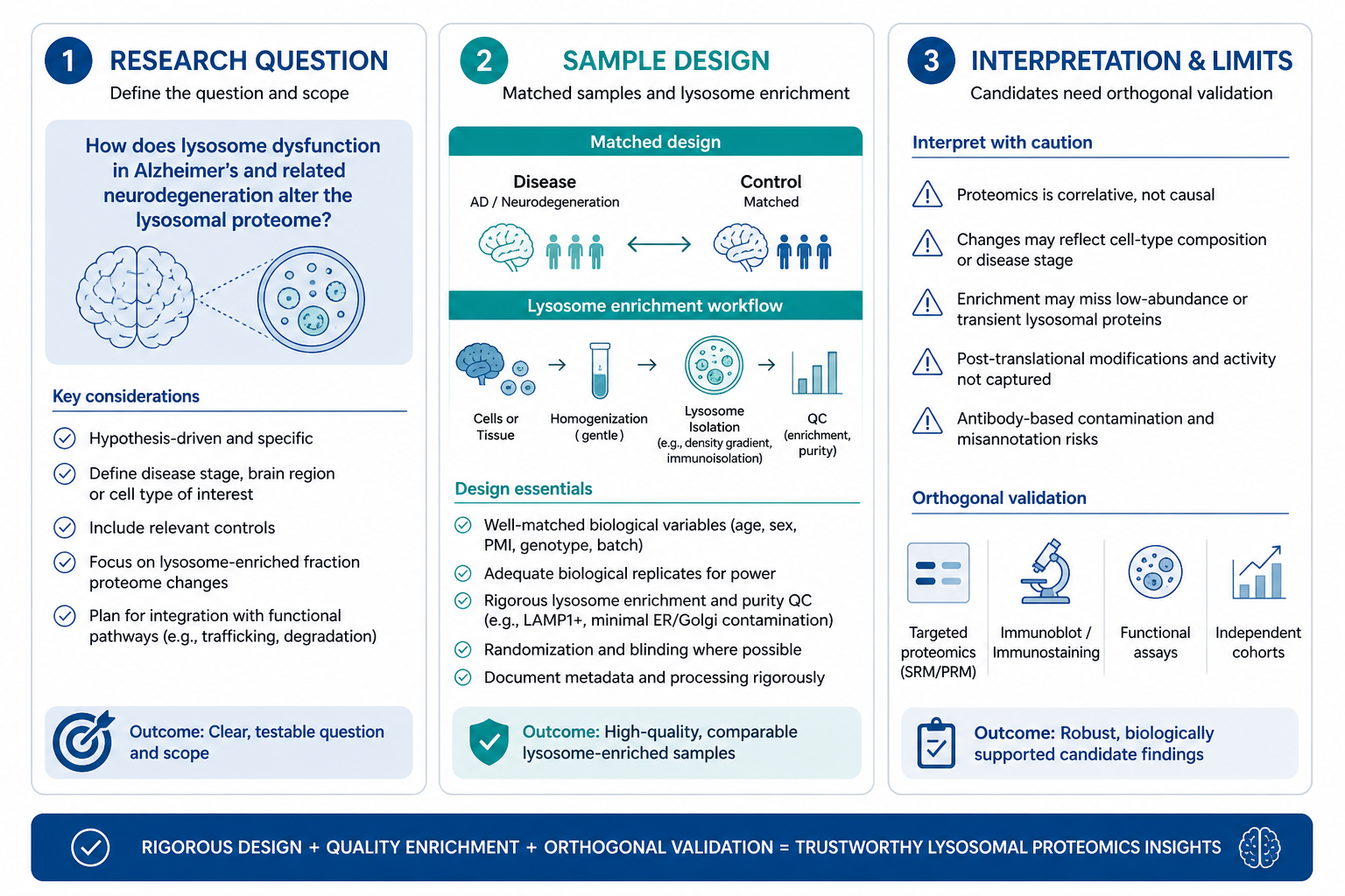
Figure 2. Strong neurodegeneration designs lock the research question, matched enrichment plan, and interpretation limits before LC-MS/MS.
Sample and QC Planning for Disease-Model Studies
Suggested planning amounts include about 1 x 10^7 cells per sample, 20 to 50 mg tissue per sample, or 20 to 50 ug protein from client-enriched lysosome fractions. Store samples at −80°C and ship on dry ice. Avoid repeated freeze-thaw and mass spectrometry-incompatible additives.
After enrichment, Western blot positivity for lysosome markers such as LAMP1 or LAMP2 is a practical readiness check before omics. Expanding pre-omics organelle-contamination panels is not recommended as a default requirement. If localization context is needed later, subcellular localization analysis can be considered after proteomics.
Low-abundance disease-relevant lysosomal proteins may still be difficult to detect. Enrichment improves detection chance but does not guarantee recovery of every priority target.
What Results Support, and What They Do Not
Research value
Lysosomal proteomics can reveal disease-associated changes in lysosome-enriched protein profiles.
It can prioritize candidates for mechanism follow-up in Alzheimer's and other neurodegeneration models.
It can support pathway-oriented discussion through standard GO, KEGG, and PPI outputs.
Under favorable conditions, cell-derived projects may reach about 1,000 or more proteins, and tissue-derived projects may reach about 3,000 or more proteins. These ranges are planning references, not fixed guarantees.
Limits that must stay explicit
Candidate proteins from discovery designs remain exploratory until orthogonal validation is completed.
Proteomics results from disease models should not be framed as diagnostic markers or clinical decision tools on their own.
Downstream experimental validation such as Western blot is not an automatic continuous package after proteomics and should be scoped separately when needed.
Lysosome colocalization imaging and TEM ultrastructural validation are outside the proteomics service path described here.
Related Services
Mitochondrial Proteomics Services
Mitochondrial Autophagy Analysis Service
What to Prepare Before Starting a Neurodegeneration Lysosomal Proteomics Project
Assemble the following before inquiry:
- disease model and one-sentence lysosomal protein claim
- species and sample type: cells, tissue, or client-enriched fractions
- group design: disease versus control, genotype, or treatment
- enrichment status and available marker QC plan
- approximate sample amount and storage history
- preferred quantification mode: Label-free, DIA, or TMT
- whether standard differential analysis plus GO, KEGG, and PPI outputs are sufficient
MtoZ Biolabs supports lysosomal proteomics analysis for neurodegeneration-focused research designs, including lysosome enrichment for suitable cell and tissue inputs, LC-MS/MS, and bioinformatics interpretation. The technical team can help determine whether an organelle-enriched proteomics design fits the current Alzheimer's or broader neurodegeneration question better than bulk proteomics.
To plan lysosomal proteomics for neurodegenerative disease research, contact MtoZ Biolabs with your model system, enrichment status, group design, and the lysosome-level protein changes you need to measure.
Frequently Asked Questions
Can lysosomal proteomics support Alzheimer's disease research?
Yes, as a research tool for lysosome-enriched protein profiling in relevant cellular or tissue models. It supports candidate discovery and comparative analysis, not standalone clinical diagnosis.
Is bulk brain or cell proteomics enough for lysosomal questions?
Not when the hypothesis depends on organelle-restricted protein changes. Lysosome enrichment proteomics is usually the better fit for those claims.
Which species are routinely suitable?
Human, mouse, and rat systems are routinely considered. Other species should be assessed separately.
Which quantification methods can be used?
Label-free, DIA, and TMT are supported for lysosome-enriched neurodegeneration study designs.
Do discovery results confirm disease biomarkers?
No. Discovery candidates require orthogonal validation and independent study design before stronger biological or translational claims.
Conclusion
Lysosomal proteomics is a practical route for neurodegenerative disease research when the question centers on lysosome-enriched protein changes rather than bulk proteome composition. In Alzheimer's and related model systems, it can inventory organelle-associated proteins, compare disease-linked profiles, and rank candidates for follow-up.
Project value depends on matched enrichment, clear research claims, and restrained interpretation. Teams designing neurodegeneration-focused lysosomal proteomics can review model systems and quantitative options with MtoZ Biolabs before locking the analytical path.
How to order?




