


How to Identify Accessory Proteins of Ion Channels by Mass Spectrometry
-
channel expression level compared with the endogenous system
-
localization at the plasma membrane or relevant organelle
-
tag accessibility and lack of obvious trafficking disruption
-
treatment timing, dose, and pathway response
-
availability of matched negative and positive controls
Introduction
Ion channels rarely act alone. Channel gating, trafficking, localization, degradation, pharmacology, and downstream signaling can all depend on accessory proteins. These partners may include regulatory subunits, scaffolding proteins, chaperones, trafficking factors, kinases, phosphatases, and membrane-associated adaptors. The challenge is that many ion channel accessory proteins are weakly bound, condition-dependent, low abundance, or sensitive to detergent extraction. A standard pull-down may recover the channel but lose relevant partners. A poorly controlled mass spectrometry experiment may also return abundant contaminants rather than a useful interaction map.
Mass spectrometry can solve this problem when the workflow is designed around membrane biology. The goal is not only to identify proteins in the same sample. The goal is to distinguish likely channel-associated partners from background proteins, expression artifacts, and extraction- dependent noise. A strong ion channel mass spectrometry project therefore starts with the bait, cell state, membrane-preserving enrichment strategy, controls, and validation plan before LC- MS/MS analysis begins.
Related Services
| Research Need | Recommended Service Direction |
| Identify ion channel-associated proteins by LC-MS/MS | MS-Based Protein-Protein Interaction Analysis Service |
| Analyze broader membrane protein interaction networks | Protein-Protein Interaction Analysis Service |
| Improve bait-centered quantification and background filtering | SILAC Based Co-IP-MS for Protein Interaction Analysis Service |
| Confirm membrane protein identity in complex samples | Membrane Protein Identification Service |
| Evaluate protein-protein interface or conformational effects | Protein-Protein Interactions Characterization Service | HDX-MS |
For projects where channel-associated partners are difficult to recover or rank, MtoZ Biolabs can help evaluate whether affinity-MS, native-cell crosslinking, membrane protein identification, HDX- MS, or a combined protein-protein interaction analysis workflow fits the research question.
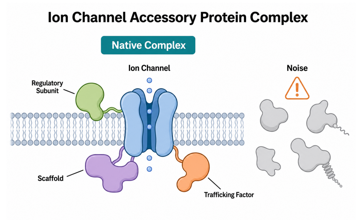
Figure 1. Ion channel accessory proteins should be separated from nonspecific background
Why Ion Channel Accessory Proteins Are Hard to Identify
Ion channels are challenging bait proteins for several reasons. Many channels are multi-pass membrane proteins with hydrophobic regions, glycosylation, subunit assembly requirements, and compartment-specific maturation. Some partners interact only during channel trafficking. Others bind after voltage change, ligand exposure, phosphorylation, drug treatment, or cellular stress. If cells are lysed with harsh detergent, a real membrane-associated complex can dissociate before enrichment.
Another problem is abundance. A regulatory protein may interact with only a small fraction of the total channel pool. A chaperone may bind during a short folding window. A scaffold may contact the channel only at a specific membrane domain. At the same time, MS analysis can detect abundant cytoskeletal proteins, heat shock proteins, ribosomal proteins, and sticky membrane- associated proteins. These proteins may be real in some contexts, but they can also appear as recurring background.
For this reason, a useful experiment should not ask, "Which proteins were detected?" It should ask which proteins are enriched with the channel under the correct condition, reproducible, control-depleted, and biologically plausible.
Step 1: Define the Channel Bait and Cellular State
The first decision is whether to study the endogenous channel or a tagged channel construct. Endogenous capture can preserve native expression, but it requires a high-quality antibody and enough channel abundance. A tagged construct can improve enrichment, but tag position, expression level, and localization must be checked. For ion channels, an N-terminal or C-terminal tag can affect trafficking, gating, or assembly, so construct validation is not optional.
Researchers should also define the cell state before sample preparation. Accessory proteins may differ between resting and stimulated cells, immature and surface-localized channels, untreated and drug-treated samples, or wild-type and mutant channels. If the goal is to identify accessory proteins involved in ligand response, voltage-dependent regulation, channel trafficking, or disease mutation effects, the sample condition must match that biology.
Practical pre-checks include:
Step 2: Choose a Membrane-Preserving Enrichment Strategy
Co-IP-MS is a common starting point for ion channel accessory protein discovery. The bait channel is enriched by antibody or affinity tag, then associated proteins are identified by MS. The method works best when the complex survives lysis, solubilization, bead capture, washing, and digestion. Mild detergent screening is often needed because detergent choice can determine whether partners stay associated or disappear.
Some projects benefit from native-cell crosslinking before lysis. A cell-permeable crosslinker can stabilize close protein contacts before detergent disrupts the membrane environment. This approach is useful for weak, transient, or condition-dependent membrane protein interactions. Crosslinking should be optimized carefully because excessive reaction strength can reduce protein recovery or increase nonspecific links.
Other workflows may combine fractionation, affinity capture, and quantitative proteomics. Surface-enriched fractions can reduce intracellular background when the target is a plasma membrane channel. Stable isotope labeling or label-free quantification can help rank candidates by enrichment over controls. The best strategy depends on channel abundance, antibody quality, detergent sensitivity, and expected interaction strength.
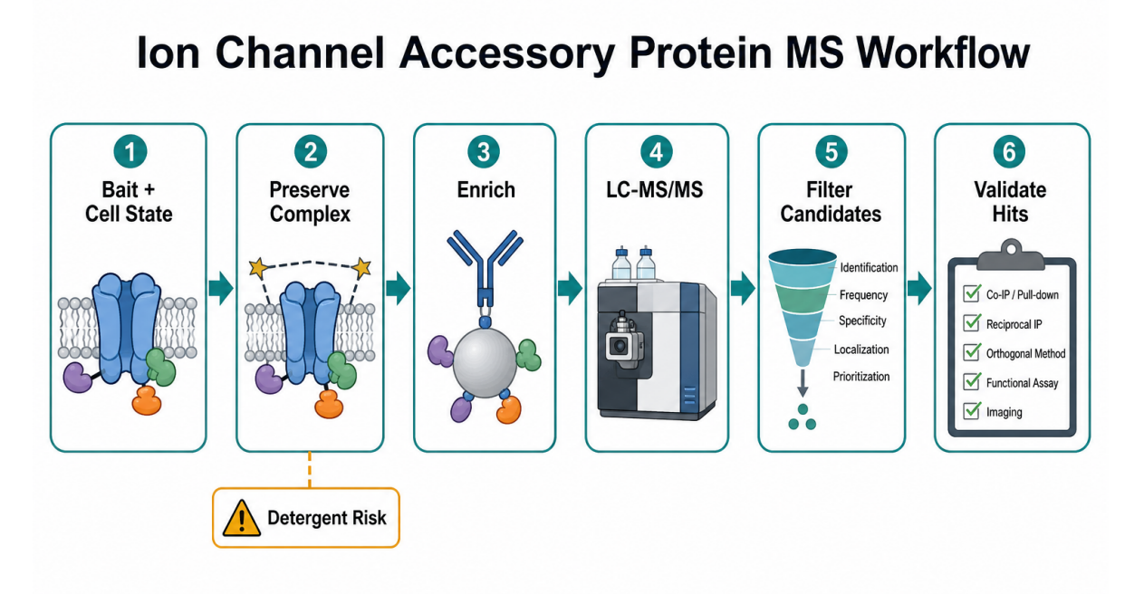
Figure 2. Workflow for identifying ion channel accessory proteins by mass spectrometry
Step 3: Build Controls That Remove Background
Controls determine whether a candidate list is interpretable. For a tagged channel, common controls include tag-only, empty-vector, unrelated membrane protein, and mock-transfected samples. For endogenous capture, IgG or isotype controls are often needed. Bead-only controls may help when resin background is high. Input lysate or membrane fraction controls show whether the channel and candidate partners were present before enrichment.
The control set should match the main failure mode:
| Risk | Useful Control | What It Tests |
| Tag-specific background | Tag-only or empty- vector control | Proteins binding the tag or expression system |
| Antibody background | IgG or isotype control | Proteins binding antibody or beads |
| Detergent-sensitive loss | Mild detergent comparison | Whether partners survive extraction |
| Condition- independent binding | Untreated or vehicle control | Whether enrichment depends on the tested state |
| Low reproducibility | Biological replicates | Whether candidates repeat across preparations |
Biological replicates are more important than technical repeats for interaction claims. A candidate detected once may be a true partner, but it should not be treated as high confidence until enrichment, reproducibility, localization, and validation support the result.
Step 4: Analyze MS Results Without Overcalling Hits
The output usually includes peptide-spectrum matches, protein identifications, quantitative values, and missing values. For ion channel mass spectrometry, the key task is to rank candidates by evidence, not by detection alone. A high-confidence accessory protein should show control- based enrichment, replicate reproducibility, plausible localization, and a connection to channel biology.
Researchers should be cautious with proteins that appear in many affinity purification experiments. Cytoskeletal proteins, chaperones, ribosomal proteins, mitochondrial proteins, and highly abundant metabolic enzymes can be true partners in selected cases. However, these proteins also appear frequently as background. Filtering should be evidence-based rather than automatic. A protein should be deprioritized because the control data, localization, or replicate behavior suggest background.
Quantitative analysis can use label-free intensity, spectral counting, TMT, SILAC, or targeted MS depending on project design. Statistical cutoffs should be paired with biological review. A modestly enriched trafficking factor may be more relevant than a highly abundant contaminant with poor localization.
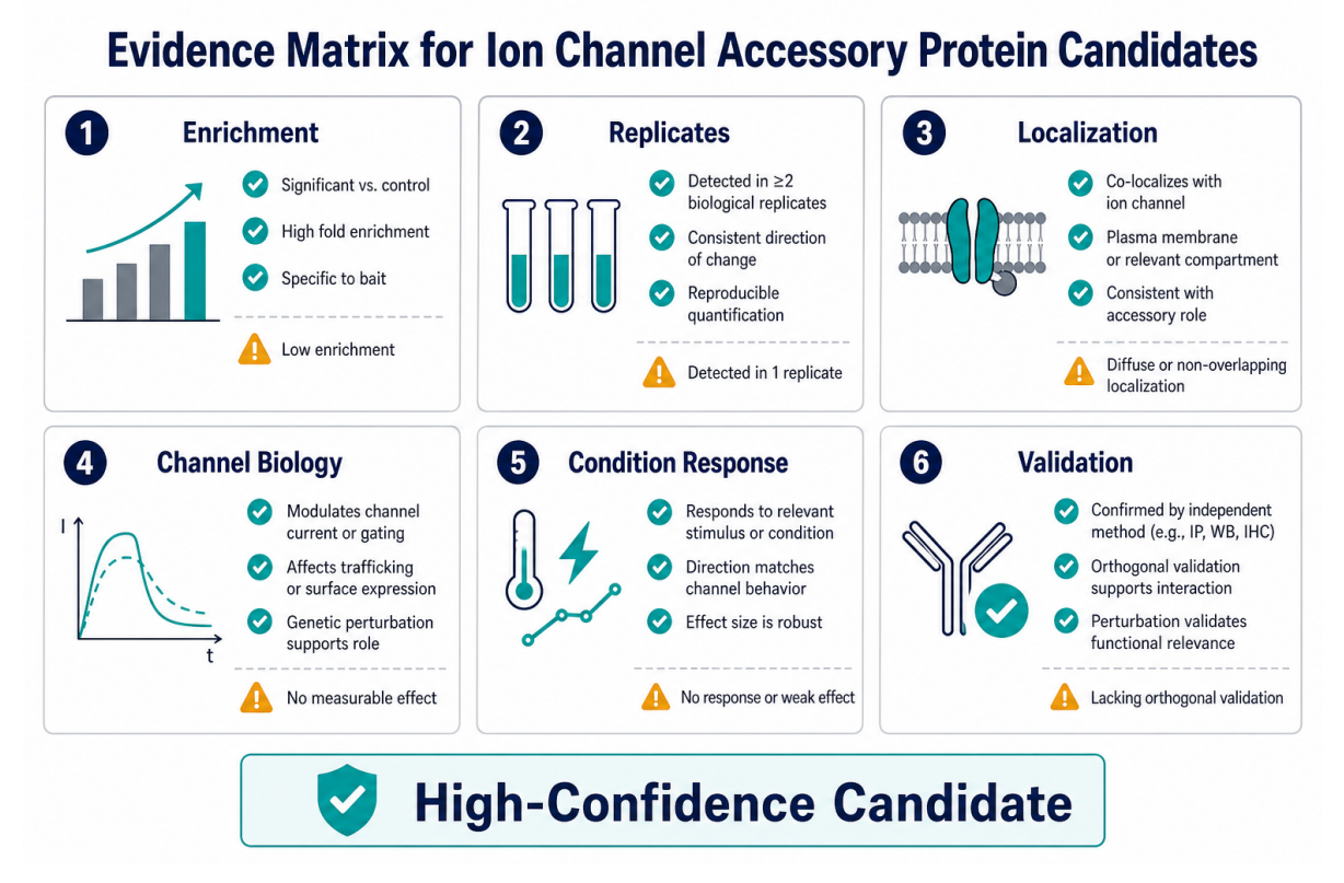
Figure 3. Evidence matrix for ranking ion channel accessory protein candidates
Step 5: Validate Candidate Accessory Proteins
Mass spectrometry identifies candidates. Validation turns candidates into stronger biological claims. The validation method should match the proposed role. If the candidate may form a channel complex, reciprocal IP, Co-IP, pull-down, or proximity assays may help. If the candidate may regulate trafficking, imaging, surface biotinylation, or fractionation can test localization effects. If the candidate may regulate function, electrophysiology or ion flux assays may be needed.
Useful validation options include:
| Candidate Role | Validation Direction |
| Complex member | Reciprocal IP, Co-IP, proximity assay |
| Trafficking factor | Imaging, surface expression assay, fractionation |
| Gating regulator | Electrophysiology, ion flux assay, perturbation |
| Scaffold or adaptor | Mutagenesis, domain mapping, pathway perturbation |
| Condition-specific partner | Treated vs control validation and time-course testing |
Validation should focus on prioritized candidates rather than every detected protein. A practical shortlist may include proteins that meet enrichment, replicate, localization, and biological criteria.
Common Mistakes to Avoid
One common mistake is using harsh lysis to improve solubility while destroying the interaction of interest. Solubilization must be balanced against complex preservation. Another mistake is relying on an overexpressed tagged channel without checking localization or expression level. A channel trapped in the ER may identify folding proteins, not surface-channel regulators.
A third mistake is treating all detected proteins as accessory proteins. Mass spectrometry is sensitive, and sensitivity can increase background. The strongest interpretation comes from controls, quantitative enrichment, biological replicates, and orthogonal validation. Researchers should also avoid making direct binding claims from affinity purification MS alone. This method supports association, while direct physical binding usually requires additional evidence.
Frequently Asked Questions
1. Can mass spectrometry identify unknown ion channel accessory proteins?
Yes. Mass spectrometry can identify candidate ion channel accessory proteins when channel enrichment, membrane preservation, controls, and replicate design are strong. Candidates should be validated before mechanistic claims are made.
2. Is affinity-MS enough for ion channel interaction discovery?
Affinity-MS is often a useful starting point, especially for stable complexes. Weak, transient, or detergent-sensitive partners may require native-cell crosslinking, optimized detergent conditions, or complementary validation methods.
3. What controls are most important?
Important controls include IgG or isotype controls, tag-only or empty-vector controls, input samples, untreated or vehicle controls, and biological replicates. The best control depends on bait design and expected background source.
4. How should candidate accessory proteins be ranked?
Candidates should be ranked by enrichment over controls, reproducibility, localization, known or plausible channel biology, condition response, and validation feasibility. Detection alone is not enough.
5. Can MS results prove that an accessory protein regulates channel function?
No. MS results can identify candidate partners and support association. Functional regulation should be tested with electrophysiology, ion flux assays, trafficking assays, perturbation experiments, or other orthogonal methods.
Conclusion
Identifying ion channel accessory proteins by mass spectrometry requires more than a sensitive MS run. The project must preserve relevant membrane complexes, control background, quantify enrichment, and validate prioritized candidates. Affinity-MS can identify stable channel-associated proteins. Native-cell crosslinking and optimized membrane workflows can help capture weak or condition-dependent partners.
For ion channel projects with low recovery, long contaminant lists, or unclear candidate ranking, contact MtoZ Biolabs to discuss whether affinity-MS, membrane protein identification, native-cell crosslinking, HDX-MS, or a combined interaction analysis workflow is appropriate for the study design.
How to order?

