


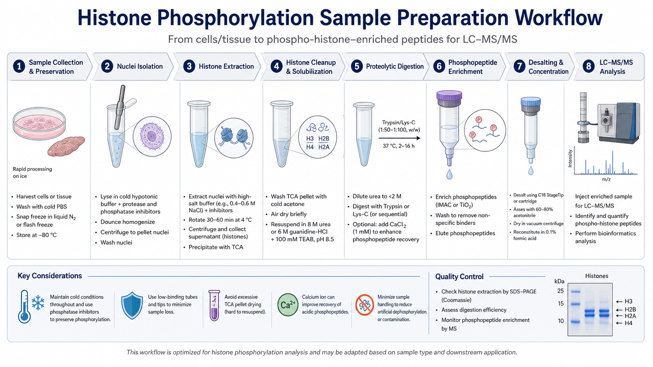
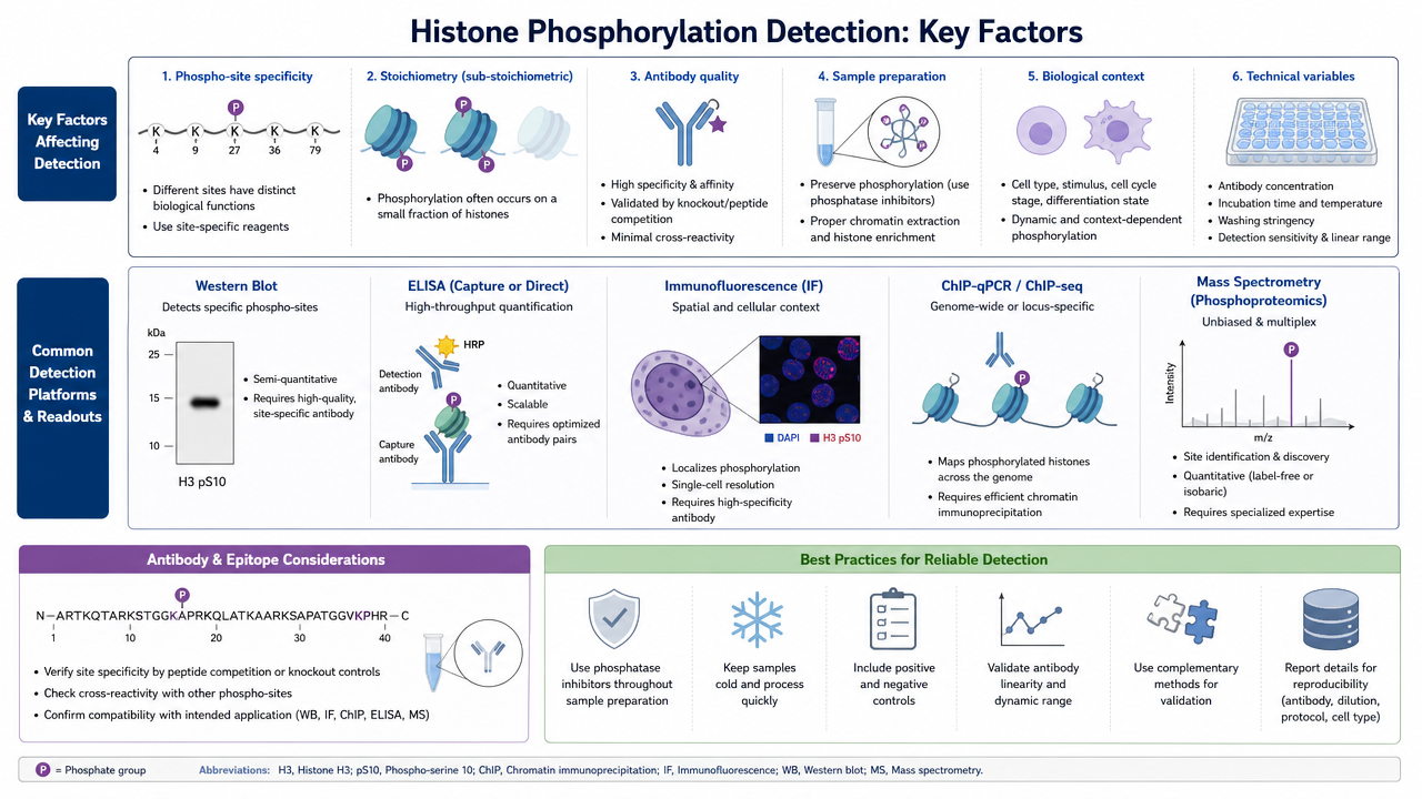
Histone Phosphorylation Detection: Sample Preparation Methods That Improve Sensitivity
- Inhibitors at lysis; work at 4°C.
- Acid extraction improves histone purity.
- Propionylation improves peptide length for MS.
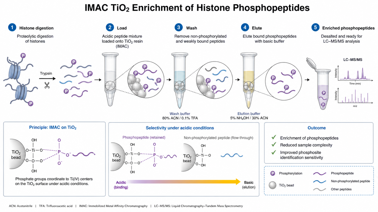
- Fractionation before IMAC or TiO2 increases depth.
- Align MS mode (HCD, ETD, DIA) with prep design.

Histone phosphorylation detection depends on sample preparation: phosphatase control, histone enrichment, derivatization, digestion, fractionation, and matched phosphopeptide enrichment.
Key Takeaways
Related Services
Histone Phosphorylation Analysis Service
Histone Phosphorylation Analysis
Serine/Threonine Phosphoproteomics Analysis Service
Histone Isolation and Enrichment Service
Acid Extraction and Derivatization
Acid extraction enriches histones; propionylation limits over-cleavage at lysines for better phosphosite localization.
MS Coordination
Use adequate input; minimize handling time; tune IMAC or TiO2 conditions.
Conclusion
Systematic sample preparation, not a single step, drives histone phosphorylation detection sensitivity.
Submit Inquiry
How to order?

