





Histone Acetylation: Key Sites, Marks, and Proteomics Analysis
Histone acetylation modifies lysine residues on histone proteins, neutralizing their positive charge and influencing chromatin interactions. Histones H3 and H4 contain multiple acetylation sites, and some histone-derived peptides may carry more than one acetylated lysine. LC-MS/MS analysis helps identify acetylated histone peptides, localize modification sites when sufficient fragment-ion evidence is available, and characterize combinations of acetylation sites within individual peptides.
For histone acetylation identification, site localization, and relative quantification, researchers can review the MtoZ Biolabs Histone Acetylation Analysis service for sample suitability and study planning.
What Is Histone Acetylation?
Lysine Acetylation on Histone Tails
Histone acetylation involves the covalent addition of an acetyl group (+42.0106 Da) to the ε-amino group of lysine. Many well-characterized acetylation sites are located within the lysine-rich N-terminal tails of histones H3 and H4. Acetylation also occurs on histones H2A and H2B and at selected residues within histone globular domains. Individual modifications are designated by the histone and lysine position, such as H3K9ac or H4K16ac.
Acetylation and Deacetylation in Histone Regulation
Histone acetyltransferases transfer acetyl groups from acetyl-CoA to lysine residues, while histone deacetylases remove these modifications (Kouzarides 2007). Their activities contribute to changes in histone acetylation patterns across cellular states and in response to experimental treatments, including deacetylase inhibition.
By neutralizing the positive charge of lysine, acetylation can reduce electrostatic interactions between histones and DNA. Acetylated lysines can also be recognized by reader domains, including bromodomains, which participate in the recruitment of chromatin-associated regulatory proteins. These mechanisms connect histone acetylation with chromatin organization and transcriptional regulation.
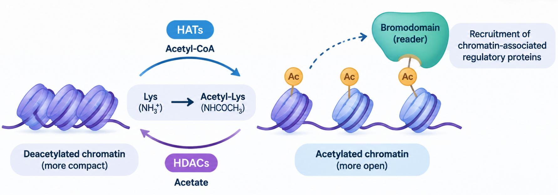
Figure 1. Histone Acetylation and Deacetylation by HATs and HDACs.
Common Histone Acetylation Sites and Their Research Contexts
Histone acetylation is commonly studied at lysine residues on histones H3 and H4. The following table summarizes frequently investigated sites and their reported associations with gene regulation and chromatin organization.
|
Acetylation Site |
Reported Research Findings |
|---|---|
|
H3K9ac |
Associated with active promoters and enhancers. |
|
H3K14ac |
Associated with active promoters and enhancers; also found at some inactive inducible promoters. |
|
H3K18ac |
Enriched near active gene transcription start sites; can stimulate MLL1/MLL4-mediated H3K4 methylation in vitro. |
|
H3K27ac |
Distinguishes active enhancers from poised enhancers; also found at active promoters. |
|
H4K5ac |
Together with H4K12ac, facilitates CENP-A deposition at centromeres. |
|
H4K8ac |
Associated with active gene promoters and NSL-complex-mediated transcription initiation. |
|
H4K12ac |
Together with H4K5ac, facilitates CENP-A deposition at centromeres. |
|
H4K16ac |
Can inhibit higher-order chromatin compaction and inter-fiber interactions in vitro. |
How Are Histone Acetylation Sites Identified by Mass Spectrometry?
Histone Peptide Preparation for Acetylation Analysis
Histone tails are rich in lysine and arginine residues. Conventional trypsin digestion can therefore generate short peptides, limiting the coverage of some histone modification sites. One established approach uses propionic anhydride to derivatize lysine residues with reactive amino groups before trypsin digestion. This blocks trypsin cleavage after these residues, directing digestion primarily to the C-terminal side of arginine and producing longer histone peptides for LC-MS/MS analysis (Garcia et al. 2007).
Unlabeled acetic anhydride is unsuitable for this purpose because it introduces the same +42.0106 Da mass shift as endogenous lysine acetylation, making the two indistinguishable by mass alone. Propionic anhydride introduces a different mass shift on derivatizable lysines, allowing endogenous acetylation to remain distinguishable (Huang et al. 2015). When endogenous histone propionylation is also an analytical target, isotopically labeled propionic anhydride can distinguish naturally occurring propionyl groups from those introduced during sample preparation (Huang et al. 2015). For projects requiring histone extraction before derivatization, see the Histone Isolation and Enrichment Service.
MS/MS Evidence for Acetylated Lysine Sites
Lysine acetylation (+42.0106 Da) and trimethylation (+42.0470 Da) differ in mass by approximately 0.0364 Da. High-resolution accurate-mass measurements can distinguish these modifications when sufficient resolving power and mass accuracy are achieved for the precursor ion. Orbitrap LC-MS/MS platforms, such as Fusion Lumos and Exploris 480, can support this distinction under suitable acquisition conditions.
MS/MS fragmentation provides additional evidence for modification identification. Zhang et al. (2004) reported an acetyllysine-derived immonium ion at m/z 126.091 and a 59 Da neutral loss corresponding to trimethylamine from trimethylated lysine. These features can help distinguish acetylated and trimethylated lysine residues under suitable fragmentation conditions, but their detection depends on peptide sequence, modification position, and fragmentation behavior. They should therefore be treated as supporting evidence rather than universal identification criteria.
Localization to a specific lysine residue requires site-determining fragment ions that distinguish competing modification positions, particularly when multiple lysines are present in the same peptide.
When modification identity or site localization cannot be confidently established, the assignment should be reported with the corresponding uncertainty rather than attributed to a specific residue without sufficient evidence. For projects that also require histone methylation identification, see the Histone Methylation Analysis Service.
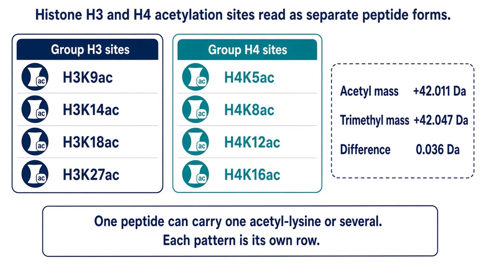
Figure 2. H3 and H4 tail lysines tracked as separate acetyl forms, and the 0.036 Da difference between acetylation and trimethylation.
What Can Histone Acetylation Proteomics Reveal?
Histone acetylation proteomics provides site-specific information about histone acetylation patterns and their changes across biological conditions. LC-MS/MS analysis can help researchers investigate:
- Histone Acetylation Sites and Patterns: Identify acetylated histone residues, including commonly studied sites on histones H3 and H4, and characterize their modification patterns.
- Changes in Acetylation Across Conditions: Compare relative acetylation levels between control and treatment groups, disease models, or different time points to identify condition-associated changes.
- Histone Acetylation Heterogeneity: Examine differences in acetylation combinations across histone peptides, helping investigate the diversity of histone modification patterns.
Together, these findings provide valuable molecular insights into histone modification patterns and their changes, supporting research into epigenetic regulation, chromatin dynamics, and the molecular mechanisms associated with gene expression.
For relative quantification of histone acetylation changes across experimental groups, researchers can review the MtoZ Biolabs Histone PTMs Quantitative Analysis Service for study planning.
Frequently Asked Questions
A drug treatment raises an H3K27ac Western blot signal. What should the peptide data add?
An H3K27ac Western blot provides information about a predefined acetylation mark. LC-MS/MS can identify additional acetylated sites on histones H3 and H4. In a quantitative study, changes in H3K27ac-associated peptide signals can be evaluated alongside total histone H3 abundance to help distinguish modification-related changes from differences in histone protein levels.
Is the acetyl peptide with the largest signal the most important site?
No. A stronger MS signal does not necessarily indicate greater biological importance or higher modification abundance, because peptide response can vary with sequence, modification state, and analytical conditions. The relevance of an acetylation site depends on the research objective, its biological context, and any reproducible changes observed across experimental groups.
Can several acetyl groups on one peptide be summed into a single percent acetylated?
No. Different acetylation patterns represent distinct modification states, and summing their peptide signals does not directly provide the percentage of acetylated histone molecules. Quantifying the fraction of molecules carrying a specific modification pattern requires a dedicated quantitative strategy with appropriate normalization and validation.
Conclusion
Histone acetylation proteomics identifies acetylated lysine residues on histones H3, H4, and other core histones through LC-MS/MS analysis. Site-specific acetylation profiles and confidently identified multi-acetylated peptides provide information about modification patterns within histone regions. These results can support investigations of chromatin regulation and help identify acetylation sites for further study.
If you are investigating histone acetylation and its changes across biological conditions, MtoZ Biolabs provides mass spectrometry-based histone PTM analysis to support acetylation site identification and relative quantitative comparisons. Explore our Histone Post-Translational Modification Analysis Service or contact our team for a free consultation about your samples and research objectives.
Reference
- T. Kouzarides (2007). Chromatin modifications and their function. Cell, 128, 693-705. https://doi.org/10.1016/j.cell.2007.02.005
- K. Zhang, P.M. Yau, B. Chandrasekhar, R. New, R. Kondrat, B.S. Imai, M.E. Bradbury (2004). Differentiation between peptides containing acetylated or tri-methylated lysines by mass spectrometry: an application for determining lysine 9 acetylation and methylation of histone H3. Proteomics, 4, 1-10. https://doi.org/10.1002/pmic.200300503
- B.A. Garcia, S. Mollah, B.M. Ueberheide, S.A. Busby, T.L. Muratore, J. Shabanowitz, D.F. Hunt (2007). Chemical derivatization of histones for facilitated analysis by mass spectrometry. Nat. Protoc., 2, 933-938. https://doi.org/10.1038/nprot.2007.106
- H. Huang, S. Lin, B.A. Garcia, Y. Zhao (2015). Quantitative proteomic analysis of histone modifications. Chem. Rev., 115, 2376-2418. https://doi.org/10.1021/cr500491u
How to order?