






Antibody Glycosylation in Autoimmune Diseases
Introduction
Autoimmune diseases are often defined by autoreactive antibodies, yet antibody specificity alone does not fully explain pathogenic potential. Two IgG preparations may recognize the same antigen yet differ in inflammatory activity because of changes in Fc glycosylation. Research in rheumatoid arthritis, systemic lupus erythematosus, inflammatory bowel disease, and related conditions has shown that disease-associated IgG populations can carry distinct glycan profiles linked to effector function, immune complex behavior, and clinical activity.
Antibody glycosylation in autoimmune diseases refers to the study and characterization of carbohydrate structures on immunoglobulins, especially IgG, in the context of autoreactive or inflammation-associated immune responses. Fc N-glycans can modulate interactions with Fc receptors, complement components, and lectin pathways that shape inflammation and tissue damage. Glycan changes may appear on total IgG, antigen-specific autoantibodies, or disease-associated immunoglobulin subsets.
For immunology and translational research teams, understanding antibody glycosylation in autoimmune diseases helps explain why glycan profiling is increasingly used alongside autoantibody titer measurement, how glycan biomarkers may reflect disease activity, and which analytical approaches are needed to study patient-derived IgG.
What Antibody Glycosylation Means in Autoimmune Disease Context
In autoimmune disease research, antibody glycosylation focuses on how glycan structures on IgG and related immunoglobulins influence immune effector pathways associated with chronic inflammation and tissue injury. The most studied site on IgG is the conserved Fc N-glycosylation position at Asn297 on each heavy chain. Glycans at this site can alter Fc receptor binding, complement activation, and signaling through lectin and cytokine networks.
Autoimmune-associated IgG glycosylation is not a single uniform pattern. Different diseases, disease stages, and antibody specificities may show distinct glycan features. Some changes reflect broad shifts in total IgG glycosylation during active inflammation. Others may be enriched on autoreactive clones or immune complexes involved in organ-specific pathology.
Antibody glycosylation therefore adds a functional layer to autoantibody analysis. Measuring antigen binding alone may miss differences in inflammatory potential that arise from Fc glycan structure.
Why IgG Glycosylation Matters in Autoimmune Diseases
IgG is the dominant immunoglobulin class in many autoimmune responses and mediates effector functions through Fc receptor and complement pathways. Small glycan differences can shift those pathways toward pro-inflammatory or anti-inflammatory outcomes.
Glycosylation matters in autoimmune disease research for several reasons.
Fc effector modulation can influence immune complex clearance, macrophage activation, and tissue inflammation even when antigen specificity is unchanged.
Disease-associated glycan shifts have been reported in multiple conditions, suggesting that glycan profiling may complement conventional serology.
Therapeutic response monitoring may benefit when glycan biomarkers track inflammation or remission more sensitively than titer alone in selected settings.
Therapeutic antibody design in immunology also depends on glycan knowledge because Fc glycosylation can tune effector function in products intended to suppress or redirect immune activity.
Glycosylation does not explain all autoimmune pathology. Genetics, T cell help, complement regulation, and tissue microenvironment all contribute. However, Fc glycan structure is now recognized as an important modifier of IgG function in autoimmunity.
Common Glycan Changes Associated with Autoimmune IgG
Research across autoimmune diseases has identified several recurring glycan features on IgG populations.
Agalactosyl IgG
Reduced terminal galactose on Fc glycans, often described as agalactosyl IgG, has been repeatedly associated with active inflammation in conditions such as rheumatoid arthritis. Agalactosyl IgG may show altered Fc receptor interaction and complement-related behavior compared with fully galactosylated forms. Galactosylation status is therefore one of the most frequently studied glycan attributes in autoimmune serology research.
Sialylation changes
Terminal sialic acid on IgG Fc glycans has been linked to anti-inflammatory signaling in some experimental contexts. Reduced sialylation has been observed in inflammatory states and may contribute to pro-inflammatory effector profiles when present on pathogenic antibody populations. Because sialylated glycoforms can be low in abundance, sensitive glycan profiling methods are often required.
Core fucosylation variation
Core fucose content can modulate Fc gamma receptor IIIa binding and antibody-dependent cellular cytotoxicity. In autoimmune research, fucosylation changes have been explored in relation to effector cell activation and disease-associated IgG subsets. Fucosylation is also relevant when comparing patient IgG with therapeutically engineered antibodies designed for modified effector function.
High-mannose and broader glycoform shifts
Changes in high-mannose or hybrid glycan content have been reported in inflammatory and autoimmune settings. These shifts may reflect B cell activation state, cytokine environment, and glycosylation enzyme regulation during disease flares. Broader glycoform profiling helps capture changes that are not limited to one terminal sugar feature.
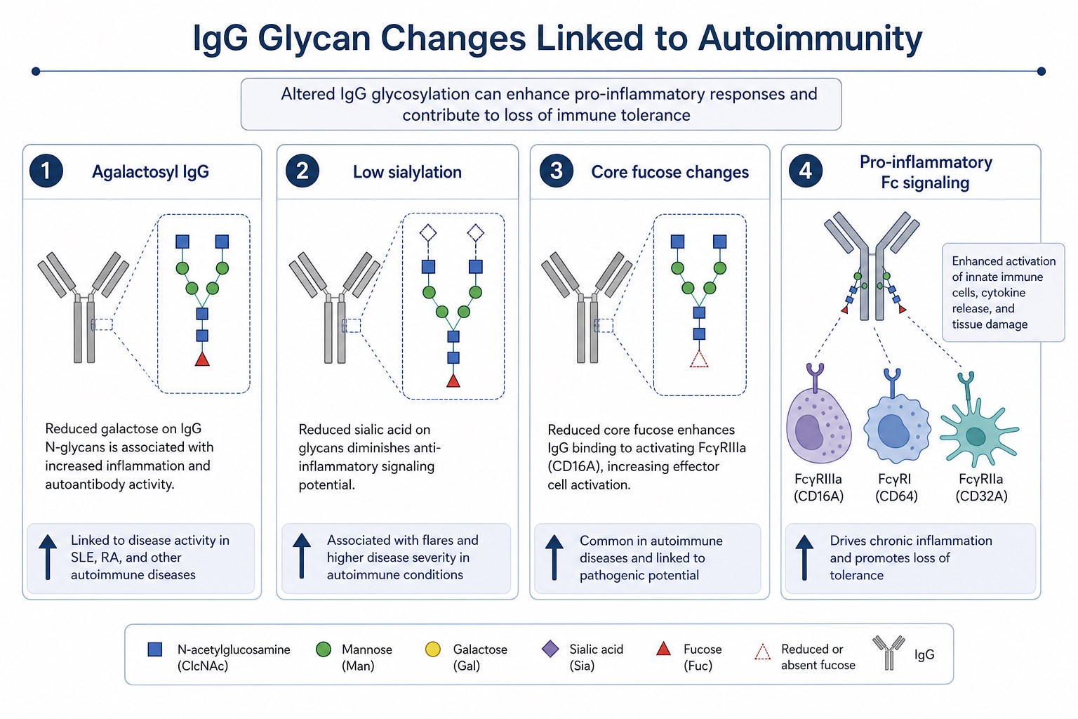
Figure 1. Autoimmune-associated IgG populations may show glycan changes such as reduced galactosylation, altered sialylation, and other Fc glycan features linked to inflammatory effector behavior.
Disease Contexts Where Antibody Glycosylation Has Been Studied
Antibody glycosylation research spans multiple autoimmune and inflammation-associated conditions.
Rheumatoid arthritis
Rheumatoid arthritis has been a major focus of IgG glycosylation research. Studies have reported increased agalactosyl IgG during active disease and changes in galactosylation with treatment response in some cohorts. Anti-citrullinated protein antibodies and other autoreactive specificities have also been examined for glycan differences that may relate to pathogenic potential.
Systemic lupus erythematosus
In systemic lupus erythematosus, IgG glycosylation changes have been explored in relation to immune complex formation, complement consumption, and renal involvement. Altered Fc glycan profiles may influence effector pathways relevant to tissue damage in lupus nephritis and other manifestations.
Inflammatory bowel disease and related conditions
Inflammatory bowel disease research has examined glycosylation of IgG and other immunoglobulin classes in mucosal and systemic immune responses. Glycan changes may reflect chronic inflammation and B cell activation in the gut immune environment.
Other autoimmune and autoinflammatory settings
IgG glycosylation has also been studied in vasculitis, myasthenia gravis, pemphigus, and other antibody-mediated conditions. The relevance of specific glycan features may vary by disease mechanism, antibody subclass distribution, and tissue target.
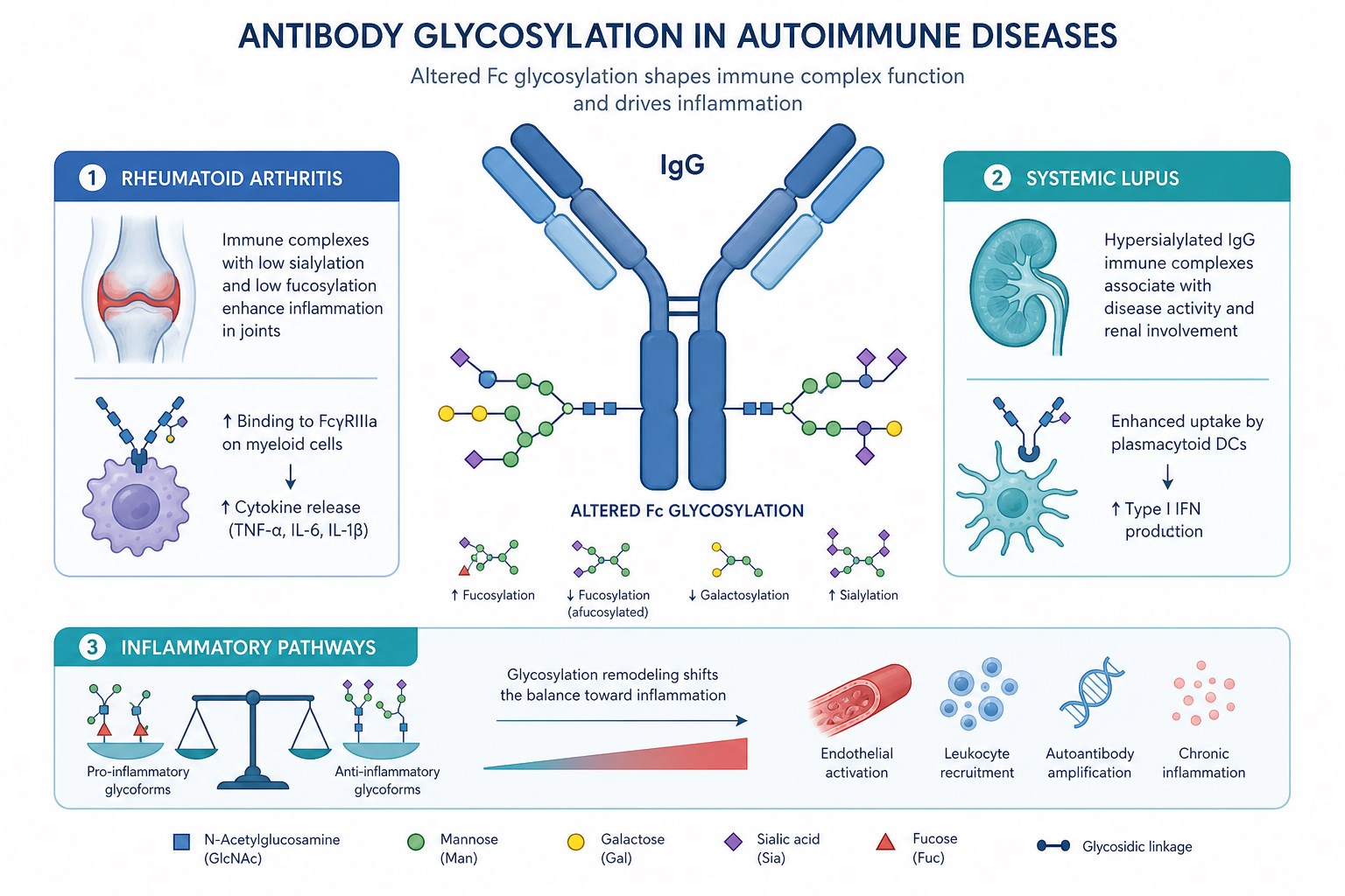
Figure 2. Antibody glycosylation research in autoimmune diseases examines IgG Fc glycan changes in conditions such as rheumatoid arthritis and systemic lupus within broader inflammatory pathways.
Mechanistic Links Between Fc Glycosylation and Autoimmune Effector Pathways
Antibody glycosylation can influence autoimmune pathology through several interconnected pathways.
Fc gamma receptor engagement on monocytes, macrophages, and neutrophils can promote phagocytosis, cytokine release, and tissue inflammation when IgG immune complexes are present. Glycan structure can modulate receptor binding strength and downstream activation.
Complement activation through the classical pathway may be affected by IgG glycan features including galactosylation status. This can influence immune complex clearance and local tissue injury.
Lectin and SIGN-related pathways have been explored in relation to sialylated IgG and inflammatory regulation. Glycan-dependent signaling may shift the balance between resolution and sustained inflammation.
Immune complex behavior including deposition, clearance rate, and cellular uptake may depend on both antibody specificity and Fc glycan context.
Mechanistic interpretation requires pairing glycan profiling with functional assays and disease-relevant models because glycan effects can vary by subclass, antigen context, and local tissue environment.
Research and Analytical Workflow for Autoimmune Antibody Glycosylation
Studying antibody glycosylation in autoimmune diseases requires careful sample design and analytical selection.
Cohort definition should document diagnosis, disease activity, treatment status, and sample type because each factor can influence IgG glycosylation. Serum and plasma are common sample matrices, although antigen-specific antibody fractions may require additional isolation when studying autoreactive subsets.
IgG enrichment or antigen-specific purification may be needed depending on whether total IgG glycosylation or disease-associated antibody populations are under study. Enrichment strategy affects interpretation and must be documented.
Glycan profiling may use released N-glycan analysis, glycopeptide LC-MS/MS, or intact IgG glycoform review. Released glycan profiling is widely used for population-level comparisons. Glycopeptide mapping provides site-specific detail when Fc assignment is required.
Functional follow-up may include Fc receptor binding assays, immune complex assays, or cell-based inflammation models when glycan differences must be linked to biological activity.
Biomarker analysis compares glycan features with clinical activity scores, treatment response, or longitudinal remission status when translational utility is the goal.
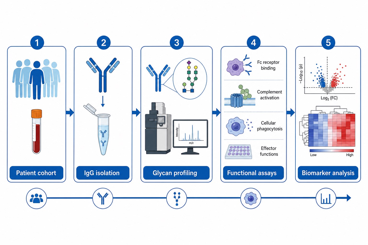
Figure 3. Autoimmune antibody glycosylation research typically moves from cohort selection and IgG preparation through glycan profiling, functional review, and biomarker analysis.
Related Services
Protein Glycosylation Analysis Service
Glycosylation Site Analysis Service
Comprehensive Glycosylation Analysis Service
Protein Drug Glycosylation Analysis Service
Research teams studying antibody glycosylation in autoimmune diseases can consult MtoZ Biolabs to review sample type, IgG preparation strategy, and the glycan profiling depth best suited to the study design.
Analytical Approaches in Autoimmune Glycosylation Research
Different research questions favor different analytical emphasis.
|
Research Question |
Common Sample Focus |
Typical Analytical Approach |
|---|---|---|
|
Total IgG glycan shift in active disease |
Serum or plasma IgG |
Released glycan profiling |
|
Fc site-specific glycan assignment |
Purified IgG |
Glycopeptide LC-MS/MS |
|
Autoantibody subset glycosylation |
Antigen-specific purification |
Targeted glycan profiling on enriched fraction |
|
Longitudinal treatment monitoring |
Serial patient samples |
Quantitative glycoform comparison |
|
Mechanistic effector linkage |
Purified IgG or immune complexes |
Glycan profiling plus functional assays |
Total IgG profiling captures population-level change but may dilute signals from rare autoreactive subsets. Antigen-specific studies provide sharper mechanistic insight but require additional purification and sample volume.
Core Research Value and Current Limitations
Core Research Value
Adds functional context to autoantibody measurement.
Glycan profiling can reveal inflammatory potential not visible from titer alone.
Supports biomarker discovery in autoimmune cohorts.
Glycan features may track disease activity or treatment response in selected settings.
Links immunology with biophysical characterization.
Glycosylation analysis connects clinical serology to Fc effector mechanism research.
Informs therapeutic antibody design in immunology.
Understanding disease-associated glycan biology supports rational Fc engineering in autoimmune therapeutics.
Enables cross-cohort comparability when methods are standardized.
Documented glycan profiling workflows improve reproducibility across studies.
Current Limitations
Cohort heterogeneity complicates interpretation.
Treatment, comorbidity, and inflammation source can confound glycan comparisons.
Antigen-specific glycan analysis is technically demanding.
Purifying autoreactive subsets at sufficient yield can be challenging.
Functional translation is not automatic.
Glycan associations in patient samples require careful validation before biomarker claims.
Subclass and isoform diversity add complexity.
IgG1, IgG2, IgG3, and IgG4 distributions differ across diseases and affect glycan interpretation.
Standardization across laboratories remains incomplete.
Method differences can affect cross-study comparison unless workflows are harmonized.
Implications for Therapeutic Development and Translational Research
Antibody glycosylation research in autoimmune diseases has implications beyond basic immunology.
Biomarker programs may incorporate IgG glycan features when early cohort data suggest association with flare, remission, or treatment response. Validation requires prospective design and standardized sample handling.
Therapeutic antibody programs in rheumatology and immunology may use glycoengineering to reduce or modulate Fc effector function when immune suppression or immune redirection is desired.
Comparability and quality review for immunology biologics still require robust glycan profiling even when the primary indication is autoimmune disease rather than oncology.
Translational teams should define whether glycan data will be used for hypothesis generation, patient stratification, or mechanistic confirmation before selecting analytical depth.
Sample and Study Design Considerations
Autoimmune glycosylation studies should address several design factors early.
- diagnosis, disease activity score, and treatment history at sampling
- sample matrix: serum, plasma, synovial fluid, or purified IgG
- whether total IgG or antigen-specific antibodies are the target analyte
- longitudinal versus cross-sectional study design
- required glycan attributes such as galactosylation, sialylation, or full Fc profiling
- need for functional assays to support mechanistic claims
- storage, freeze-thaw, and processing consistency across cohorts
Poor sample documentation is a common source of irreproducible glycan associations in autoimmune research.
Frequently Asked Questions
1. Why is antibody glycosylation studied in autoimmune diseases?
IgG glycosylation can modulate Fc effector function and inflammatory activity, providing functional context beyond autoantibody specificity or titer.
2. Which glycan change is most commonly linked to rheumatoid arthritis research?
Increased agalactosyl IgG, reflecting reduced terminal galactose on Fc glycans, is one of the most frequently reported glycan features in rheumatoid arthritis studies.
3. Does low IgG sialylation relate to inflammation?
Reduced sialylation has been associated with pro-inflammatory IgG effector profiles in research settings, although disease context and assay design strongly influence interpretation.
4. Can glycan profiling be used as a biomarker in autoimmunity?
Glycan biomarker utility is an active research area. Some cohorts show associations with disease activity or treatment response, but validation requirements remain substantial.
5. What samples are used to study autoimmune antibody glycosylation?
Serum and plasma are common starting materials. Purified total IgG or antigen-specific antibody fractions may be used when deeper subset analysis is required.
6. Which analytical methods are used for autoimmune IgG glycosylation studies?
Released glycan profiling, glycopeptide LC-MS/MS, and intact IgG glycoform analysis are commonly applied depending on whether global or site-specific glycan data are needed.
Conclusion
Antibody glycosylation in autoimmune diseases examines how Fc glycan structures on IgG and related immunoglobulins modulate inflammatory effector pathways in chronic autoreactive conditions. Glycan features such as agalactosylation, sialylation changes, and broader Fc glycoform shifts have been reported across rheumatoid arthritis, systemic lupus erythematosus, and other antibody-mediated diseases, adding functional depth to conventional autoantibody analysis.
Reliable research in this area depends on well-defined cohorts, appropriate IgG preparation, glycan profiling methods matched to the study question, and functional validation when mechanistic claims are made. As translational immunology continues to link glycan structure with disease activity and therapeutic design, standardized analytical support becomes increasingly important. Research teams studying antibody glycosylation in autoimmune diseases can contact MtoZ Biolabs to review sample strategy and glycan profiling scope for their program.
How to order?




