






Why Some Mitochondrial Membrane Proteins Are Hard to Detect by LC-MS/MS
Some mitochondrial membrane proteins are hard to detect by LC-MS/MS because they are hydrophobic, often low in relative abundance, and yield few chromatography-friendly tryptic peptides. In mitochondrial proteomics, that means absence from an identification table is not automatic proof that the protein was missing from the biology. It may simply have been difficult to extract, digest, ionize, or retain as usable peptide evidence.
For mitochondrial protein analysis, the practical takeaway is to set detection expectations early, especially for multi-pass transmembrane proteins, and to confirm sample amount and study goals before treating non-detection as a biological conclusion. Teams targeting difficult membrane proteins can share the protein list, sample type, and approximate amounts with MtoZ Biolabs before the LC-MS/MS plan is locked.
Why Membrane Proteins Behave Differently in Bottom-Up Proteomics
Most mitochondrial proteomics workflows are bottom-up: proteins are extracted, digested into peptides, separated by liquid chromatography, and measured by tandem mass spectrometry. Soluble matrix proteins often fit that path well. Inner- and outer-membrane proteins frequently do not.
Three chemical realities drive the gap:
- Hydrophobic transmembrane segments resist clean solubilization and can aggregate or stick to surfaces during sample handling.
- Transmembrane helices often contain fewer convenient trypsin cleavage sites, so digestion may return long, sticky, or sparse peptides.
- The peptides that are produced can be poorly soluble or poorly retained in standard reverse-phase separations, which weakens detection even when the parent protein was present.
As a result, coverage of membrane proteins is usually uneven. Some membrane-associated proteins appear readily; deeply embedded multi-span proteins may remain under-represented.
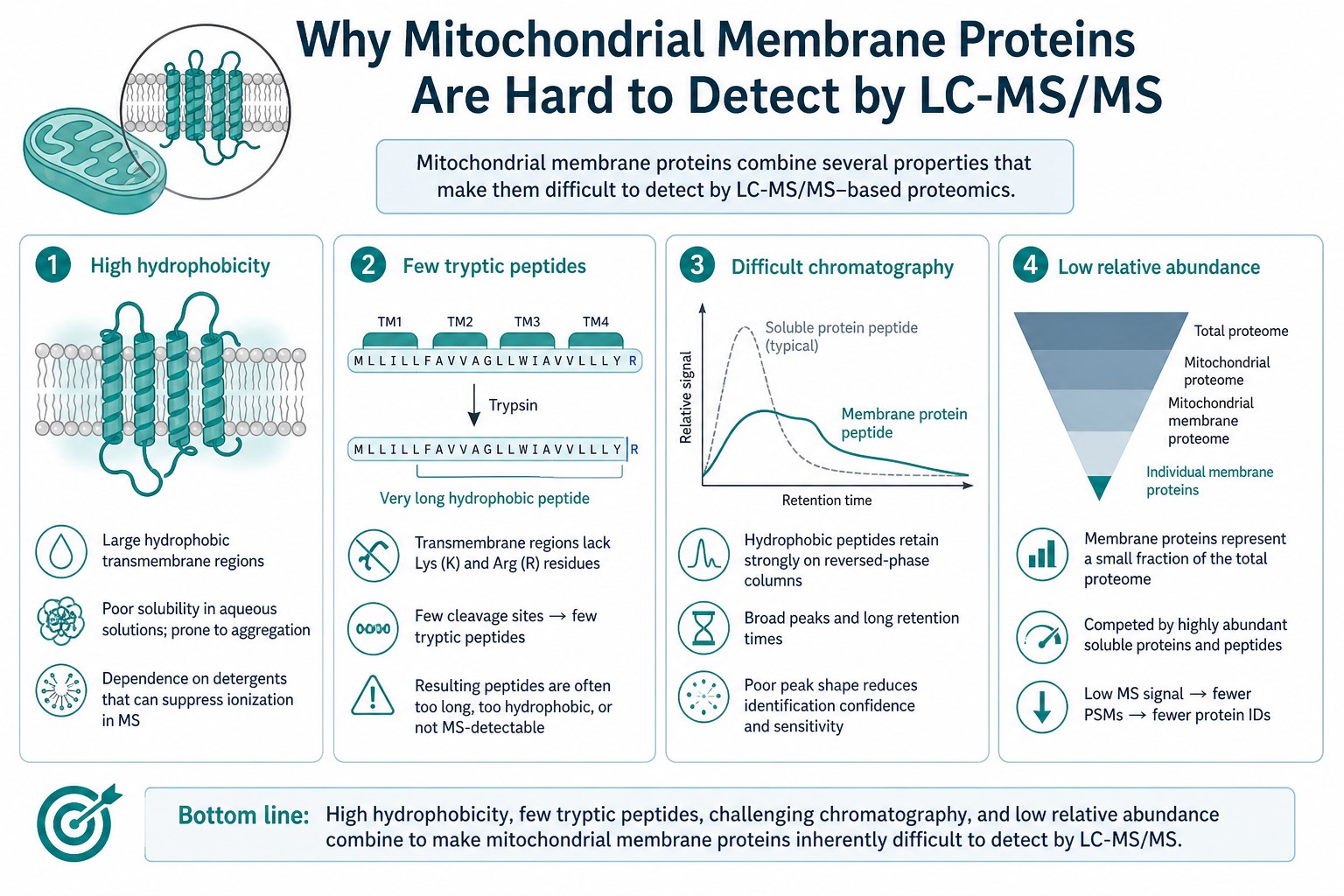
Figure 1. Hydrophobicity, limited tryptic peptides, and difficult chromatography all reduce membrane-protein detectability.
Abundance and Background Effects Inside Mitochondrial Preparations
Detection is competitive. In many mitochondrial preparations, abundant matrix and peripheral proteins occupy a large share of the peptide signal. Lower-abundance membrane proteins then compete for digestion, chromatography capacity, and MS sampling time.
This matters for interpretation:
- A strong signal for abundant soluble proteins does not guarantee equal visibility for membrane channels, transporters, or respiratory-complex membrane subunits.
- Comparative mitochondrial proteomics can still work when the same membrane proteins are quantified consistently across groups, even if absolute coverage of the membrane proteome is incomplete.
- Non-detection in one condition is weak evidence of biological loss unless method sensitivity, input amount, and replicate behavior support that reading.
Sample input remains one of the few levers that can be planned directly. Planning references are about 5×10^7 cells, about 200 mg animal tissue, or extracted mitochondrial protein of at least about 50 µg with about 80-100 µg commonly planned, at a concentration commonly planned around 1 µg/µL. These anchors improve the chance of usable evidence; they do not create a promised detection list for every membrane target.
What LC-MS/MS Can and Cannot Resolve for Membrane Targets
LC-MS/MS mitochondrial proteomics can still contribute useful membrane-related evidence when expectations are realistic.
It can:
- Detect many membrane-associated or membrane-anchored proteins when they yield usable peptides.
- Quantify detectable membrane proteins across designed groups by DDA or DIA paths.
- Rank candidates for follow-up when a membrane protein is observed consistently enough to compare.
It cannot reliably:
- Guarantee detection of every mitochondrial membrane protein of interest.
- Convert non-detection into proof of absence without supporting method context.
- Replace functional assays such as respiration, membrane potential, ROS, or enzyme activity panels, which sit outside this proteomics service scope.
Acquisition options for this service line include Orbitrap Exploris 480, timsTOF Pro, and Orbitrap Astral. DDA data are commonly processed with MaxQuant or Proteome Discoverer; DIA data with Spectronaut or DIA-NN. Mode choice should follow whether the study needs flexible discovery or consistent cohort quantification. Neither mode erases the chemistry of difficult transmembrane peptides.
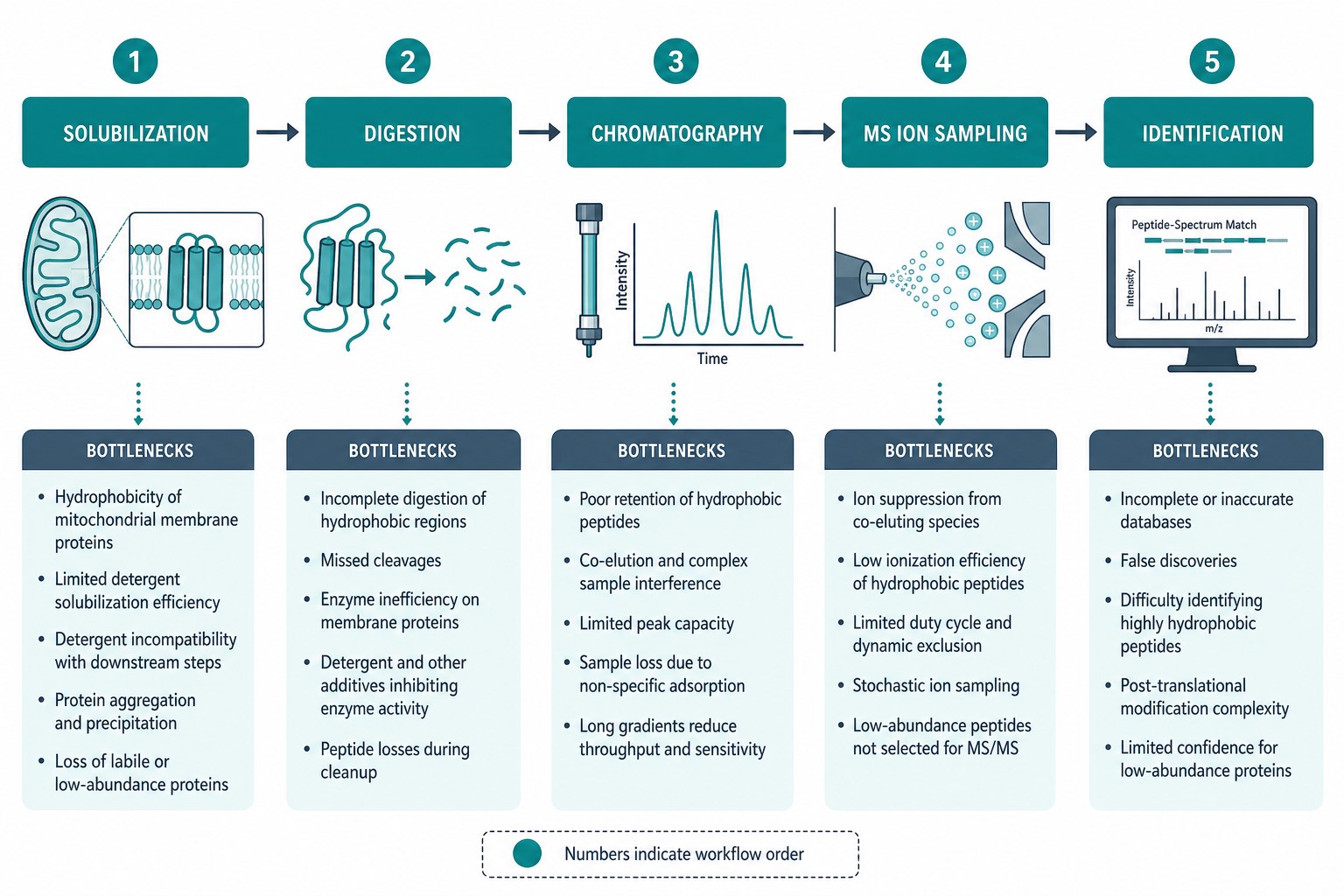
Figure 2. Solubilization, digestion, chromatography, and ion sampling each can drop membrane proteins before identification.
How to Plan When Membrane Proteins Are Central to the Question
If mitochondrial membrane proteins sit at the center of the claim, confirm feasibility before treating the project like a routine inventory.
Practical planning steps:
- List the priority membrane proteins and whether the claim needs detection, quantification, or both.
- Confirm sample class and amount against planning guidance, and keep amounts matched across groups.
- State whether mitochondria are already extracted and how protein concentration was measured.
- Decide whether the endpoint is broad mitochondrial proteome context or a narrower membrane-focused comparison.
- Keep orthogonal validation ready for critical membrane targets that are known to be analytically difficult.
Borderline targets or unusual preparations should be reviewed case by case. Planning guidance for turnaround is about 4 weeks, but difficult membrane-protein goals may need extra feasibility discussion before a start date is firm.
|
Challenge |
Why detection suffers |
Planning response |
|---|---|---|
|
Hydrophobicity |
Poor solubilization and losses on surfaces |
Confirm input amount and preparation quality early |
|
Few useful tryptic peptides |
Sparse or sticky peptides |
Treat non-detection cautiously |
|
Low relative abundance |
Signal buried by abundant proteins |
Use matched groups and realistic sensitivity expectations |
|
Difficult chromatography |
Weak retention or recovery of hydrophobic peptides |
Do not equate absence with biological loss |
|
Function questions |
Activity is not a proteomics readout |
Plan separate functional assays if needed |
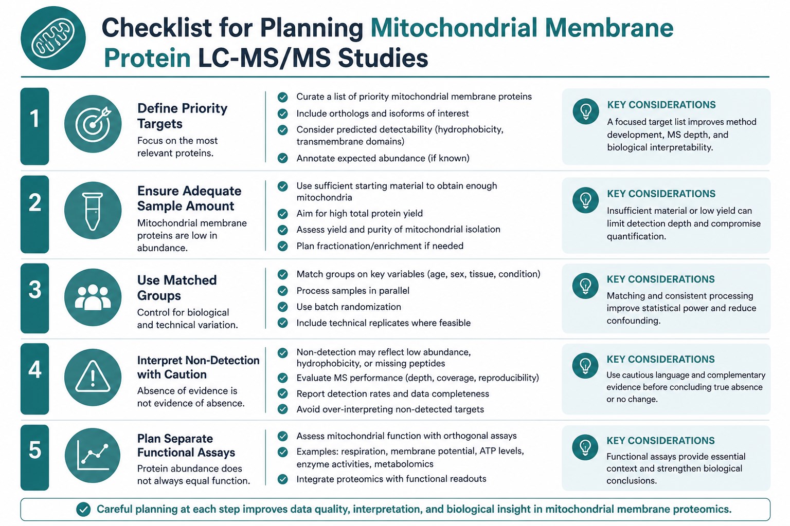
Figure 3. Define priority membrane targets, confirm input, and separate detection claims from functional claims before LC-MS/MS.
When requesting a membrane-focused review, send the priority protein list, sample type and amounts, extraction status, and whether the claim is detection, quantification, or pathway-level remodeling. MtoZ Biolabs can help judge whether a standard mitochondrial proteomics path is likely to give usable evidence for those targets.
Related Services
Teams weighing mitochondria-focused and membrane-focused options can review the services below while detection expectations are still open.
Mitochondrial Proteomics Service
The main route for mitochondrial protein analysis when membrane-protein detection questions sit inside a mitochondrial proteome study.
Consider this when the study priority is membrane-protein recovery more broadly rather than mitochondria-only context.
Subcellular Proteomics Service
Use this when membrane-protein questions span multiple subcellular fractions rather than a single mitochondrial preparation.
Frequently Asked Questions
1. Why are some mitochondrial membrane proteins hard to detect by LC-MS/MS?
Because they are hydrophobic, often lower in relative abundance, and may yield few chromatography-friendly tryptic peptides.
2. If a membrane protein is not identified, is it absent?
Not necessarily. Non-detection can reflect extraction, digestion, or peptide behavior rather than biological absence.
3. Can better instruments solve the problem completely?
High-end platforms help, but they do not erase transmembrane chemistry. Input quality and peptide behavior still matter.
4. How much sample should I plan?
Planning references are about 5×10^7 cells, about 200 mg tissue, or about 50 µg protein minimum for extracted mitochondria, with about 80-100 µg commonly planned.
5. Does mitochondrial proteomics measure membrane potential?
No. Membrane potential and related functional assays are outside this proteomics scope.
6. What should I send if membrane proteins are the priority?
Priority protein names, sample type and amounts, extraction status, and whether you need detection, quantification, or both.
Conclusion
Hard-to-detect mitochondrial membrane proteins are usually an analytical chemistry problem before they are a biological conclusion. Hydrophobicity, limited useful peptides, and competition from abundant proteins all reduce LC-MS/MS visibility.
If those limits are acknowledged at the design stage, mitochondrial proteomics remains useful for the membrane proteins that are detectable and comparable, without forcing non-detection to carry more meaning than it should. Teams prioritizing difficult membrane targets can review detection expectations with MtoZ Biolabs before mitochondrial protein analysis begins.
How to order?




