






Why Intact Mass Analysis Can Miss Minor Variants
- Intact Mass Analysis Service
- Intact Mass Analysis of Antibodies Service
- Peptide Mapping Service
- Charge Heterogeneity Analysis Service
- SEC and RPLC Based Protein Purity Analysis Service
- Comprehensive Glycosylation Analysis Service
- High-Resolution LC-MS Molecular Weight Identification Service
- Biological Products Analysis Service
- Is the concern clipped forms, oxidation, deamidation, glycoform shifts, conjugation heterogeneity, or aggregates?
- What abundance level must be detected for the decision at hand?
- Is the goal release support, comparability, stability monitoring, or early clone screening?
- Which orthogonal methods are already available if intact MS is inconclusive?
- use LC-MS instead of direct infusion when multiple intact species are expected
- increase separation selectivity when glycoforms or clipped forms are suspected
- acquire enough signal across charge states for reliable deconvolution
- compare new lots against a reference material at the profile level, not only by one mass value
- document whether the method targets main species confirmation or variant profiling
- reversed-phase or other LC modes tuned for intact protein resolution
- SEC when size-related variants are the primary concern
- enriched fraction collection before MS when a specific variant class is suspected
- charge-based prefractionation when acidic or basic variants are likely relevant
- overlay of deconvoluted profiles from replicate injections
- comparison to a reference lot or control material
- annotation of major and minor features when confidently resolved
- clear statement of detection limits or unresolved regions
- recommended orthogonal assay when minor species remain ambiguous
- whether main mass confirmation is sufficient
- whether low-level variant profiling is required
- which variant classes are in scope
- whether quantitative comparison is needed
- which follow-up assays are acceptable if intact results are inconclusive
Introduction
A biologics team submits an antibody lot for intact mass review expecting a clean confirmation of the main species. The report shows a dominant mass that matches the expected product, yet SEC, cIEF, or bottom-up mapping later reveals clipped forms, low-level oxidation, or minor glycoform shifts that were not clearly resolved at the intact level. A comparability study may conclude that two lots are equivalent by intact MS, while charge variant analysis later shows a small acidic species increase that matters for release review.
These outcomes are common and do not always mean the intact measurement failed. Intact mass analysis is efficient for confirming the main product mass and screening major mass-shifting changes. It is less reliable as a standalone tool for detecting every low-abundance variant, especially when variants are closely spaced in mass, buried under the main envelope, or present below the practical detection threshold of the selected method.
Understanding why low-level species are missed helps teams set realistic reporting goals, choose the right follow-up assays, and avoid over-interpreting a single intact mass result.
Related Services
For projects where low-abundance variant detection is critical, MtoZ Biolabs can help define whether intact MS alone is sufficient or whether peptide-level mapping, charge heterogeneity review, or enriched separation is needed.
Common Situations Where Low-Level Variants Are Missed
Several recurring scenarios explain why intact workflows under-report low-level species.
1. Dominant Main Species Masking
When the main antibody or fusion protein contributes most of the ion current, minor clipped, oxidized, or partially glycosylated forms may sit on the shoulder of the main deconvoluted peak rather than appear as distinct resolved masses.
2. Closely Spaced Glycoforms
Glycoform detection at the intact level depends on resolving mass differences that may be only tens of daltons apart across a broad charge envelope. Without sufficient separation or resolution, minor glycoforms merge into the main profile.
3. Low-Abundance Product-Related Impurities
A variant present at a few percent or below may not generate enough signal for confident annotation, especially in direct infusion workflows on heterogeneous samples.
4. Co-Eluting Intact Species in LC-MS
LC coupling helps, but closely eluting variants can still enter the mass analyzer together and produce overlapping charge states that deconvolute into one broad mass distribution.
5. Small Mass Shifts from Localized Modifications
A single oxidation, deamidation, or conjugation event may shift mass only slightly relative to the full biologic. That shift can be hard to distinguish from natural isotopic breadth or deconvolution artifacts.
6. Over-Interpretation of a Single Average Mass
Reporting only one deconvoluted mass number can hide unresolved heterogeneity. A result that appears consistent with expectation may still contain unresolved minor components.
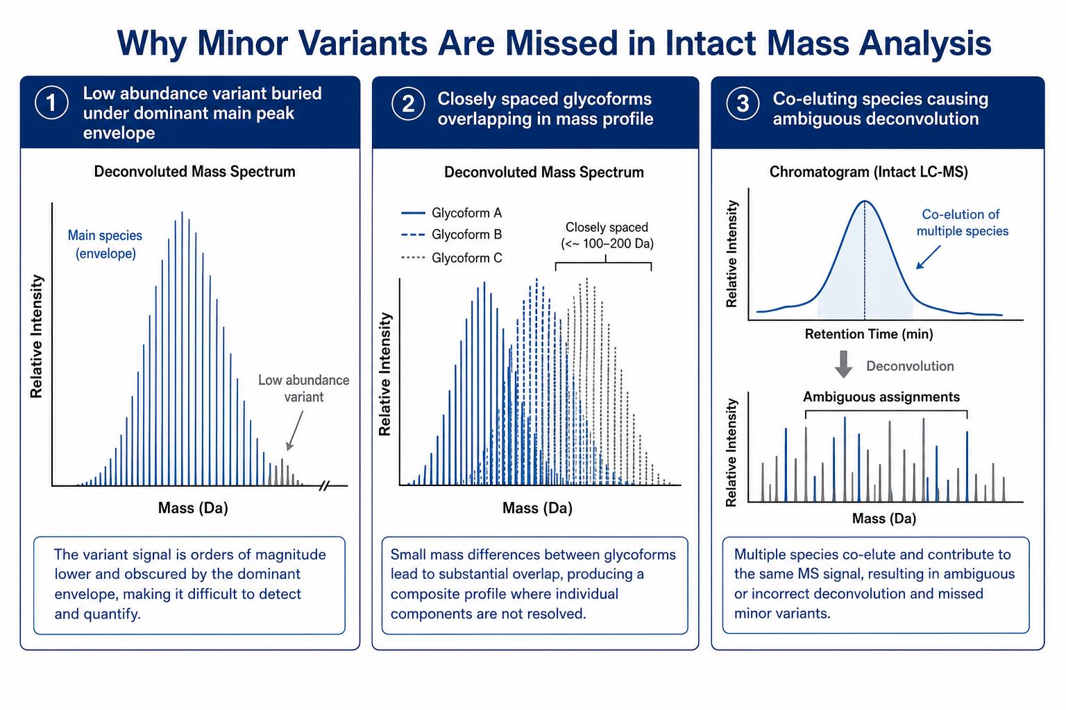
Figure 1. Resolution limits, low abundance, and deconvolution overlap are common reasons subdominant species are not resolved at the intact level
Root Causes Behind the Detection Gap
Most missed low-level species trace back to a combination of physical, methodological, and reporting factors.
1. Resolution and Mass Spacing
Intact protein spectra contain many charge states spread across a wide m/z range. Deconvolution quality depends on charge state spacing, signal-to-noise ratio, and freedom from interfering ions. When two intact species differ by a small mass increment, the deconvoluted profiles may overlap unless chromatographic separation or higher resolving power separates them first.
2. Abundance and Ion Suppression
Low-abundance forms compete for ionization with the main product. In complex buffers or less purified material, suppression can reduce the visible signal of low-level species below the annotation threshold. What is biologically present is not always what the spectrum reports with confidence.
3. Method Choice
Direct infusion intact MS is fast and useful for relatively clean samples, but it offers less physical separation than LC-MS. A method chosen for speed or limited sample amount may not support low-level variant profiling even when main species confirmation is successful.
4. Deconvolution and Data Processing Limits
Deconvolution algorithms assume reasonably well-resolved charge envelopes. Noise, adducts, incomplete desalting, or co-eluting impurities can create artifact peaks or broaden true species. Analysts may conservatively report only the dominant feature when minor shoulders are ambiguous.
5. Reporting Scope Mismatch
A project may request intact mass confirmation but interpret the result as full variant profiling. If the method was not designed for low-level species detection, absence of annotated subdominant peaks does not prove absence of those species in the sample.
Step 1: Define What Counts as a Low-Level Variant for Your Project
Before troubleshooting a missed peak, define the variant type and decision threshold that matter for the study.
Useful planning questions include:
A stability study tracking a 2 percent acidic variant increase needs a different workflow than early construct screening that only requires main mass confirmation.
Step 2: Review Sample Quality and Preparation
Low-level variant detection starts with sample condition. Impure, aggregated, or poorly desalted material increases background and weakens deconvolution.
|
Review Point |
What to Check |
Why It Affects Low-Level Variant Detection |
|---|---|---|
|
Purity |
Main product dominance by SEC or SDS-PAGE when available |
Impurities add competing intact signals |
|
Buffer and salts |
Desalting or buffer exchange before MS |
Excess salt reduces intact ion quality |
|
Aggregation |
Visible SEC or light scattering concerns |
Aggregates can distort intact profiles |
|
Concentration |
Suitable for selected LC or infusion method |
Too little signal hides low-level species |
|
Storage history |
Freeze-thaw, oxidation exposure, clipping risk |
Degradation may create variants that need targeted follow-up |
Share biologic type, buffer composition, purity estimate, and the decision the result must support before analysis begins.
Step 3: Match the Intact Method to the Detection Goal
If low-abundance variants are part of the decision, the intact workflow must be built for separation and profile review rather than a single mass readout.
Practical method adjustments include:
Intact MS is often excellent for the first goal and only conditionally sufficient for the second.
Step 4: Improve Separation Before Mass Measurement
Physical separation is one of the most effective ways to expose minor intact species that co-migrate in a bulk measurement.
Separation options may include:
Better separation reduces overlapping charge envelopes and gives deconvolution a cleaner input.
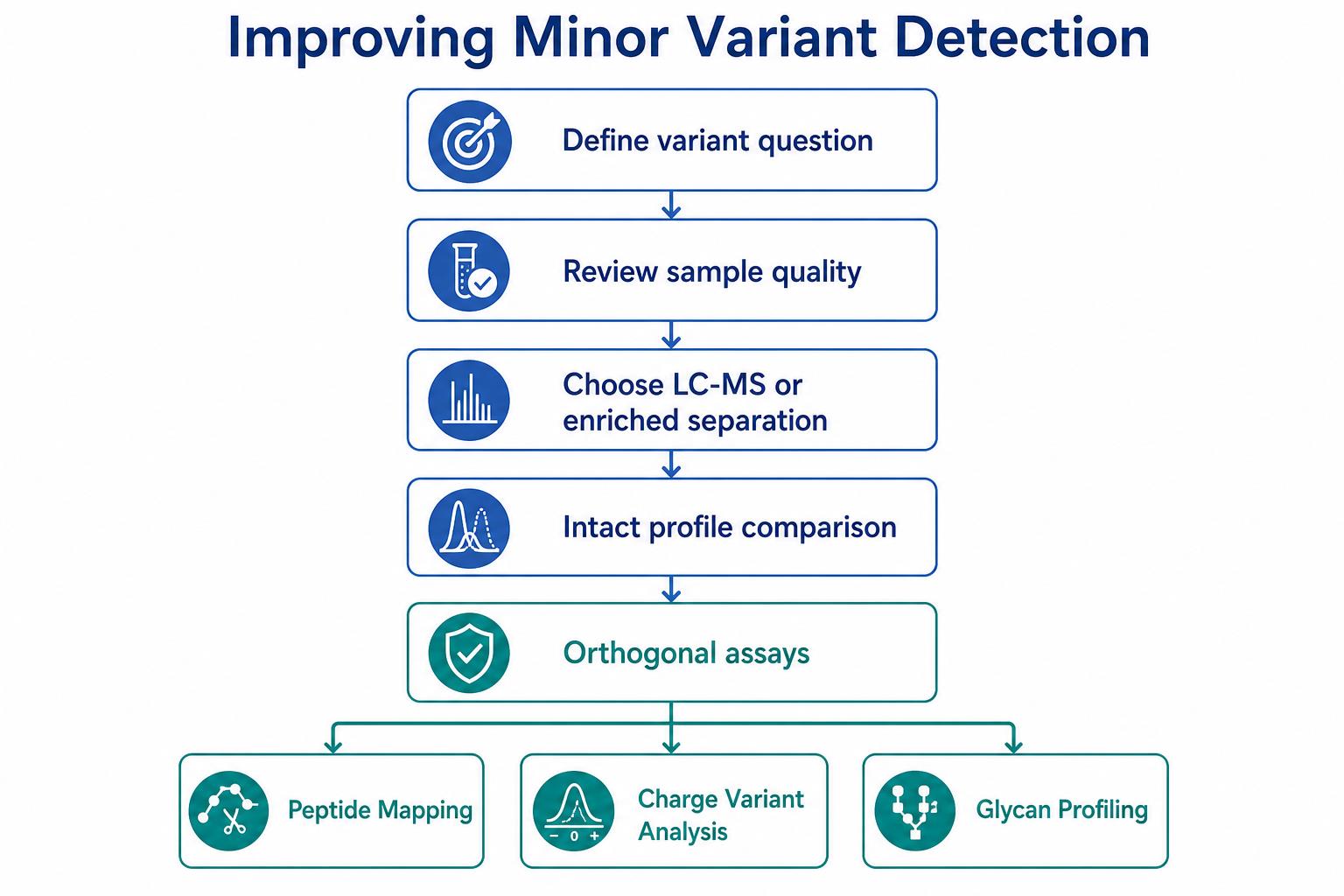
Figure 2. Improved sample prep, LC separation, and orthogonal assays help recover subdominant species missed at the intact level
Step 5: Add Orthogonal Assays When Intact MS Is Not Enough
When intact measurement cannot resolve the variant question, the next assay should match the variant class.
|
If the suspected variant is... |
A useful follow-up may be... |
|---|---|
|
Clipped or truncated forms |
Bottom-up peptide workflow, N-terminal or C-terminal analysis |
|
Localized oxidation or deamidation |
Bottom-up mapping with modification search |
|
Glycoform differences |
Glycan profiling or glycoform-sensitive intact method with optimized separation |
|
Charge microheterogeneity |
Charge heterogeneity analysis or cIEF |
|
Size-related impurities |
SEC with UV or multi-detector review |
|
Conjugation heterogeneity |
ADC-specific intact workflow plus peptide-level confirmation |
Bottom-up mapping is often the most direct route to localize a small mass shift that intact MS only hints at.
Step 6: Validate Findings With Profile-Level Comparison
Do not rely on a single deconvoluted mass number when low-level heterogeneity matters. Compare intact profiles between reference and test material, review peak shoulders, and note unresolved heterogeneity explicitly.
A useful validation package may include:
If a subdominant species is biologically important but not intact-resolved, the validation outcome should say so rather than imply full clearance.
Step 7: Set Reporting Expectations Before Submission
The fastest way to avoid disappointment is to align method scope with the decision upfront.
Researchers should specify:
A well-designed biologics characterization plan treats intact MS as one layer in a broader evidence stack, not as a universal impurity detector.
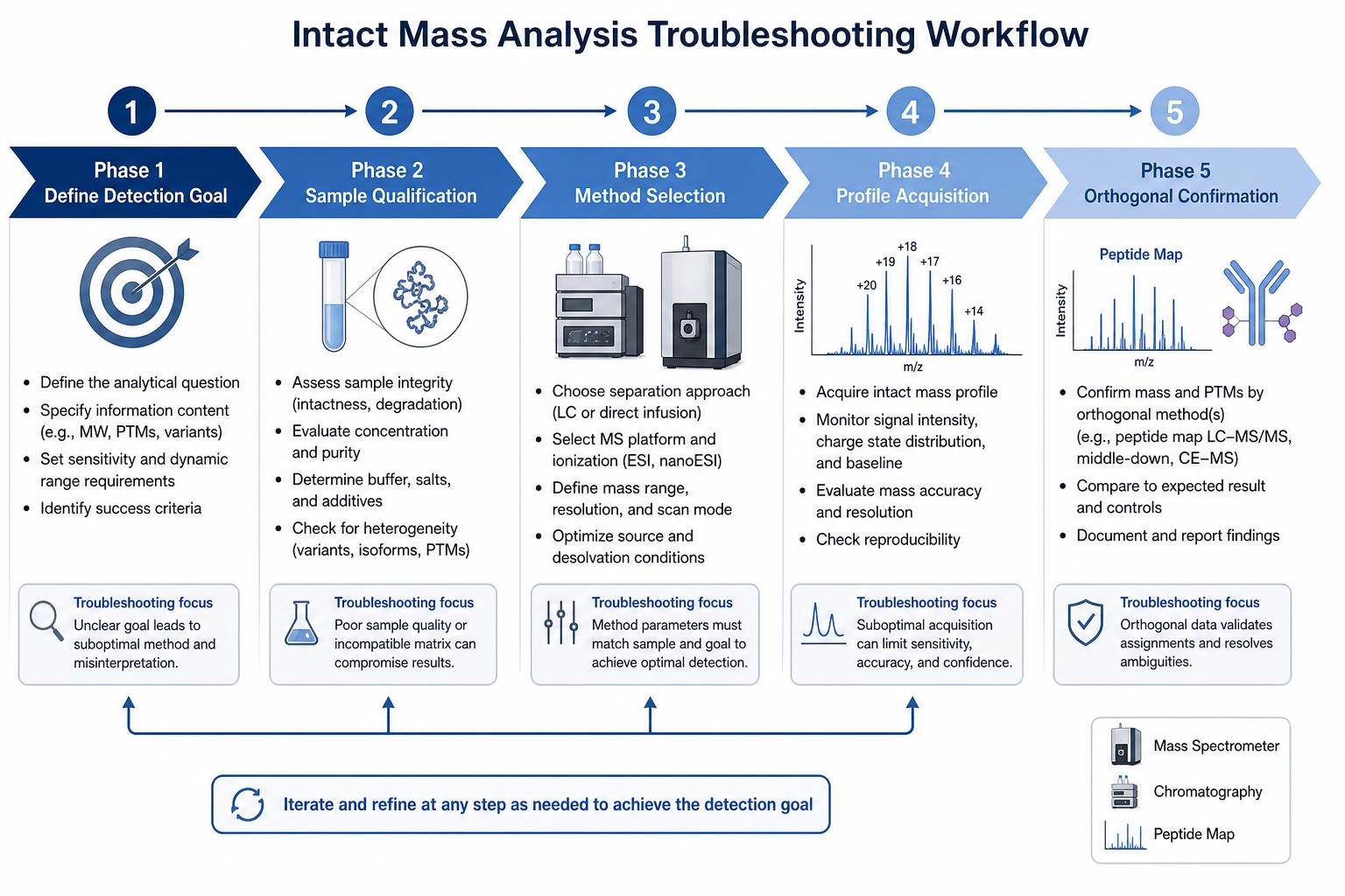
Figure 3. A practical workflow moves from variant definition through sample review, method selection, and orthogonal confirmation
Expected Outcomes After Corrective Action
When the workflow is adjusted appropriately, teams should expect clearer outcomes tied to the original question.
|
Scenario |
Realistic Outcome After Adjustment |
|---|---|
|
Main mass confirmation only |
Dominant species mass reported with defined comparison to expected biologic mass |
|
Minor glycoform review |
Improved separation may resolve major glycoforms while minor ones still may need glycan analysis |
|
Low-level clipping concern |
LC-MS or bottom-up mapping may reveal truncated forms not visible by bulk intact infusion |
|
Comparability decision |
Profile comparison plus orthogonal charge or peptide data supports a stronger lot review |
|
Stability monitoring |
Trend analysis across time points may require a more sensitive assay than initial screening |
The goal is not to force intact MS to do everything. The goal is to use it where it is strong and escalate when low-abundance variants drive the decision.
Key Cautions
Do not treat absence of a subdominant peak as proof of absence when the method was not designed for low-level profiling. Do not compare lots using only one average mass value when heterogeneity is expected. Do not use direct infusion results alone to settle glycoform or clipping questions on complex antibody material. Do not skip sample purity review when deconvolution profiles look noisy or broad. Do not delay bottom-up mapping or charge variant analysis when the business decision depends on a variant class that intact MS cannot reliably resolve.
Low-level variant questions are often solvable, but the solution may require a different assay rather than repeating the same intact measurement under the same conditions.
Frequently Asked Questions
1. Why can intact mass analysis miss low-abundance variants?
Low-abundance variants produce weaker ion signals and may be hidden under the main species envelope. Without sufficient separation, resolution, or signal, they may not be confidently annotated.
2. Can better deconvolution alone solve the problem?
Better data processing helps, but it cannot fully recover overlapping species that were never separated physically or resolved in the original spectrum.
3. When should peptide-level mapping be used after intact MS?
Bottom-up mapping is appropriate when a small mass shift is suspected but not localized, or when clipping, oxidation, deamidation, or sequence-level confirmation is required.
4. Is LC-MS always better than direct infusion for low-level variants?
LC-MS usually helps when multiple intact species are present, but it is not automatic guarantee of subdominant species detection. Separation chemistry and reporting depth still matter.
5. What information should be shared before requesting low-level variant support?
Provide biologic type, buffer conditions, purity estimate, suspected variant class, reference material if available, and the decision the data must support.
Conclusion
Intact mass analysis is a valuable tool for whole-molecule mass confirmation and major variant screening in biologics characterization. It can miss minor variants when species are low in abundance, closely spaced in mass, co-eluting, or affected by sample quality and processing limits. Teams that define the variant question early, match method design to detection goals, and add orthogonal assays such as bottom-up mapping or charge variant analysis are more likely to reach a supportable answer.
If low-level variant detection is central to your release, comparability, or stability decision, contact MtoZ Biolabs to review whether intact mass analysis alone is sufficient or whether an expanded biologics characterization workflow is needed.
How to order?




