






What Makes Plant Extracellular Vesicle Proteomics Challenging?
Plant extracellular vesicle proteomics is challenging because EV-enriched fractions can be scarce, contain co-isolated non-vesicle material, and still need to support meaningful protein identification or quantitative comparison. The main pressure points are EV enrichment quality, co-isolated background, low protein input, and limited identification depth.
None of these challenges makes the experiment impossible. They do mean that the project should be planned as a low-input, contamination-aware workflow rather than treated like standard tissue proteomics. If EV preparation quality and protein input are not considered before digestion, the resulting protein list can be difficult to interpret even when LC-MS/MS performance is strong.
Plant extracellular vesicles should also be distinguished from vesicle-like fractions obtained directly from disrupted plant tissues or juices. When extracellular origin has not been established, such preparations should not automatically be interpreted as bona fide EV fractions.
Why Plant EV Proteomics Is More Challenging Than Tissue Proteomics
Tissue proteomics usually starts from a relatively abundant protein pool. Plant EV studies often begin with much less protein because vesicles must first be recovered from extracellular sources such as apoplastic fluid or culture supernatant and then enriched before proteomic analysis.
This creates a mismatch between scientific ambition and sample reality. Researchers may want broad coverage of vesicle-associated proteins, while the preparation contains limited protein together with soluble proteins, membrane fragments, aggregates, or other co-isolated material.
Comparative plant EV studies add another challenge. Differences in clarification, enrichment, concentration, storage, or handling between experimental groups can appear as biological differences in the final protein dataset.
Plant EV proteomics therefore depends on controlling three practical variables: what enters the EV-enriched fraction, how much protein reaches digestion, and how confidently the resulting proteins can be interpreted.
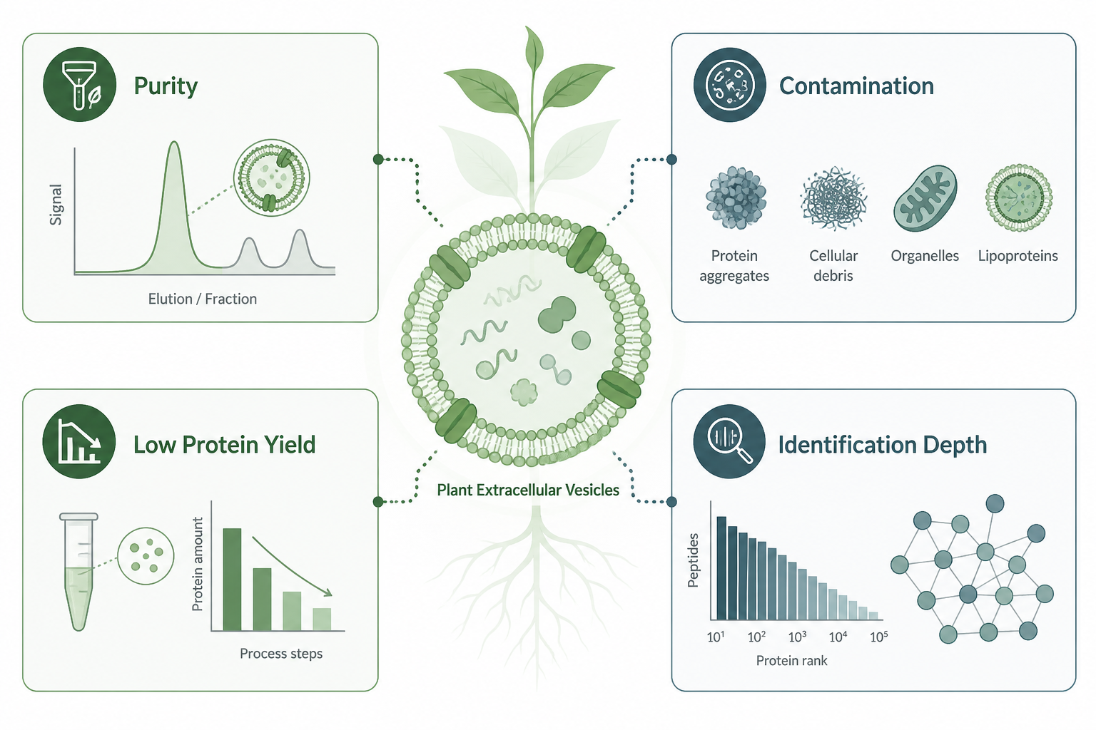
Figure 1. EV enrichment quality, co-isolated background, low protein input, and identification depth are major factors that shape plant EV proteomics.
Challenge 1: Vesicle Purity Is Easy to Overstate
A common assumption is that an enriched pellet or particle fraction represents a pure EV sample. In practice, extracellular preparations may also contain protein aggregates, membrane fragments, soluble proteins, and other particles that co-isolate during enrichment.
For this reason, EV preparation quality cannot be judged from a protein list alone. A longer identification list does not necessarily indicate a cleaner preparation. It may also reflect greater background entering digestion.
Characterization data, candidate EV-associated markers, and matched isolation records provide more useful context than protein counts alone. Proteomic data should therefore be interpreted as coming from an EV-enriched preparation unless additional evidence supports a stronger localization claim.
For comparative studies, enrichment quality should also be treated as a design variable. If resistant and susceptible lines, or treated and control plants, are processed using different isolation conditions, some apparent differential proteins may reflect preparation differences rather than EV biology.
Challenge 2: Co-Isolated Background Can Look Like Cargo
Contamination in plant EV proteomics is not limited to laboratory contaminants. A more important concern is biological carryover, including abundant soluble proteins, intracellular proteins released through tissue damage, membrane fragments, and other material that remains in the EV-enriched fraction.
This matters because EV studies are often interpreted in terms of cargo composition or selective loading. Proteins detected in an enriched fraction may be biologically interesting, but their presence alone does not confirm exclusive localization within extracellular vesicles.
Practical ways to reduce this risk include consistent clarification and enrichment across groups, minimizing tissue damage during extracellular fluid collection, and maintaining comparable storage and freeze-thaw histories.
Degraded, visibly contaminated, or repeatedly freeze-thawed material should be avoided. Acceptance requirements for infectious, regulated, or unusual plant materials should be confirmed before shipment.
Challenge 3: Low Protein Input Can Limit Study Design
Low protein input can become a major constraint in plant EV proteomics, particularly when the extracellular preparation or EV recovery is limited.
Even when EV enrichment appears successful, the protein available for digestion may be modest. This can reduce flexibility for repeated analysis, additional fractionation, or large multi-group comparisons.
Starting material requirements depend on the plant source, extracellular preparation, isolation strategy, and expected EV recovery. A standard tissue-proteomics amount should not be assumed to provide sufficient EV protein. The required starting material should therefore be confirmed before sample collection.
A realistic first study often favors a focused comparison with matched handling and a clearly defined biological question. Expanding immediately to many genotypes, treatments, or time points before EV recovery is understood can increase the risk of uneven protein input across groups.
Challenge 4: Identification Depth Is Easy to Misinterpret
Researchers may expect tissue-like proteome coverage from an EV-enriched fraction that contains much less protein and a different background composition. A shorter identification list does not automatically mean the project failed.
Identification depth should be interpreted together with protein input, EV preparation quality, and the biological question. A smaller, well-characterized candidate list may be more informative than a much larger list dominated by co-isolated background proteins.
Conversely, a long protein list without information about the isolation process can create false confidence about EV cargo coverage.
DDA and DIA can both support plant EV proteomics. The acquisition strategy should be selected according to sample characteristics, protein input, quantitative objectives, required data consistency, and overall study design. An appropriate mass spectrometry platform can then be selected according to the finalized analytical plan.
No acquisition strategy can compensate for a poorly controlled or heavily contaminated EV preparation.
| Challenge | Why It Appears in Plant EV Work | Practical Response |
| EV enrichment uncertainty | Non-vesicle material can co-isolate with EVs | Match isolation procedures and avoid overclaiming EV cargo |
| Co-isolated background | Soluble proteins, debris, and membrane material may enter the fraction | Keep collection and handling consistent across groups |
| Low protein input | EV recovery may provide limited protein for digestion | Keep the first comparison focused and confirm starting material requirements |
| Limited identification depth | Protein input and preparation quality constrain coverage | Interpret depth together with isolation and sample context |
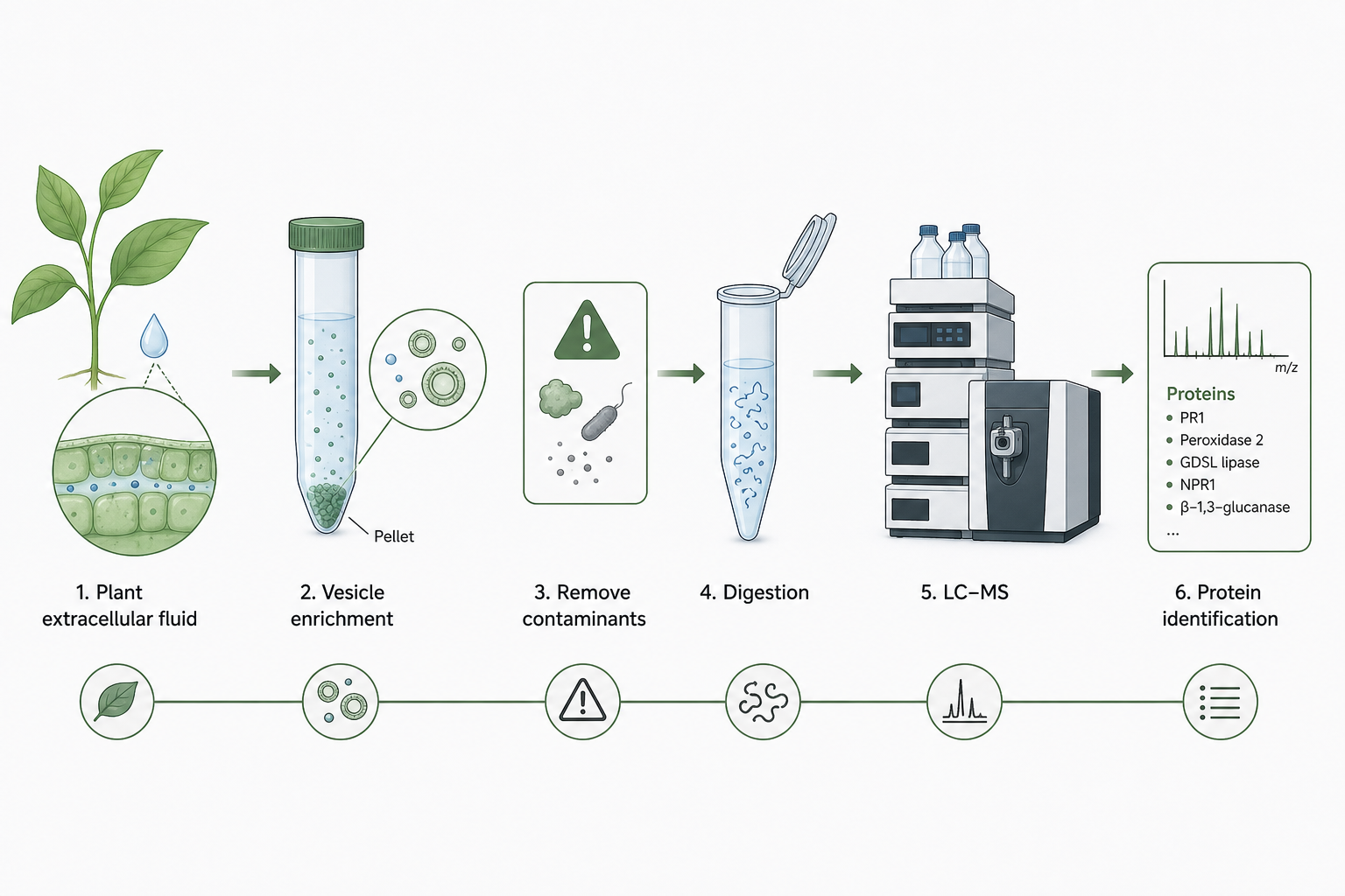
Figure 2. Plant EV proteomics moves from an EV-enriched preparation to digestion and LC-MS/MS while controlling co-isolated background and limited protein input.
How to Plan Around These Challenges
A workable plant EV proteomics project should answer several questions before digestion begins.
First, define the intended claim. Is the project intended to characterize an EV-enriched preparation, compare EV-associated protein patterns between groups, or support a stronger localization hypothesis that will require additional evidence?
Second, decide how EV preparation will be matched across groups, including clarification, enrichment, concentration, storage, and freeze-thaw history.
Third, confirm whether enough starting material can be collected consistently to support the planned number of experimental groups.
Fourth, define how the final protein list will be interpreted. Proteins identified in an EV-enriched fraction should generally be treated as candidate EV-associated proteins rather than proof of exclusive vesicle localization.
Proteomic analysis may include protein identification or quantification followed by functional annotation, pathway enrichment, and protein interaction context depending on the analytical scope. These analyses can help organize candidate proteins, but they do not replace evidence about EV origin or localization.
If EV isolation itself is still uncertain, upstream plant-derived vesicle isolation support may need to be evaluated before committing the preparation to proteomics. If the research question later expands into broader extracellular communication or multi-omics interpretation, those questions are better treated as additional study modules rather than forcing one limited EV preparation to answer every biological claim.
Before You Start a Plant EV Proteomics Project
Before sample preparation:
-
Define whether the material represents an extracellular EV-enriched fraction or another plant-derived vesicle preparation.
-
Write the intended biological claim in one sentence.
-
Match EV collection, clarification, enrichment, and storage across experimental groups.
-
Confirm starting material requirements according to the plant source and isolation strategy.
-
Keep the first comparison focused when EV recovery or protein input is uncertain.
-
Avoid degraded, contaminated, or repeatedly freeze-thawed material.
-
Select the acquisition strategy after sample characteristics, protein input, and quantitative objectives are clear.
-
Interpret identified or differential proteins as candidates until additional evidence supports stronger EV localization or functional claims.
If these points are still open, they should be addressed before the EV preparation is committed to digestion. MtoZ Biolabs can review the plant source, EV preparation status, group design, and proteomics objective to evaluate a suitable analytical strategy.
Related Services
Plant-derived Exosome Isolation and Development Service
Extracellular Vesicles Proteomics Services
Frequently Asked Questions
1. What makes plant extracellular vesicle proteomics challenging?
Plant EV-enriched fractions may contain limited protein together with co-isolated background material. EV preparation quality, protein input, experimental consistency, and identification depth therefore need to be considered together.
2. Does a longer protein list mean higher EV purity?
No. A longer protein list may reflect greater analytical depth, but it can also reflect more co-isolated background. EV preparation quality cannot be determined from protein counts alone.
3. Why can protein input be limiting in plant EV proteomics?
EVs must first be recovered from an extracellular source and enriched before proteomic analysis. Depending on the plant source and isolation strategy, the resulting protein amount may be limited.
4. Can plant EV proteomics confirm that a protein is exclusive vesicle cargo?
Not by itself. Detection in an EV-enriched fraction supports candidate association. Stronger claims about exclusive localization require additional evidence.
5. How should starting material be planned for plant EV proteomics?
Starting material should be planned according to the plant source, extracellular preparation, EV isolation strategy, expected recovery, and proteomics objective. Requirements should be confirmed before collection rather than inferred from standard tissue-proteomics amounts.
6. What information should be shared before starting a project?
Share the plant source, EV preparation or isolation method, comparison groups, intended biological claim, approximate available material, and expected proteomics output. These details help identify technical risks before analysis begins.
Conclusion
Plant extracellular vesicle proteomics is challenging because EV enrichment quality, co-isolated background, low protein input, and identification depth all affect the same limited preparation. Reliable projects are usually those that define the EV source clearly, match isolation across groups, keep the initial comparison realistic, and interpret protein identifications according to the limitations of the preparation.
Plant EV proteomics can identify and compare proteins associated with an EV-enriched fraction, but proteomic detection alone should not be treated as proof of exclusive vesicle localization or biological function.
To evaluate a plant EV proteomics project before digestion, contact MtoZ Biolabs with the plant source, EV preparation context, comparison groups, and expected analytical objective.
How to order?




