Sodium dodecyl sulfate-polyacrylamide gel electrophoresis (SDS-PAGE) is frequently employed in protein expression analysis and other scientific research endeavors. It facilitates obtaining electrophoretic profiles of protein samples, which, when combined with mass spectrometry-based protein identification services, allows for analysis or purification of samples, particularly for proteomic analysis of complex biological samples.
MtoZ Biolabs offers protein separation services based on either 1D SDS-PAGE or 2D SDS-PAGE according to your requirements. For 1D SDS-PAGE protein separation, we follow our optimized standard operating procedure using the Bio-Rad Mini-PROTEAN® Tetra gel system. Protein samples (5-20 µg) are diluted to a certain concentration with sample buffer and heated (5 min in boiling water bath). The appropriate concentration gel is selected based on the analysis purpose, and the sample is loaded onto the gel wells. Electrophoresis buffer is added, and separation is performed under a constant voltage of 80 V for 15 min followed by 120 V for 60 min. After separation, the gel is removed from the cassette and stained with coomassie brilliant blue or silver stain.
2D SDS-PAGE protein separation involves 1D isoelectric focusing using IPG (pH 3-10) gel, followed by 2D SDS-PAGE for molecular weight separation. Samples are initially purified using ultrafiltration, and the solution system is replaced with 2D lysis buffer (30 mM Tris-HCl, pH 8.8, containing 7 M urea, 2 M thiourea, and 4% CHAPS) for isoelectric focusing. After focusing, the gel strip is placed gel-side up on dry filter paper to remove excess moisture, mineral oil, and residual samples. It is then swelled in equilibration buffer for 15 min and further equilibrated in equilibrium buffer for another 15 min. The gel strip is then placed on a glass plate of the gel and sealed with low-melting agarose. The gel is transferred to the electrophoresis chamber, starting with low voltage, increasing as the sample migrates out of the IPG strip, and stopped when the bromophenol blue indicator reaches the bottom edge. After removing the gel cassette, staining is performed using coomassie brilliant blue, silver stain, or fluorescent stain. Gel images are scanned using Typhoon TRIO (GE Healthcare) and analyzed using Image Quant software (version 6.0, GE Healthcare) and DeCyder 5.0 (GE Healthcare).
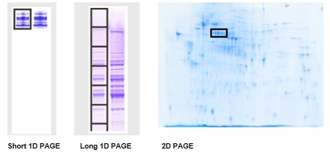
Figure 1. MtoZ Biolabs 2D SDS-PAGE Example
Sample Submission Requirements
1. Liquid or Solid Samples are Acceptable
2. For 1D SDS-PAGE, Generally 2-30 µg of Protein is Required, for 2D SDS-PAGE, Generally 50-1000 µg of Protein is Required
3. Proteins Stained with Coomassie Brilliant Blue, Silver Stain, or Fluorescent Stain are Acceptable


