






PhIP-Seq vs Traditional Phage Display for Epitope Mapping
Introduction
Epitope mapping projects often begin with the same practical question: which peptide regions are recognized by antibodies in a sample, and how should that recognition be measured with enough specificity to support downstream validation? A vaccine team may need to localize antibody binding across an antigen after immunization. An antibody discovery group may need to define peptide targets before lead optimization. An autoimmune or infectious disease program may need to map serum antibody reactivity across many candidate regions rather than a single predefined antigen.
Traditional phage display and PhIP-Seq both use phage-displayed peptides, but they answer different epitope mapping questions. Traditional phage display epitope mapping relies on iterative biopanning, clone enrichment, and sequence validation of selected binders. PhIP-Seq combines antibody immunoprecipitation with next-generation sequencing to quantify enrichment across large peptide libraries in parallel. One workflow is often stronger when binder development or focused antigen enrichment is the goal. The other is often stronger when broad peptide-level epitope profiling across many samples is required.
Understanding how each platform captures epitope information helps teams choose the right mapping strategy before library design, sample use, and validation planning are fixed.
What Epitope Mapping Requires from a Display Platform
Epitope mapping asks which peptide sequences are recognized by antibodies under defined experimental conditions. In most peptide-based workflows, the readout depends on three linked features: how peptides are displayed, how binding is captured, and how positive signals are reported.
Peptide display provides the physical basis for binding. Each phage particle presents a peptide on its surface while carrying the DNA sequence that encodes it. That genotype-phenotype linkage is essential because binding must be traceable back to sequence identity.
Binding capture determines which displayed peptides remain after incubation with antibody-containing samples or purified antibody reagents. Immunoprecipitation, biopanning, and washing conditions all influence which clones survive enrichment.
Signal reporting determines whether results are interpreted as selected clones, enriched peptide populations, or quantitative read counts across a library. This reporting layer is where traditional phage display and PhIP-Seq diverge most clearly.
For linear epitope mapping, both platforms can represent short peptide sequences. For conformational or modified epitopes, neither platform alone may be sufficient, and protein-level or structural methods may still be required.
Core Principles of Traditional Phage Display Epitope Mapping
Traditional phage display epitope mapping is built on repeated rounds of binding enrichment against a target antigen or peptide library. The workflow usually begins with a phage-displayed peptide library incubated with antibody or antigen-coated surfaces. Bound phage are recovered, amplified, and subjected to additional selection rounds until dominant clones emerge.
Each round of biopanning increases the proportion of peptide clones that bind the target under the chosen conditions. After enrichment, individual clones are picked and sequenced by Sanger or targeted sequencing to identify the displayed peptide sequence. When the library is tiled across a protein, overlapping enriched peptides can help localize a linear epitope region.
The strength of this approach lies in clone-level selection. Researchers can recover specific peptide-displaying phage, confirm binding behavior, and in some workflows move selected clones into binder development or follow-up binding assays. For projects focused on one antigen or a narrow target set, iterative enrichment can produce interpretable peptide candidates with direct clone traceability.
Traditional phage display epitope mapping is less optimized for parallel quantification across very large libraries and many sample groups. Results are often dominated by the strongest enriched clones rather than a full quantitative profile of all peptide binders in the library.
Core Principles of PhIP-Seq Epitope Mapping
PhIP-Seq epitope mapping uses the same phage display principle but changes the readout from clone picking to sequencing-based enrichment analysis. A peptide library is incubated with serum, plasma, or another antibody-containing sample. Antibody-bound phage are immunoprecipitated, DNA is extracted, and next-generation sequencing quantifies representation of each peptide-encoding clone.
Because every displayed peptide carries a trackable DNA barcode, antibody binding events can be converted into read counts. Input library sequencing and negative controls support normalization and background filtering. Enriched peptides are mapped back to protein coordinates when the library is tiled or proteome-derived.
PhIP-Seq is designed for parallel epitope screening across large peptide spaces. A single experiment can profile many peptide targets simultaneously and compare enrichment patterns across case-control groups, vaccine time points, or treatment cohorts. For linear epitope discovery, adjacent tiled peptides with shared enrichment often provide stronger mapping evidence than isolated single-peptide signals.
PhIP-Seq does not usually deliver individual selected clones ready for immediate binder development in the same way traditional biopanning does. Its value is in broad, quantitative epitope profiling and candidate region identification at library scale.
Standard Workflow Comparison
Traditional phage display epitope mapping typically follows a clone-centered path. Library construction or selection, target incubation, washing, elution, bacterial amplification, and repeated panning rounds lead to clone isolation and sequence confirmation. Validation may include peptide synthesis, ELISA, or binding assays on selected sequences.
PhIP-Seq epitope mapping follows a population-centered path. Library incubation, immunoprecipitation, DNA recovery, sequencing, read mapping, normalization against input, and enrichment analysis produce a ranked peptide or tiled-region output. Validation usually moves to peptide arrays, targeted ELISA, or orthogonal binding assays on prioritized candidates.
The difference is not only throughput. Traditional phage display emphasizes recovering binders. PhIP-Seq emphasizes measuring antibody recognition patterns across a defined library with quantitative comparison.
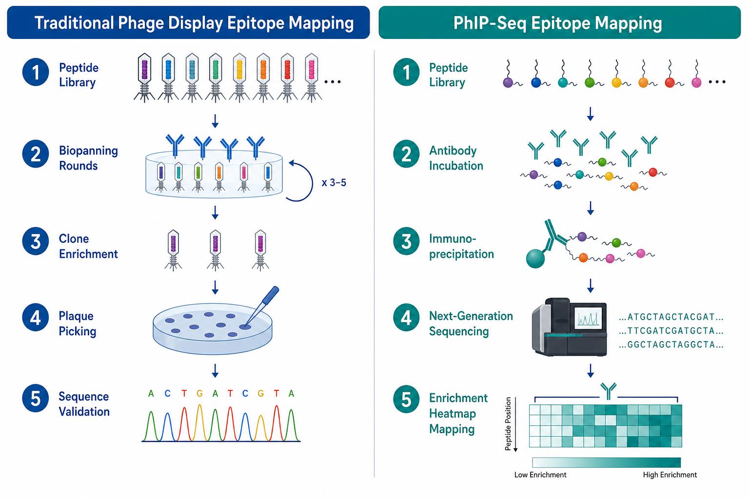
Figure 1. Traditional phage display epitope mapping uses iterative biopanning and clone validation, while PhIP-Seq uses immunoprecipitation and sequencing-based enrichment mapping.
Related Services
PhIP-Seq Antibody Analysis Service
Antibody Epitope Mapping Service
Peptide Array-Based Epitope Mapping Service
High-Throughput Peptide Epitope Mapping Service
Researchers planning epitope mapping can consult MtoZ Biolabs to review sample type, library design, and whether clone-based phage display enrichment or sequencing-based PhIP-Seq profiling better matches the project goal.
Method Comparison for Epitope Mapping
Both platforms can support linear epitope mapping, but they differ in library use, output type, and downstream utility. The table below summarizes common technical differences relevant to project design.
|
Comparison Dimension |
Traditional Phage Display Epitope Mapping |
PhIP-Seq Epitope Mapping |
|---|---|---|
|
Primary readout |
Enriched clone sequences after panning |
Sequencing read enrichment across library peptides |
|
Library scale |
Often focused on one antigen or smaller target set |
Suited to large tiled or proteome-scale libraries |
|
Sample comparison |
Usually sequential or assay-by-assay |
Supports parallel comparison across many samples |
|
Epitope localization |
Strong when tiled library enrichment is analyzed clone by clone |
Strong when tiled enrichment patterns are quantified across regions |
|
Binder recovery |
Selected clones can support downstream binder workflows |
Focuses on epitope discovery rather than clone recovery |
|
Quantitative profiling |
Limited for full-library representation |
Designed for count-based enrichment analysis |
|
Typical validation |
Peptide synthesis, ELISA, binding assay on selected clones |
Peptide array, targeted ELISA, orthogonal mapping on prioritized peptides |
Core Technical Advantages and Current Limitations
Traditional Phage Display
Clone-level traceability.
Enriched phage clones can be recovered, sequenced, and tested individually, which supports focused epitope confirmation and binder-oriented follow-up.
Iterative enrichment control.
Multiple panning rounds allow stringency tuning against a defined target antigen or antibody reagent.
Established selection workflow.
Biopanning remains a practical route when the project centers on one antigen and selected peptide binders are the main deliverable.
Limited library-wide quantification.
Results often reflect dominant clones rather than a complete quantitative map of all peptide binders in the library.
Lower natural fit for large cohort comparison.
Comparing many serum samples across a broad peptide library is usually slower and less standardized than sequencing-based profiling.
Conformational epitope constraints.
Displayed peptides primarily represent linear sequences, so conformation-dependent epitopes may remain unresolved without complementary assays.
PhIP-Seq
Parallel peptide profiling.
Large peptide libraries can be screened in pooled format with count-based readout across many targets at once.
Quantitative enrichment analysis.
Sequencing read counts support normalization, background filtering, and group-level comparison across samples.
Cohort-compatible epitope discovery.
Case-control, vaccine time course, and treatment comparison designs can be analyzed within one library framework.
Less direct clone deliverable.
The workflow prioritizes epitope region discovery over recovery of individual binding clones for immediate downstream development.
Library-dependent detection boundary.
Peptides absent from the library cannot be reported, so library design defines the mapping space.
Linear epitope emphasis.
Short displayed peptides may not represent glycan-dependent, lipid-associated, or fully conformational epitopes without additional methods.
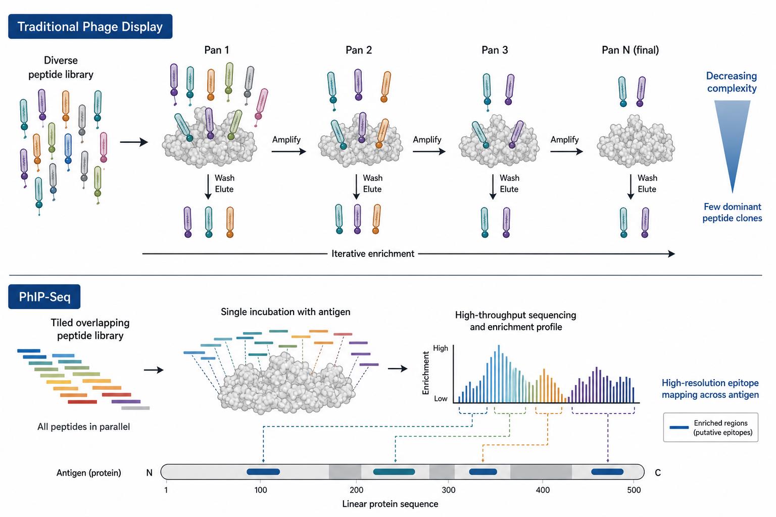
Figure 2. Traditional phage display often converges on enriched clones through iterative panning, while PhIP-Seq maps enrichment across tiled peptide regions in parallel.
Applications in Epitope Mapping Projects
Epitope mapping needs vary by sample type, antigen knowledge, and validation plan. The following application patterns are common in project planning.
Monoclonal antibody epitope localization.
When a purified antibody must be mapped against a defined antigen, traditional phage display with a tiled peptide library can enrich binding clones and localize a linear region through overlapping peptide sequences.
Serum or plasma antibody epitope profiling.
When polyclonal antibody reactivity must be screened across many peptide targets or sample groups, PhIP-Seq provides a scalable route to enrichment-based epitope discovery and comparative profiling.
Vaccine-induced epitope mapping.
Post-immunization samples often require comparison across time points or responder groups. PhIP-Seq supports parallel analysis of peptide enrichment patterns after vaccination.
Focused antigen epitope discovery.
Traditional phage display remains useful when the target antigen is fixed and the goal is to recover peptide binders through repeated selection and clone validation.
Candidate epitope prioritization before validation.
PhIP-Seq can generate a ranked peptide or tiled-region list that is then confirmed by peptide array, ELISA, or protein-level binding assays.
Teams evaluating either platform should define whether the immediate need is clone recovery, broad peptide profiling, or cohort comparison before library construction begins.
Sample and Project Design Considerations
Epitope mapping quality depends on sample suitability and library design as much as on the display platform itself. Common planning factors include:
- Antibody source: purified monoclonal antibody, serum, plasma, or hybridoma supernatant
- Target definition: single antigen, tiled protein region, pathogen panel, or proteome-derived library
- Mapping goal: clone recovery, linear epitope localization, or cohort-level profiling
- Control design: input library, no-antibody control, and matrix-matched background controls for PhIP-Seq
- Validation plan: peptide synthesis, peptide array, ELISA, or protein binding follow-up
- Sample quality: hemolysis, storage history, and repeated freeze-thaw can increase background in both workflows
These factors support feasibility review but do not replace project-specific consultation before library selection and sample submission.
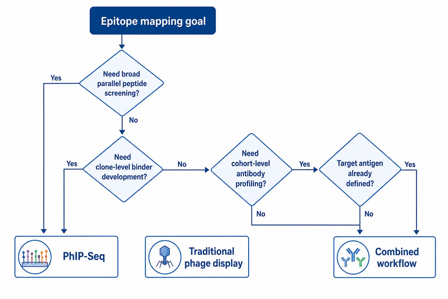
Figure 3. Method selection depends on whether the project requires broad parallel peptide profiling, clone-level binder recovery, or cohort-based epitope comparison.
Frequently Asked Questions
1. What is the main difference between PhIP-Seq and traditional phage display for epitope mapping?
Traditional phage display epitope mapping uses iterative biopanning and clone sequencing to recover peptide binders against a target. PhIP-Seq uses immunoprecipitation and next-generation sequencing to quantify peptide enrichment across large libraries in parallel.
2. Which method is better for serum antibody epitope profiling?
PhIP-Seq is often preferred when serum or plasma samples must be compared across many peptide targets or sample groups because the readout is sequencing-based and supports parallel enrichment analysis.
3. Can traditional phage display map linear epitopes on a defined antigen?
Yes. A tiled phage-displayed peptide library can be used in biopanning against a monoclonal antibody or antigen target, and enriched clone sequences can localize linear epitope regions through overlapping peptides.
4. Does PhIP-Seq replace clone picking in phage display?
Not entirely. PhIP-Seq replaces clone picking as the primary readout for large-scale epitope profiling, but traditional phage display remains useful when selected clones or binder recovery are central to the project.
5. Do both methods detect conformational epitopes?
Both methods primarily represent linear peptides on phage surfaces. Conformational, glycan-dependent, or structurally complex epitopes usually require complementary protein-level or structural assays.
Conclusion
PhIP-Seq and traditional phage display both leverage phage-displayed peptides for epitope mapping, but they convert antibody binding into different types of evidence. Traditional phage display epitope mapping is built around iterative enrichment and clone-level sequence recovery, which supports focused antigen work and binder-oriented follow-up. PhIP-Seq epitope mapping is built around immunoprecipitation and sequencing-based enrichment, which supports broad peptide profiling and sample-group comparison at library scale.
The appropriate choice depends on whether the project requires clone recovery, tiled linear epitope localization on a defined antigen, or parallel antibody epitope profiling across many peptides and samples. Neither platform resolves every epitope type alone, and validation by peptide array, ELISA, or orthogonal binding assays remains important for both workflows. Researchers planning epitope mapping can contact MtoZ Biolabs to review sample type, library strategy, and the validation path best suited to the mapping goal before experiments begin.
How to order?




