


PhIP-Seq vs Protein Microarrays for Antibody Profiling
Introduction
Choosing an antibody profiling method is often a project-defining decision. A serum cohort may need broad immune recognition screening, but the team may not know whether peptide-level discovery or folded protein recognition matters more. A vaccine study may need response breadth across time points. An autoimmune project may need candidate antigens for validation. In each case, the method determines what antibody signal can be detected and what evidence can be claimed.
PhIP-Seq and protein microarrays are both used for antibody profiling, but they are not interchangeable. PhIP-Seq uses a phage display peptide library and sequencing readout to identify enriched peptide targets. Protein microarrays immobilize proteins, protein fragments, or antigens on a surface and detect antibody binding by signal intensity. One method is often stronger for broad peptide-scale discovery. The other can be stronger when recognition of prepared protein antigens is the main question.
The best choice depends on target type, epitope biology, sample availability, throughput, data output, and validation. If your team is deciding between PhIP-Seq antibody profiling, protein microarray antibody profiling, or a staged strategy, MtoZ Biolabs can review the sample set and research goal before the first screening run.
Related Services
| Research Need | Recommended Service |
| Need broad antibody reactivity profiling from serum or plasma | PhIP-Seq Antibody Analysis Service |
| Need peptide-level epitope discovery after screening | Antibody Epitope Mapping Service |
| Need targeted validation of candidate peptide regions | Peptide Array-Based Epitope Mapping Service |
| Need broader peptide epitope screening support | High-Throughput Peptide Epitope Mapping Service |
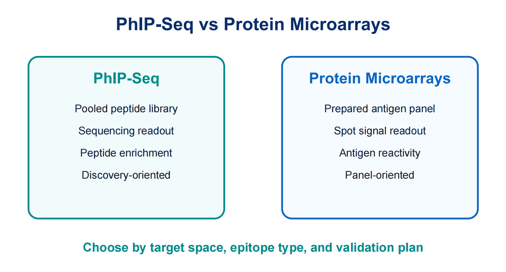
Figure 1. PhIP-Seq and protein microarrays answer different antibody profiling questions.
What Each Method Measures
PhIP-Seq measures antibody binding to peptides displayed by phage clones. After serum or plasma antibodies bind the library, antibody-bound phage are immunoprecipitated and sequenced. The readout is peptide enrichment. This makes PhIP-Seq useful for large peptide spaces, candidate linear epitopes, and cohort-level antibody reactivity patterns.
Protein microarrays measure antibody binding to immobilized proteins, domains, fragments, or antigen preparations. Binding is usually detected through fluorescence or another labeled reagent. The readout is signal intensity at each printed spot. This makes protein microarrays useful when the target set is defined and the question depends on prepared protein antigens.
The distinction is important. PhIP-Seq is not a direct protein-binding assay, and protein microarrays are not usually sequencing-based peptide discovery methods. A PhIP-Seq hit can point to a peptide region or motif. A microarray hit can point to an antigen or construct. Validation should match that evidence type.
Comparison Dimension 1: Target Coverage
Target coverage is one of the clearest differences in PhIP-Seq vs protein microarrays. PhIP-Seq can screen very large peptide libraries. A library may represent viral proteomes, human proteome fragments, pathogen panels, allergens, tumor-associated proteins, or custom tiled antigens. Because the readout is sequencing-based, large libraries can be handled in pooled format.
Protein microarrays are limited by what can be printed, immobilized, stored, and detected on the array surface. Arrays can cover many proteins, but each antigen must be prepared in a usable form. This is powerful for defined panels when proteins are biologically relevant and well controlled.
For discovery projects where the antigen space is broad or poorly known, PhIP-Seq often offers a wider starting point. When candidate antigens are already known, protein microarrays may provide a more direct panel.
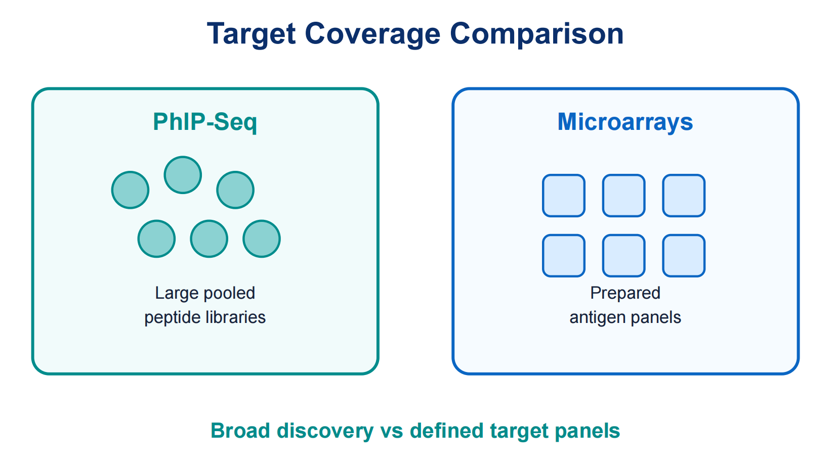
Figure 2. PhIP-Seq favors broad pooled peptide libraries, while protein microarrays depend on prepared antigen panels.
Comparison Dimension 2: Epitope and Antigen Resolution
PhIP-Seq is strongest when the relevant antibody recognition can be represented by linear peptides or short motifs. Dense tiling can help localize candidate peptide regions. This is useful for epitope discovery, cross-reactive motif screening, and narrowing a broad antigen response into smaller candidate regions.
Protein microarrays can preserve more antigen-level context when proteins or domains are folded and displayed properly. This can help when antibodies recognize larger protein regions. However, immobilization, purification, folding, and surface chemistry can affect whether the relevant structure is preserved.
Neither method captures every epitope type. PhIP-Seq may miss conformational, glycan- dependent, lipid-associated, or structure-dependent epitopes. Protein microarrays may miss epitopes that are hidden, denatured, absent, or altered by immobilization. Method choice should follow expected antibody biology.
Comparison Dimension 3: Sample Use, Throughput, and Cohorts
Both methods can support cohort studies, but the workflow differs. PhIP-Seq can profile many samples against the same pooled phage display peptide library. This helps compare enrichment patterns across disease groups, vaccine time points, exposure histories, or treatment cohorts.
Protein microarrays can also process many samples, but cost and logistics depend on array format, antigen panel size, detection, and replicate design. When the same defined panel is tested repeatedly, microarrays can be efficient. When the target space remains broad, PhIP-Seq may scale more naturally.
Sample quality matters for both approaches. Hemolysis, contamination, inconsistent storage, and repeated freeze-thaw cycles can increase background or weaken signal. Matched controls and balanced batch design are more important than simply increasing sample number.
Comparison Dimension 4: Data Output and Interpretation
PhIP-Seq produces sequencing count data that are converted into peptide enrichment values. Analysis usually accounts for library input, read depth, negative controls, replicate behavior, and background binding. Results may be interpreted as enriched peptides, tiled regions, summaries, or cohort-level patterns.
Protein microarrays produce spot-level signal intensity data. Analysis considers local background, spot quality, replicate spots, sample dilution, secondary antibody signal, and normalization across slides or batches. Results may be interpreted as antigen reactivity, relative signal, or group-level differences.
Both methods require careful statistics. A high sequencing read count does not automatically prove a meaningful PhIP-Seq hit. A bright microarray spot does not automatically prove a relevant antigen. Controls, replicate consistency, and independent validation are needed for both.
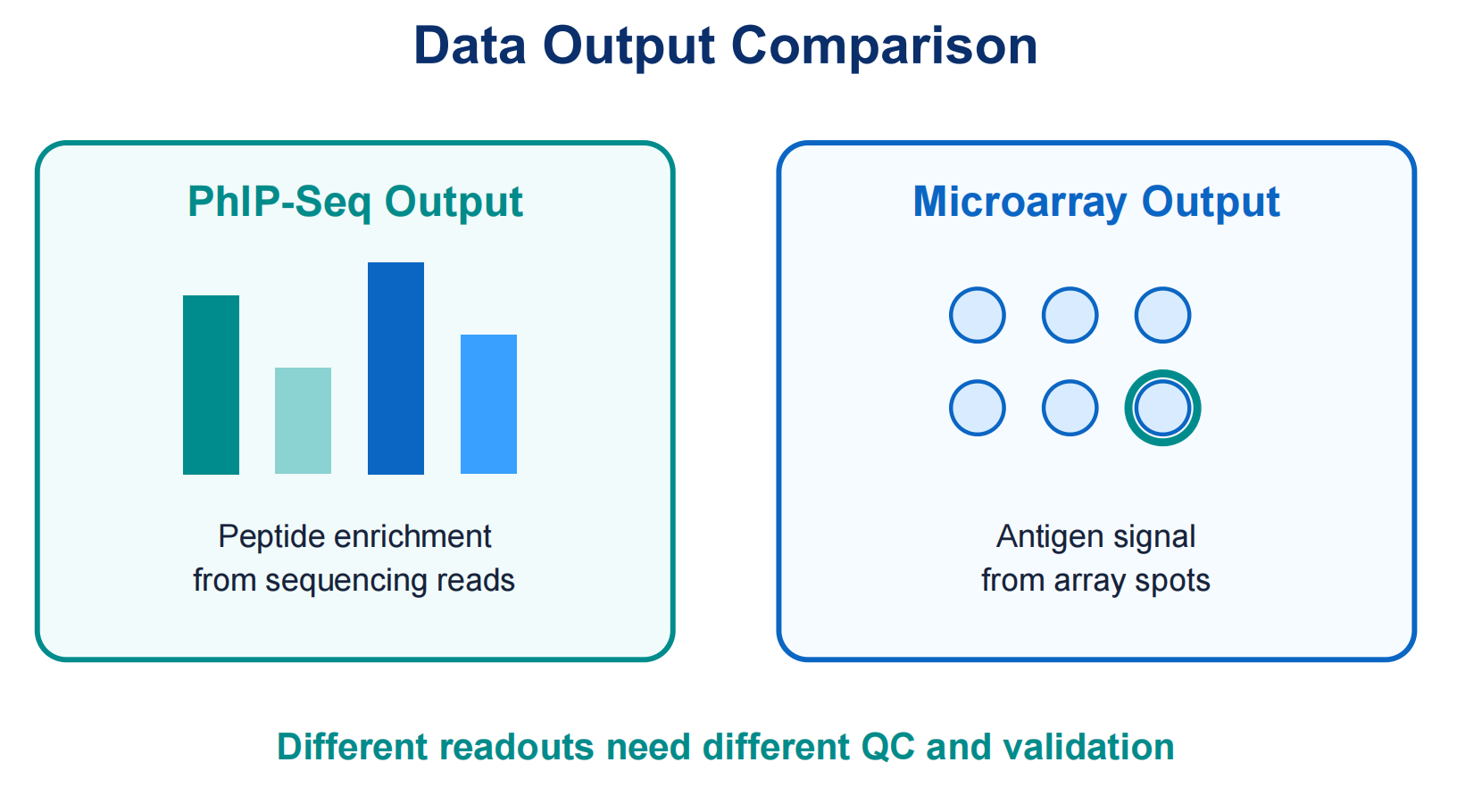
Figure 3. PhIP-Seq produces peptide enrichment data, while protein microarrays produce spot-level antigen signal.
Comparison Dimension 5: Strengths and Limitations
PhIP-Seq is strong for broad discovery, peptide-level localization, pooled library screening, and comparison across many peptide targets. Its limitations include dependence on library design, weaker representation of conformational epitopes, and sensitivity to immunoprecipitation background.
Protein microarrays are strong for defined antigen panels, direct antigen-level screening, and studies where prepared protein constructs are central. Their limitations include antigen production needs, folding or immobilization artifacts, surface background, and limited coverage when the antigen space is large.
The comparison is not about which method is universally better. The practical question is which evidence type is more useful next. If the goal is to discover unknown peptide regions, PhIP-Seq may be preferred. If the goal is to compare binding against known protein antigens, a protein microarray may be more direct.
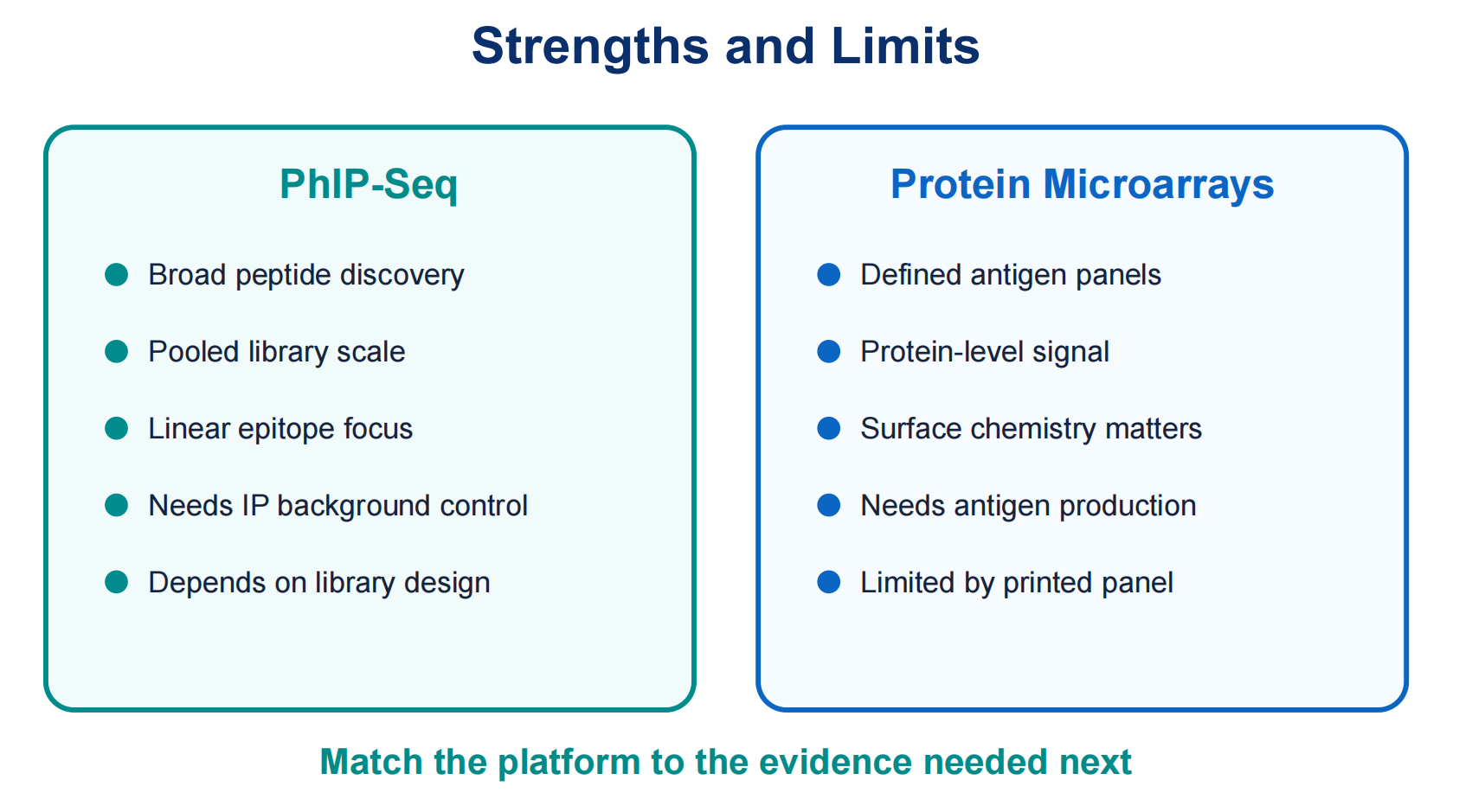
Figure 4. The right method depends on whether peptide discovery or defined antigen screening is the priority.
When to Choose PhIP-Seq
Choose PhIP-Seq when the project needs broad antibody profiling across many peptide targets, especially when the antigen space is large or uncertain. It fits pathogen-wide peptide screening, serum antibody repertoire profiling, linear epitope discovery, cross-reactive motif exploration, and cohort-level immune mapping.
PhIP-Seq is also useful when sequencing-based quantification and pooled library scale matter. A well-designed library can support many samples while maintaining consistent target representation across disease groups, vaccine time points, or exposure histories.
PhIP-Seq is less suitable when the main signal depends on intact protein conformation, glycosylation, lipids, or complex antigen structure. In those cases, protein microarrays or orthogonal binding assays may be needed.
When to Choose Protein Microarrays
Choose protein microarrays when candidate antigens are already defined and the question is whether samples contain antibodies against those prepared proteins or domains. This can support focused antigen panels, autoantigen screening, allergen panels, pathogen panels, or comparison across known targets.
Protein microarrays can also help when antigen-level signal is easier to communicate than peptide-level enrichment. A translational study may need to compare antibody reactivity to known proteins before targeted validation.
Researchers should confirm that printed proteins represent the biology of interest. If folding, modification, construct boundaries, or immobilization affect binding, microarray results need careful follow-up.
When a Combined Strategy Makes Sense
Some projects benefit from both methods. PhIP-Seq can identify candidate peptide regions across a broad library. Protein microarrays, peptide arrays, ELISA, or other assays can then validate selected targets. The reverse can also work: a protein microarray may identify antigen-level candidates, followed by peptide-level mapping.
This staged approach is useful when discovery and validation need different evidence types. PhIP- Seq can generate hypotheses at scale, while follow-up assays test candidates in a focused format. For sample-limited projects, early planning helps preserve material.
Practical Decision Rules
Use PhIP-Seq when target discovery, peptide-level resolution, and broad pooled screening are the priorities. Use protein microarrays when known antigen panels, protein-level signal, and direct comparison across defined targets are the priorities. Use both when the project needs broad discovery followed by focused confirmation.
Researchers should also consider sample volume, expected epitope type, library or antigen availability, cohort size, validation budget, and reporting needs. A method that does not support the next decision can create more work than clarity.
For uncertain projects, early technical review can align the method with sample type, biological question, and validation plan. This is important when serum volume is limited, cohorts are difficult to recollect, or the project must support publication or translational decisions.
Frequently Asked Questions
1. Is PhIP-Seq better than protein microarrays for antibody profiling?
Not universally. PhIP-Seq is often better for broad peptide-level discovery, while protein microarrays are often better for defined antigen panels. The best method depends on target space and epitope biology.
2. Which method is better for epitope mapping?
PhIP-Seq can identify candidate linear peptide regions when the library is designed for that purpose. Protein microarrays usually provide antigen-level information unless paired with fragment arrays or peptide mapping.
3. Which method works better for conformational epitopes?
Protein microarrays may be more relevant when folded proteins are available, but immobilization can still alter structure. PhIP-Seq peptide libraries are stronger for linear or motif-like epitopes.
4. Can the same serum samples be tested by both methods?
Often yes, if enough sample volume is available and handling conditions are compatible. Using both methods can provide complementary evidence, but the study should be planned early.
5. Do hits from either method require validation?
Yes. Important hits should be validated with independent assays such as peptide arrays, ELISA, Western blotting, or targeted immunoassays before use as biomarkers or mechanistic conclusions.
Conclusion
PhIP-Seq vs protein microarrays is not a simple competition between two antibody profiling platforms. PhIP-Seq is often stronger for broad peptide-scale discovery and antibody repertoire profiling. Protein microarrays are often stronger for defined antigen panels and protein-level binding questions. The right choice depends on target space, epitope type, sample constraints, and the decision the data must support.
For teams comparing methods for serum antibody profiling, epitope discovery, vaccine response studies, or autoantibody screening, method selection should happen before sample submission. To choose a workflow that fits your project, contact MtoZ Biolabs for technical review of sample readiness, target strategy, and validation options.
How to order?

