


Micelles Structure Characterization Service | Cryo-EM
Micelles are spherical nanostructures spontaneously formed by surfactants or amphiphilic molecules in aqueous environments, typically consisting of a hydrophobic core and a hydrophilic shell. They are widely used for encapsulating hydrophobic drugs and improving drug solubility and stability. Micelles structure characterization is a high-resolution structural analysis service based on cryogenic electron microscopy (Cryo-EM), specifically designed to characterize the structural features of micelle-based nanocarriers. This technique involves rapidly cooling micelle samples to liquid nitrogen temperature to avoid structural damage caused by drying or staining, followed by low-dose electron beam imaging to faithfully preserve and visualize the nanostructure in its native state.
Micelles structure characterization service is widely applied in drug delivery systems, self-assembled nanomaterials, surfactant structure optimization, and biomembrane modeling. Cryo-EM enables researchers to assess micelle stability, drug-loading capacity, and particle size uniformity, providing essential structural insights for the development, formulation screening, and quality control of micelle-based nanomedicines.
Services at MtoZ Biolabs
Based on the high-end cryogenic electron microscopy platform, the micelles structure characterization service based on Cryo-EM provided by MtoZ Biolabs enables rapid freezing and in situ fixation of micelle samples under ultra-low temperature conditions, achieving high-resolution imaging without the need for staining. This service provides critical data on micelle particle size distribution, morphological features, hydrophobic core and hydrophilic shell structure, and aggregation state. Through automated image acquisition and structural analysis, the results deliver nanometer-scale resolution and high structural fidelity, making the service ideal for structural research and quality assessment of drug delivery systems, self-assembled nanostructures, and surfactant-based micellar formulations.
Analysis Workflow
1. Sample freezing fixation
Micelle samples are rapidly cooled to liquid nitrogen temperature to form vitreous ice, preserving their native structure and preventing aggregation or collapse.
2. Cryo-EM Image Acquisition
Low-dose, high-resolution imaging is performed using a cryogenic transmission electron microscope to avoid radiation damage to the sample.
3. Image Preprocessing and Particle Identification
Images are denoised, corrected, and analyzed to identify and extract target micelle regions.
4. Structural Analysis and Measurement
Micelle particle size, morphology, core–shell structure, and aggregation state are analyzed to assess stability and uniformity.
5. Data Delivery and Report Generation
Original images, structural parameters, and a professional analysis report are provided to support scientific publication and formulation development.
Service Advantages
1. High Structural Fidelity
Micelles are rapidly vitrified at ultra-low temperatures to preserve their native structure and prevent deformation caused by drying or staining.
2. No Staining and Low Damage
Low-dose, stain-free imaging minimizes radiation damage and external interference, enhancing image clarity and data reliability.
3. High Resolution
With the support of advanced Cryo-EM platforms, nanometer-scale resolution allows clear visualization of micelle core–shell structures and aggregation patterns.
4. Broad Compatibility
Suitable for structural analysis of drug-loaded micelles, surfactant-based micelles, and various self-assembled nanostructures.
Applications
1. Drug Delivery System Optimization
The micelles structure characterization service can be used to analyze micelle particle size, core–shell structure, and aggregation state during drug loading, helping to evaluate encapsulation efficiency and stability.
2. Structural Evaluation of Nanomedicines
Supports morphological observation and structural consistency assessment of novel polymeric micelles, targeted micelles, and other nanocarriers.
3. Surfactant Performance Analysis
The micelles structure characterization service can reveal the self-assembly behavior of surfactants in aqueous environments and assess how different formulations influence micelle structure.
4. Development of Self-Assembled Materials
Enables visualization of micelles formed from amphiphilic molecules or supramolecular systems, guiding the design and optimization of advanced nanomaterials.
5. Quality Control and Scale-Up
The micelles structure characterization service can be applied in industrial production to monitor batch-to-batch consistency and microstructural integrity of micelles, enhancing process stability and product reliability.
Case Study
1. Structural Characterization of Amphiphilic Homopolymer Micelles Using Light Scattering, SANS, and Cryo-TEM
This study aims to characterize the structure of amphiphilic homopolymer micelles formed from poly(N-isopropylacrylamide) (PNIPAM) using light scattering, small-angle neutron scattering (SANS), and cryogenic transmission electron microscopy (cryo-TEM). The subject of investigation is PNIPAM polymers with hydrophobic end-group ligands, which self-assemble into spherical micelles in aqueous solution. Cryo-TEM imaging clearly reveals the micelles’ core–shell structure and allows extraction of their radial density profiles. The results show that the micelles consist of a dense hydrophobic core and a gradually diluted hydrophilic corona, with PNIPAM chains primarily distributed in the outer region. The study demonstrates that cryo-TEM effectively preserves and visualizes the nanostructural features of micelles, serving as a powerful tool for analyzing the self-assembly behavior of amphiphilic polymers.
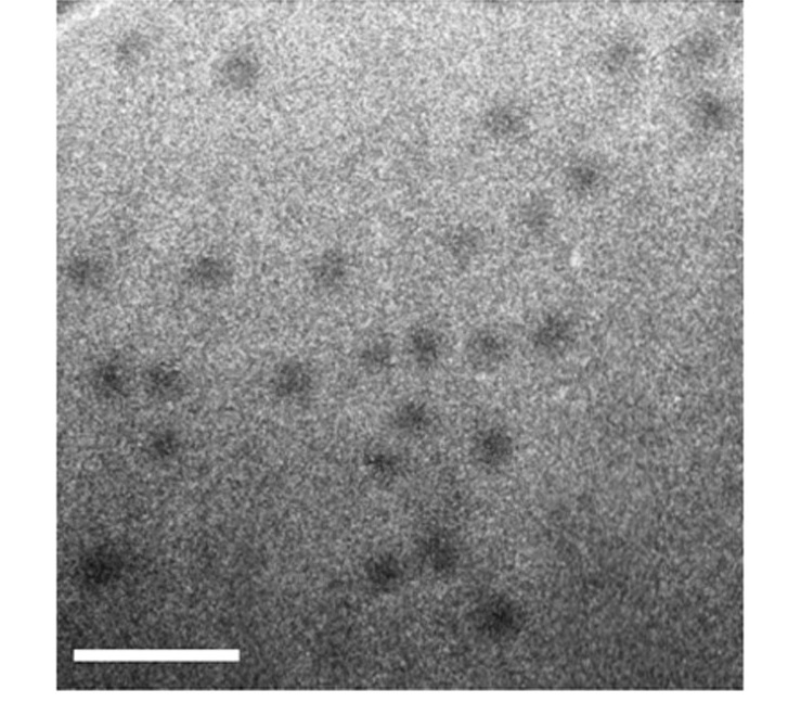
Patterson, J P. et al. Macromolecules, 2013.
Figure 1. Cryo-TEM Micrograph of Micelles.
2. Structure and Dynamics of Poly(oxyethylene) Cholesteryl Ether Wormlike Micelles: Rheometry, SAXS, and Cryo-TEM Studies
This study aims to characterize the structure of poly(N-isopropylacrylamide) (PNIPAM) homopolymer micelles using cryogenic transmission electron microscopy (cryo-TEM), with a focus on analyzing the micellar core–corona architecture and radial density distribution. The subject is end-functionalized PNIPAM homopolymers that, despite containing less than 5% hydrophobic ligands by mass, can self-assemble into well-defined spherical micelles in aqueous solution. The authors performed cryo-TEM imaging and applied grayscale analysis to extract the micelles’ radial density profiles, revealing a gradually decreasing corona density formed by PNIPAM chains. These findings are consistent with small-angle neutron scattering (SANS) results, which show similar structural features. The study concludes that cryo-TEM provides direct, detailed insights into micellar density distribution and serves as a powerful complementary tool to scattering techniques like SANS for investigating solution-assembled nanostructures.
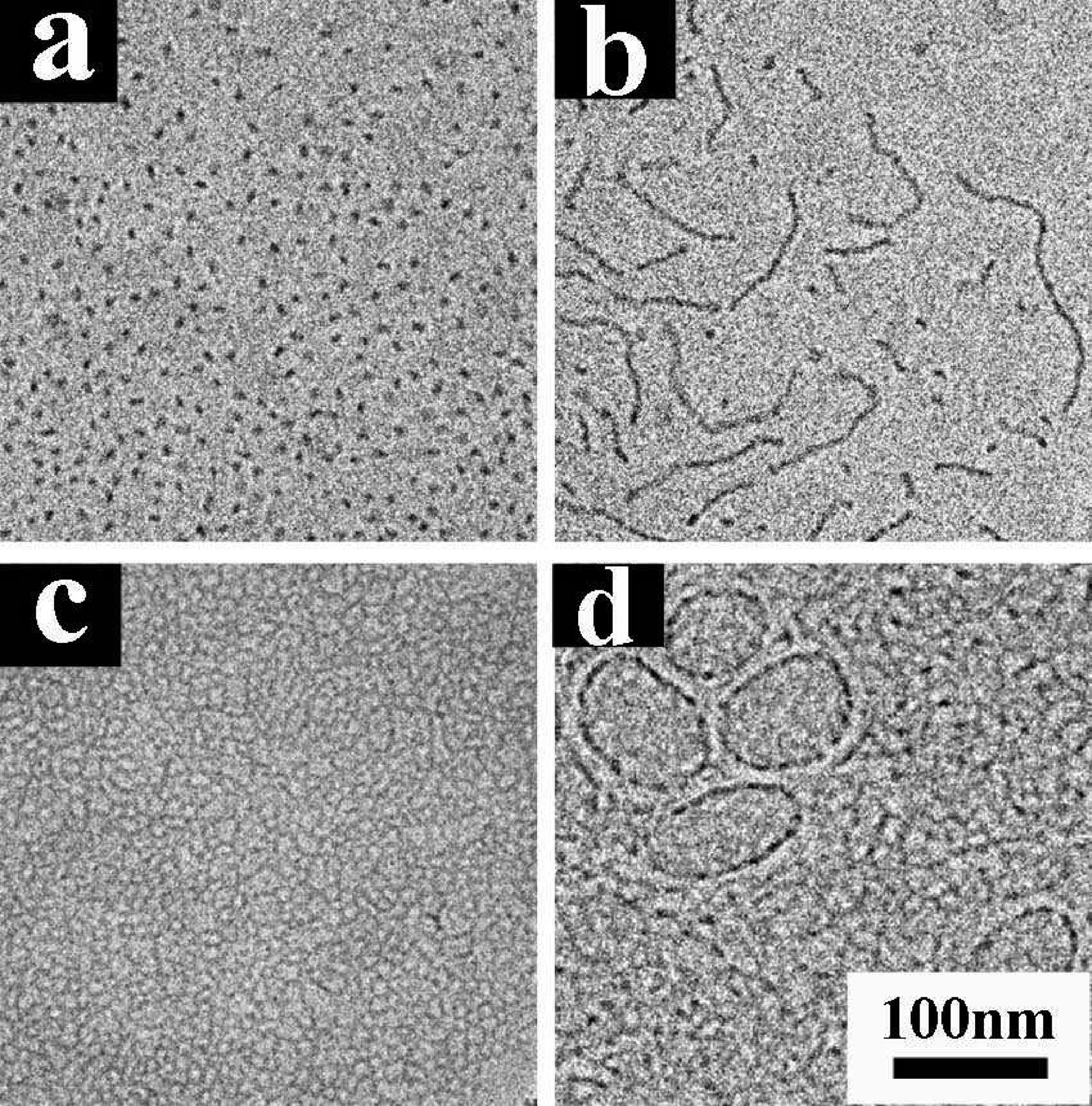
Shrestha, R G. et al. Langmuir, 2011.
Figure 2. Cryo-TEM Micrographs of Micelles Formed by Different Conditions.
FAQ
Q1: Why Is Cryo-EM Suitable for Micelle Structural Analysis?
A1: Cryo-EM allows in situ imaging without damaging micelle structures, avoiding structural deformation caused by drying or staining. It accurately reveals micelle size, core–shell architecture, and aggregation state, making it especially suitable for fine-structured and environmentally sensitive micelle systems.
Q2: How Does Cryo-EM Differ from Conventional TEM?
A2: Conventional TEM typically relies on negative staining, which may introduce structural compression or artifacts. In contrast, Cryo-EM uses rapid vitrification to preserve the native morphology of micelles, providing higher structural fidelity and enabling more accurate characterization.
How to order?

