


Membrane Proteins Structure Characterization Service | Cryo-EM
- Structure-based screening and optimization of membrane-targeting drugs
- Mechanistic studies of GPCRs, ion channels, and other membrane protein families
- Structural and inhibitory analysis of viral transmembrane proteins
- Interaction studies between antibodies/ligands and membrane proteins
- Conformational change analysis of transporters, transmembrane enzymes, and related proteins
Membrane proteins represent one of the most functionally complex and diverse classes of proteins in living organisms. They play essential roles in signal transduction, substance transport, cell recognition, and metabolic regulation. Statistics indicate that over 60% of drug targets are membrane proteins, including G protein-coupled receptors (GPCRs), ion channels, transmembrane enzymes, and transporters. However, due to their high hydrophobicity, structural instability, and low expression levels, traditional crystallographic approaches face significant challenges in studying their structures.
With the rapid development of cryo-electron microscopy (Cryo-EM), researchers are now able to visualize membrane proteins in near-native states, overcoming the limitations of crystallization and conformational heterogeneity. Cryo-EM has thus become a key technique for elucidating the three-dimensional structures of membrane proteins.
Leveraging an advanced Cryo-EM platform and an experienced structural biology team, MtoZ Biolabs provides end-to-end Membrane Proteins Structure Characterization Service based on Cryo-EM. We support researchers in exploring membrane protein mechanisms and advancing structure-based target research and drug development.
Technical Principles
Cryo-EM is a low-temperature imaging technique that captures biological samples in vitreous ice, avoiding structural artifacts caused by traditional staining or dehydration processes. For membrane proteins, Cryo-EM enables high-resolution imaging and 3D reconstruction while preserving their native conformations.
In recent years, single particle analysis (SPA) has become the most widely used Cryo-EM technique for structural analysis, particularly suited for heterogeneous and difficult-to-crystallize membrane proteins. By classifying and aligning a large number of particle images, researchers can reconstruct the 3D structure at sub-nanometer resolution, providing structural insights for functional studies and drug discovery.
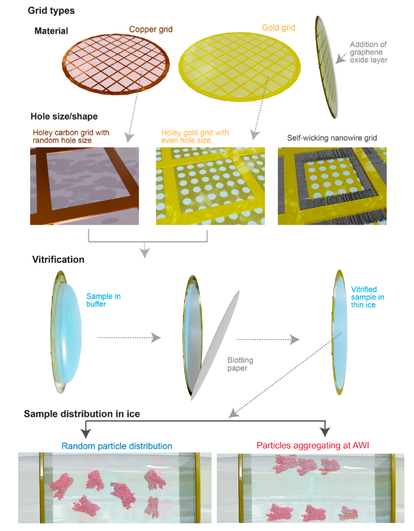
Piper, SJ. et al. Chem Rev. 2022.
Figure 1. Overview of the Steps Involved in Single-Particle Cryo-EM on Membrane Proteins
Analysis Workflow
MtoZ Biolabs’ Membrane Proteins Structure Characterization Service covers the full workflow from protein evaluation to structural modeling, including:
1. Sample Pre-assessment and Quality Control
Clients provide expressed or purified membrane protein samples, which undergo initial quality checks including SDS-PAGE, dynamic light scattering (DLS), and concentration determination.
2. Cryo Sample Preparation
Samples are vitrified in liquid nitrogen, forming thin ice films that preserve the protein’s solution-state conformation.
3. Data Collection
High-quality single-particle images are collected using 300 kV cryo-transmission electron microscopy.
4. Image Processing and 3D Reconstruction
This includes motion correction, particle picking, 2D classification, 3D reconstruction, and map refinement.
5. Data Delivery
A comprehensive data package is provided, including raw cryo-EM images, image processing reports, 3D density maps, and other relevant deliverables.
Service Advantages
1. No Crystallization Required: Eliminates the dependence on crystallization, suitable for proteins that are hard to crystallize.
2. Conformational Insight: Capable of capturing multiple conformational states, ideal for studying protein dynamics.
3. Low Sample Consumption: Requires less sample than X-ray crystallography, reducing experimental cost.
4. High-Resolution Imaging: Enables near-atomic resolution structure analysis, supporting drug-binding site identification and structure-based optimization.
5. Integrated Workflow: Provides a standardized, one-stop solution from sample evaluation to 3D modeling.
Applications
Membrane Proteins Structure Characterization Service is applicable to a wide range of research scenarios, including:
Case Study
1. Cryo-EM Analysis of a Membrane Protein Embedded in the Liposome
This study presents a practical workflow for cryo-EM three-dimensional structural analysis of membrane proteins (MPs) embedded in liposomes. Using AcrB as a model, the researchers combined optimized proteoliposome preparation, graphene-supported cryo-sample preparation, and efficient particle selection strategies to successfully resolve the trimeric structure of AcrB at 3.9 Å resolution. The study found that AcrB maintains its conformation across different membrane curvatures, supporting the applicability of this approach for analyzing MPs whose functions depend on membrane electrochemical gradients or curvature. Membrane Proteins Structure Characterization Service enables structural analysis of samples in liposome-reconstituted environments, utilizing efficient particle processing and cryo-EM reconstruction techniques. It is well-suited for studying membrane proteins with soluble domains or functions influenced by membrane environments.
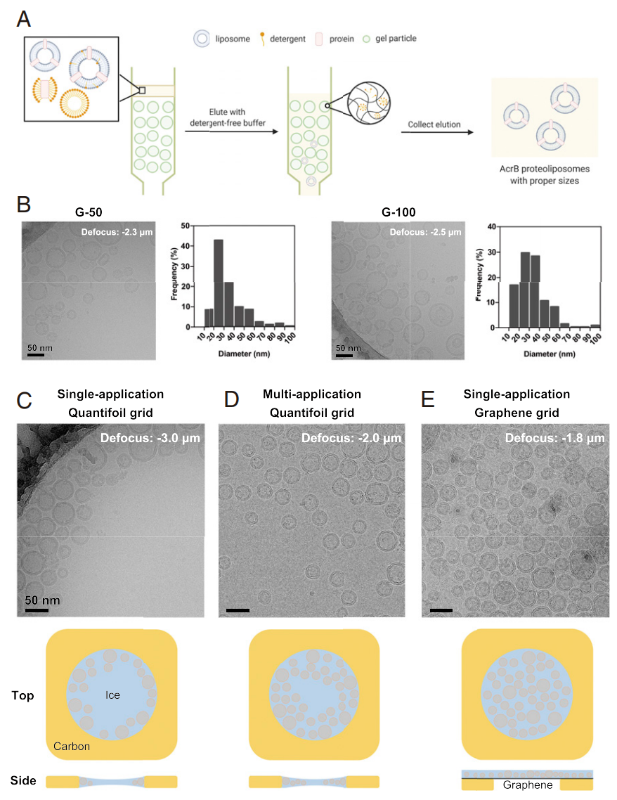
Yao, X. et al. Proc Natl Acad Sci U S A. 2020.
Figure 2. Optimization of Proteoliposome Cryo-Sample Preparation
2. A Methodology to Simultaneously Solve Cryo-EM structures of Membrane Proteins
This study introduces an iterative strategy called “Build and Retrieve” (BaR), which enables the identification and cryo-EM structural determination of multiple membrane proteins and their complexes from heterogeneous, impure samples. The method was successfully applied to inner and outer membrane proteins from E. coli crude membranes and lysates, resolving structures of proteins under 100 kDa and with abundances below 10%, demonstrating cryo-EM’s capability in high-resolution structure analysis within complex mixtures. Membrane Proteins Structure Characterization Service enables structure separation and analysis of multiple membrane proteins from heterogeneous or impure samples through advanced reconstruction strategies, making it suitable for studying low-abundance or small membrane proteins.
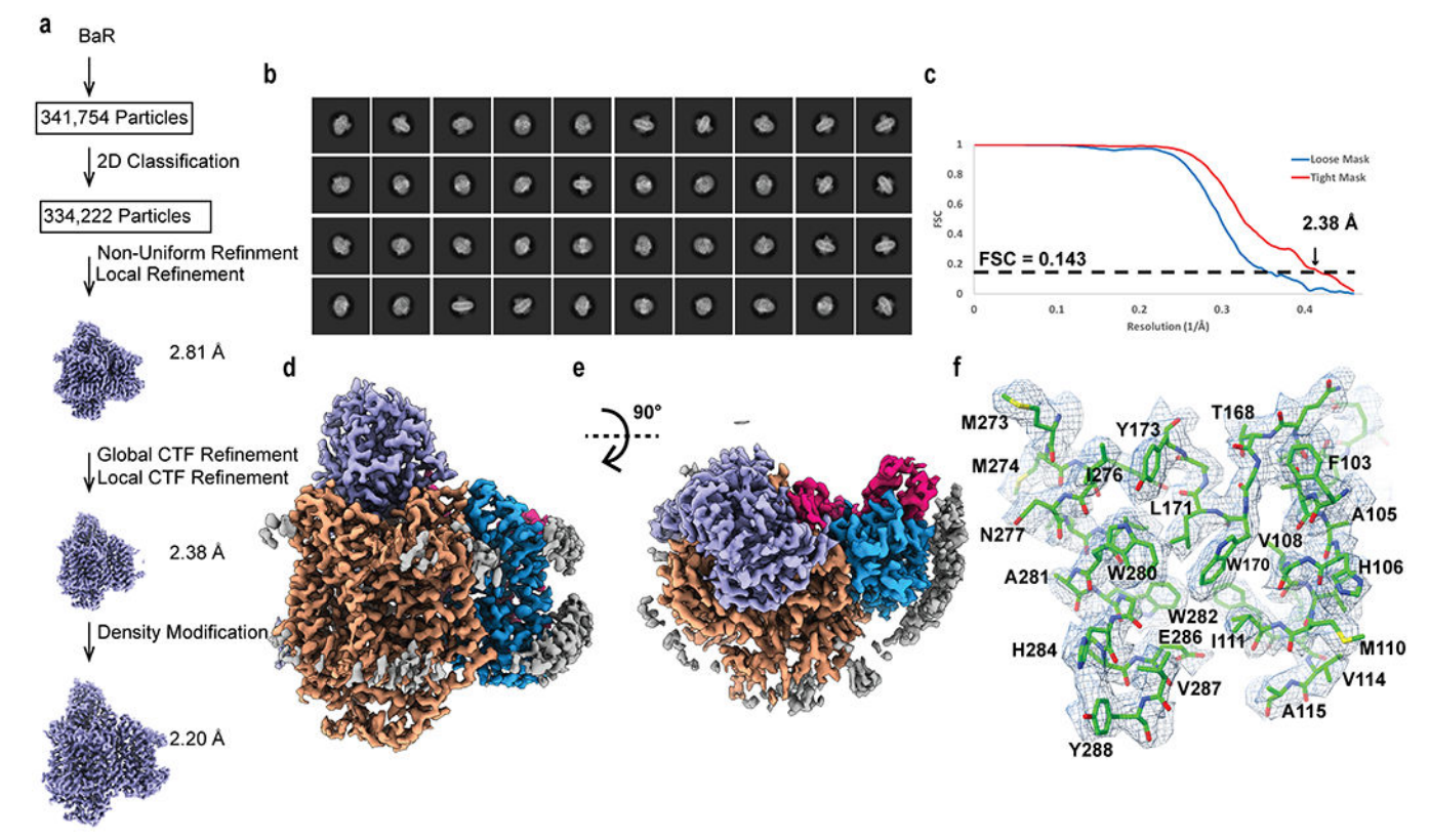
Su, CC. et al. Nat Methods. 2021.
Figure 3. Cryo-EM Analysis of the E. coli Cytochrome bo3 Complex
FAQ
Q1: What are the specific requirements for membrane protein samples?
A1: We recommend samples with >90% purity and a concentration ≥1 mg/mL. Buffers should be free of glycerol and high salt. We also offer consultation for optimized sample preparation protocols.
Q2: Can I provide only the expression vector and have MtoZ Biolabs handle expression and purification?
A2: Yes. We can assist with expression system setup and protein production as needed.
Q3: Do you support structure analysis of protein-drug or protein-antibody complexes?
A3: Yes. We can help design co-complex preparation strategies tailored to your structural goals.
MtoZ Biolabs is committed to delivering high-quality Membrane Proteins Structure Characterization Service. Our Cryo-EM platform empowers membrane protein research at structural depth. For more service details, feel free to contact our technical team.
How to order?

