



Tandem mass tag (TMT) reagents are isobaric chemical tags that label peptide amines so every channel shares the same precursor mass at MS1 but releases channel-specific reporter ions after fragmentation. In practice, digested samples are tagged, pooled, and analyzed by liquid chromatography-tandem mass spectrometry (LC-MS/MS). Quantification reads the relative intensities of those low m/z reporter peaks at MS2 or, on some workflows, MS3 such as synchronous precursor selection MS3 (SPS-MS3), not ordinary MS1 precursor areas. Each reporter mass acts as a channel fingerprint inside one closed multiplex plex. The valid output is relative same-peptide comparison across labeled channels in that plex, not absolute protein concentration without matched standards (Thompson et al. 2003).
Researchers who already have defined samples, comparison groups, or a specific project objective can also review the MtoZ Biolabs TMT Quantitative Proteomics Analysis Service for project-specific feasibility and analysis planning.
TMT Reporter Ions at a Glance
Stage |
What you observe |
Role in TMT quantification |
|---|---|---|
MS1 |
One combined precursor for the same labeled peptide |
Identification and selection; not the primary TMT ratio readout |
MS2 |
Peptide fragment ions plus reporter ions near the low m/z region |
Default reporter-intensity ratios across channels |
MS3 (optional) |
Further isolation of a fragment before reporter readout |
Can reduce co-isolation mixing at MS2 (Ting et al. 2011; McAlister et al. 2014) |
Software |
Reporter peak areas summed to peptide and protein values |
Builds the relative quantitative matrix for the plex |
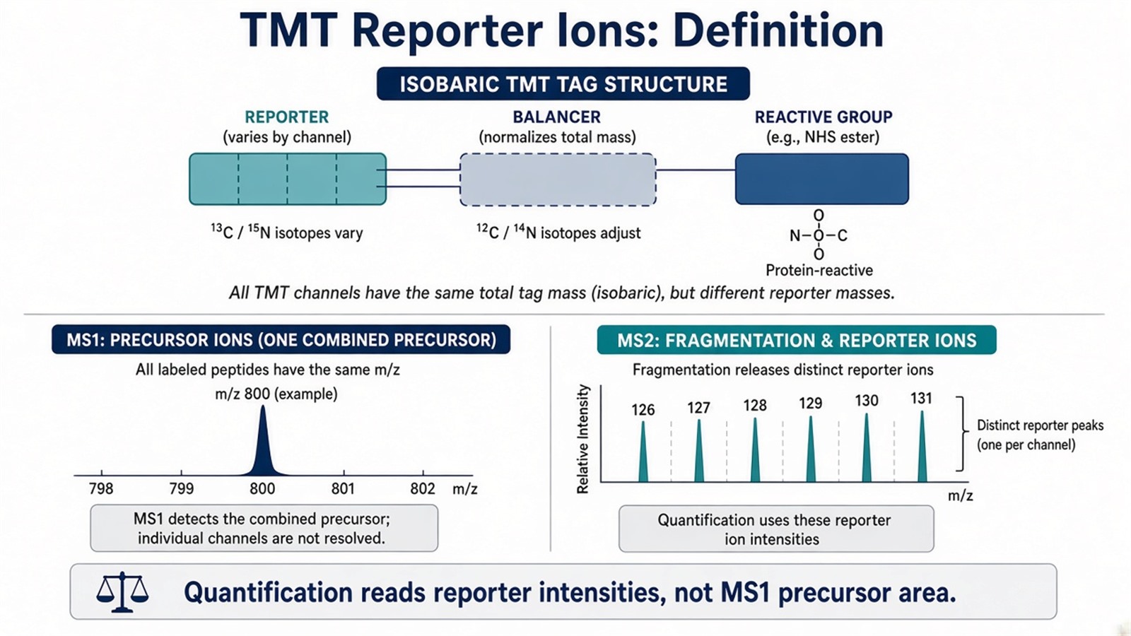
Figure 1. TMT quantification is defined by reporter-ion intensities released after fragmentation, while isobaric design keeps channels merged at MS1.
Reporter ions are the quantitative signal; the mass balancer keeps tags isobaric until fragmentation.
Mass Spectrometry Analysis Path From Labeling to Ratios
TMT reporter analysis follows a fixed mass spectrometry sequence.
1. Digestion and channel labeling
Proteins are digested, usually with trypsin, then each sample receives one TMT channel. Incomplete labeling or unequal loads before pooling become ratio artifacts that later acquisition cannot remove.
2. Pooling and LC-MS/MS acquisition
Labeled peptides are mixed and injected once per plex fraction. Data-dependent acquisition (DDA) is the common mode for reading TMT reporters because precursors are selected and fragmented in real time. Data-independent acquisition (DIA) is an unlabeled acquisition strategy; it is not a substitute definition for TMT reporter readout.
3. Reporter extraction and protein summarization
Search software assigns peptide sequences to fragment spectra, integrates reporter-ion peaks per channel, and rolls peptide evidence into protein-level relative ratios. Tools such as Proteome Discoverer and MaxQuant are widely used, but the scientific claim still depends on plex design, FDR control, and QC rather than software name alone.
For the full experimental sequence beyond reporter readout, researchers can refer to A Detailed Workflow of TMT-Based Quantitative Proteomics. For labeling chemistry detail, see Mechanism of TMT Labeling in Mass Spectrometry.
MS2 Reporter Readout and Co-Isolation Limits
At MS2, the instrument isolates a precursor window and fragments all ions inside it. If more than one peptide co-isolates, reporter ions from different peptides can mix. That co-isolation interference compresses fold changes toward unity and is a core limit of MS2-only TMT quantification in complex digests (Ting et al. 2011).
Mitigations include narrower isolation windows where instrument settings allow, offline fractionation to simplify precursors, and MS3 or SPS-MS3 routes that re-isolate a fragment before reporter integration (McAlister et al. 2014). These steps change how cleanly reporters are read; they do not fix wrong channel maps, incomplete labeling, or absolute concentration claims.
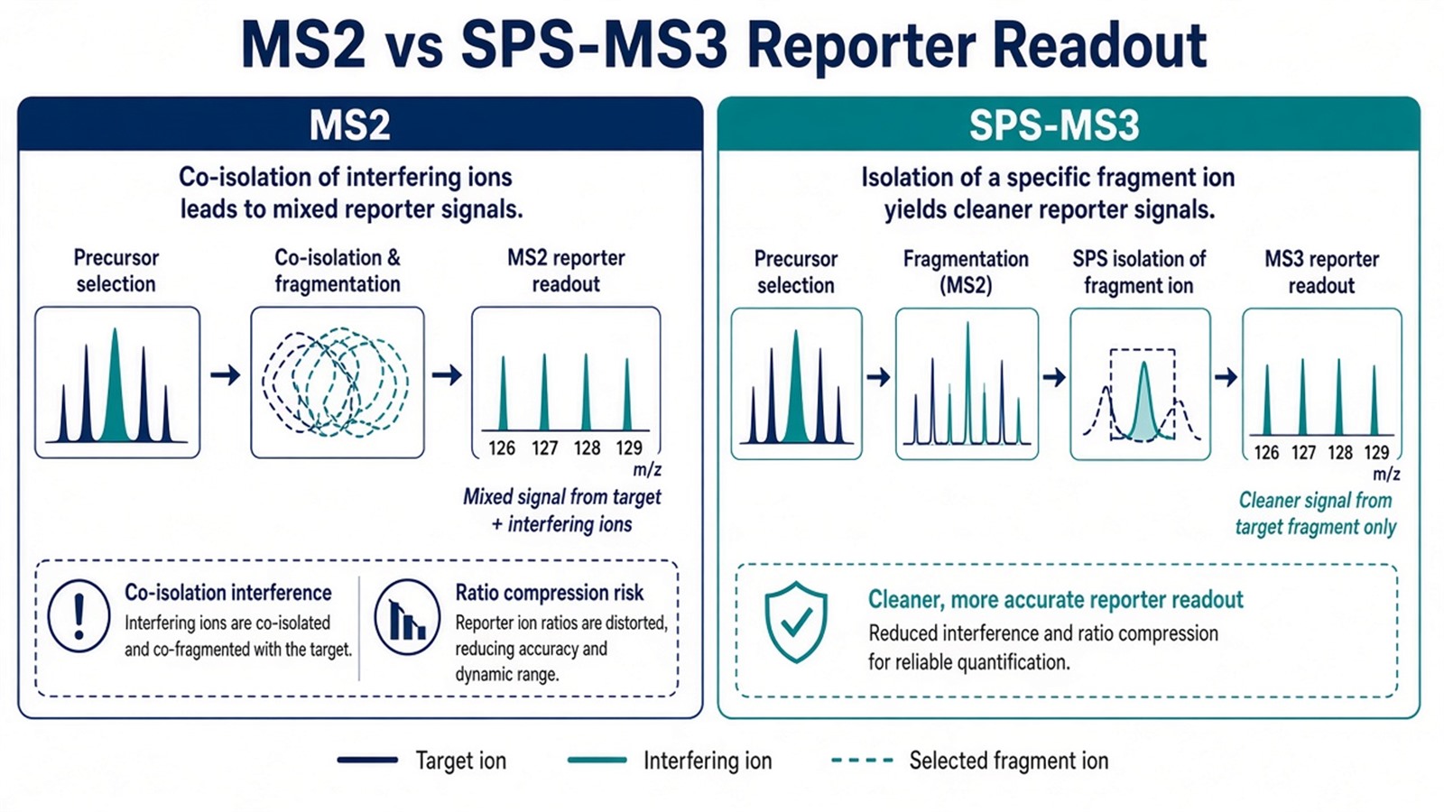
Figure 2. TMT reporter analysis must account for co-isolation at MS2; MS3 strategies can reduce mixed reporter signal but do not replace sound plex design.
For acquisition-focused mitigation detail, see TMT MS3 and SPS-MS3: How to Mitigate Ratio Compression and Enhance Quantitative Accuracy and How Can SPS-MS3 Be Utilized to Optimize TMT Workflows and Minimize Ratio Compression?. For why compressed ratios stay relative, see How Does Ratio Compression Arise in TMT-Based Quantitative Proteomics and Why Is It Limited to Relative Quantification?.
Judgment Criteria: When TMT Reporter Quantification Fits
Study need |
TMT reporter workflow often fits |
Reassess before labeling |
|---|---|---|
Claim |
Relative ratios across channels in one plex |
Absolute concentration or diagnostic cutoff |
Design |
Closed channel map before mixing |
Open enrollment after the plex is pooled |
Chemistry |
Clean amine labeling at N-termini and lysines |
Blocking buffers or failed digestion |
Readout plan |
DDA with MS2 or planned MS3 reporter integration |
Expecting unlabeled DIA to read TMT reporters |
Scale |
Groups and replicates fit one kit |
Sample count forces many plexes without bridging |
Prefer TMT reporter quantification when every planned comparison can share one labeled run and the question is relative same-run discovery. Prefer label-free quantification (LFQ) or unlabeled DIA when samples must stay independent injections. Prefer stable isotope-labeled standard (SIS) or AQUA-style assays when defined absolute units are required.
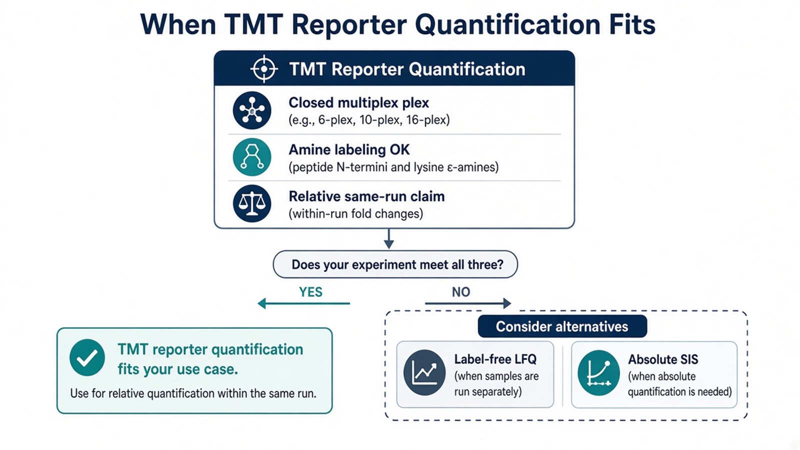
Figure 3. Reporter-ion TMT fits closed relative multiplex designs; change strategy when enrollment, chemistry, or the quantitative claim differs.
For method selection against unlabeled routes, see TMT vs Label Free Quantitative Proteomics and DDA vs. DIA vs. TMT in LC-MS/MS Proteomics: How to Choose the Right Strategy. For design trade-offs, see Advantages and Disadvantages of TMT-Based Quantitative Proteomics Analysis.
Application Boundaries and Typical Uses
Reporter-ion TMT supports multi-condition treatment panels, genotype contrasts, dose-response arms, and time-course designs that fit one commercial multiplex kit, because channels are compared inside the same acquisition path. It suits biomarker discovery lists that need relative ranking within a frozen plex, not standalone clinical cutoffs.
Weak reporter signal can follow low input, ion suppression, or heavy fraction complexity. Those are experimental limits to plan around with loading, cleanup, and fractionation, not reasons to treat reporter ratios as absolute amounts.
Diagnostic decision rules, regulatory assay claims, and cross-plex absolute stoichiometry sit outside ordinary TMT reporter reporting unless a separately validated framework is in place.
Once the channel map, fragmentation level (MS2 versus MS3), and relative-ratio claim are defined, the project can be evaluated as a TMT reporter-ion study. Researchers can review the MtoZ Biolabs TMT Quantitative Proteomics Analysis Service for sample evaluation, feasibility assessment, and workflow planning.
Frequently Asked Questions
1. What is a TMT reporter ion?
It is the low m/z fragment-specific signal released from an isobaric TMT tag after peptide fragmentation. Its intensity carries relative abundance for that channel.
2. Why are TMT peptides isobaric at MS1?
Balancer groups offset channel mass differences so the same peptide from every tag shares one precursor mass until fragmentation.
3. Are reporter-ion ratios the same as MS1 peak areas?
No. TMT quantification is defined by reporter peaks after fragmentation, not by comparing MS1 precursor areas across unlabeled runs.
4. Does MS3 change what a reporter ion is?
No. MS3 changes where reporters are measured to reduce interference. The reporter masses and relative readout principle stay the same.
5. Can TMT reporter data report absolute protein amount?
Not by default. Reporters support relative comparison inside one plex unless matched standards and calibration are added in a separate absolute assay design.
Reference
- A. Thompson, J. Schaefer, K. Kuhn, S. Kienle, J. Schwarz, G. Schmidt, T. Neumann, C. Hamon (2003). Tandem mass tags: a novel quantification strategy for comparative analysis of complex protein mixtures by MS/MS. Anal. Chem., 75, 1895-1904. https://doi.org/10.1021/ac0262560
- L. Ting, R. Rad, S.P. Gygi, W. Haas (2011). MS3 eliminates ratio distortion in isobaric multiplexed quantitative proteomics. Nat. Methods, 8, 937-940. https://doi.org/10.1038/nmeth.1714
- G.C. McAlister, D.P. Nusinow, M.P. Jedrychowski, M. Wuhr, E.L. Huttlin, B.K. Erickson, R. Rad, W. Haas, S.P. Gygi (2014). MultiNotch MS3 enables accurate, sensitive, and multiplexed detection of differential expression across cancer cell line proteomes. Anal. Chem., 86, 7150-7158. https://doi.org/10.1021/ac502040v
