


Immunofluorescent Labeling Service
Immunofluorescent labeling is an imaging technique that utilizes fluorescently labeled antibodies to recognize specific proteins or molecular targets, and is commonly used to detect the expression and distribution of target molecules within cells or tissues. The basic principle involves the specific recognition of the target antigen by the primary antibody, followed by the binding of a fluorescent dye-conjugated secondary antibody to enable visual detection of the target. In subcellular localization studies, immunofluorescence helps researchers analyze the spatial distribution of target proteins within organelles such as the nucleus, mitochondria, endoplasmic reticulum, and lysosomes, thereby revealing their functional mechanisms.
The immunofluorescent labeling service is widely used in fields such as cell biology, oncology, neuroscience, and immunology, and is particularly suitable for studying subcellular protein localization. This service can be used to analyze organelle function, study the spatial dynamics of signaling pathways, reveal localization characteristics of disease-associated proteins, and validate the effects of candidate drugs on target protein distribution, thereby supporting fundamental research and scientific discovery.
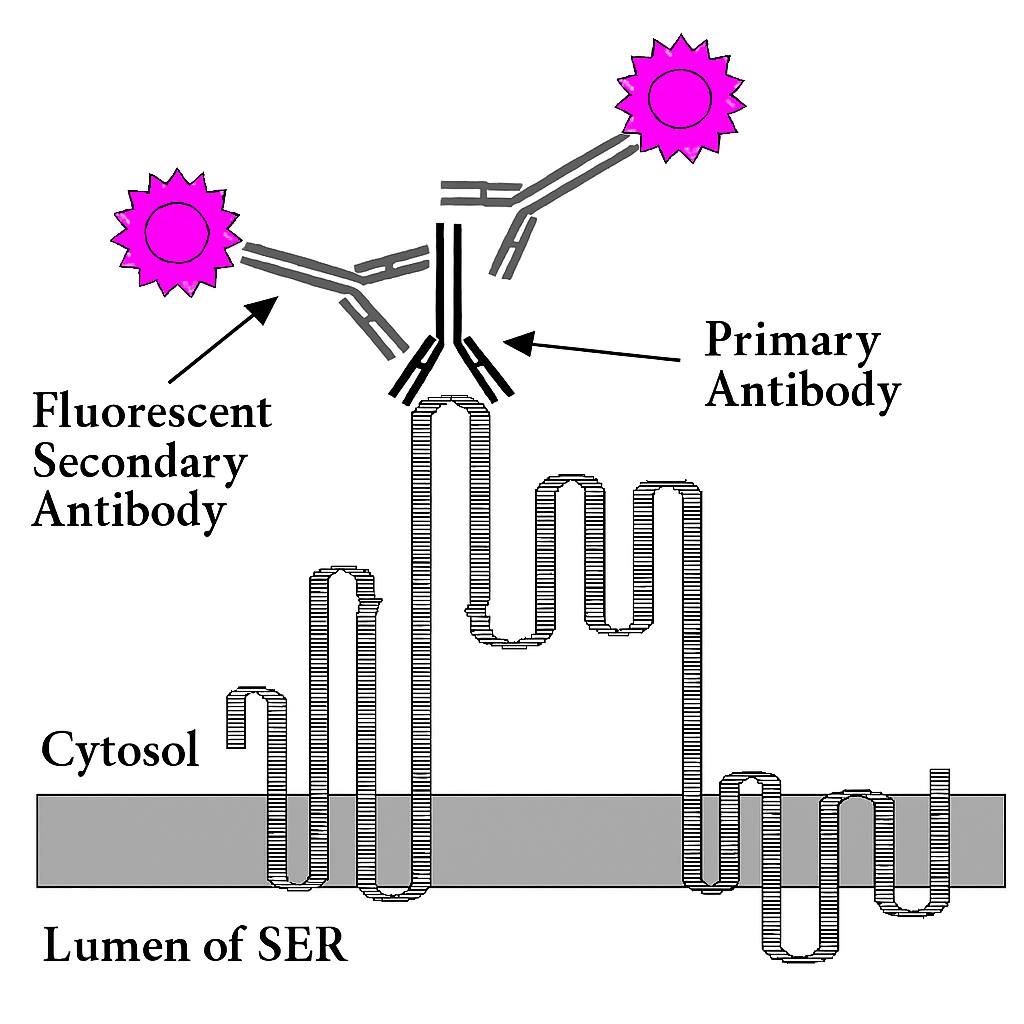
Figure 1. The Working Principle of Immunofluorescence Labeling.
Services at MtoZ Biolabs
Based on an advanced technical platform, MtoZ Biolabs has launched the immunofluorescent labeling service focusing on subcellular localization studies of proteins. By labeling target proteins with highly specific antibodies and combining them with organelle-specific fluorescent dyes, this service enables precise multi-channel co-localization analysis. It can be used to analyze the distribution of proteins in structures such as the nucleus, mitochondria, endoplasmic reticulum, and Golgi apparatus, providing high-resolution imaging data and quantitative localization information. This supports target validation, mechanistic studies, and functional annotation. Immunofluorescent labeling mainly includes two types: direct immunofluorescence and indirect immunofluorescence.
1. Direct Immunofluorescence
The fluorescent dye is directly conjugated to the primary antibody. After the labeled primary antibody binds to the target antigen, the fluorescent signal can be detected using a fluorescence microscope, enabling the visualization and localization of the target molecule. This method is simple, highly specific, and reduces non-specific background interference; however, its sensitivity is relatively low and is more suitable for the localization of high-abundance proteins.
2. Indirect Immunofluorescence
An unlabeled primary antibody is first used to recognize the target antigen, followed by detection with a dye-conjugated secondary antibody that binds to the primary antibody. This method enhances the fluorescent signal through secondary antibody amplification, making it more suitable for detecting low-abundance proteins or performing multiplex labeling experiments. However, the procedure involves additional steps and requires careful control of cross-reactivity.
Analysis Workflow
1. Sample Fixation and Permeabilization
Cells are chemically fixed and permeabilized to allow antibodies to access intracellular and subcellular structures.
2. Antibody Labeling
Specific primary antibodies are selected based on the target protein. Either a fluorescently labeled secondary antibody is used for indirect labeling, or a fluorophore-conjugated primary antibody is applied for direct labeling.
3. Co-staining and Microscopy Imaging
Optional fluorescent dyes are used for staining. Confocal or fluorescence microscopy is performed to capture images that reveal the spatial distribution of target proteins within organelles.
4. Image Analysis and Localization Interpretation
Fluorescent signals are analyzed using imaging software to determine the localization of proteins in subcellular structures such as the nucleus, mitochondria, and Golgi apparatus.
Service Advantages
1. Advanced Technology Platform
Equipped with state-of-the-art technology to ensure clear imaging and specific signals, meeting the demands of high-precision subcellular localization analysis.
2. Experienced Technical Team
Our team is well-versed in processing various cell types and tissue samples, with expertise in antibody selection and labeling strategies to ensure high experimental success rates.
3. Customized Labeling Strategies
Flexible design of direct or indirect immunofluorescent labeling protocols based on target protein, research needs, and fluorescence channel configuration—balancing sensitivity and throughput.
4. One-Stop Service
Comprehensive workflow including sample preparation, antibody labeling, fluorescence imaging, and data output, streamlining the process and improving research efficiency.
Applications
1. Subcellular Localization Studies
By labeling specific proteins, researchers can accurately determine their spatial distribution within organelles (such as the nucleus, mitochondria, endoplasmic reticulum, and Golgi apparatus), revealing functional roles and dynamic changes.
2. Cell Cycle and Proliferation Analysis
In combination with markers of cell cycle-related proteins, the immunofluorescent labeling service can be used to study the status and proliferation activity of cells at different stages, and is widely applied in cancer biology research.
3. Drug Effect Evaluation
By observing the expression and localization changes of target proteins before and after drug treatment, this service enables the assessment of candidate drugs’ effects on cellular structure and function, supporting efficacy and toxicity screening.
4. Disease Mechanism Exploration
The immunofluorescent labeling service can be applied to compare differences in protein localization and expression patterns between normal and diseased cells, providing insights into the potential mechanisms of abnormal signaling pathways in disease contexts.
FAQ
Q1: Is it Possible to Perform Co-Labeling of Multiple Proteins?
A1: Yes. We support multiplex immunofluorescent labeling using various fluorescent dyes to enable co-localization studies of multiple targets. However, it is essential to ensure that there is no spectral overlap between fluorescence channels.
Q2: Can the Service Analyze Localization to Specific Subcellular Organelles?
A2: Yes. We provide customized labeling solutions for commonly studied organelles such as mitochondria, nucleus, endoplasmic reticulum, Golgi apparatus, and lysosomes, allowing accurate subcellular localization analysis of target proteins.
Deliverables
1. Comprehensive Experimental Details
2. Materials, Instruments, and Methods
3. Data Analysis, Preprocessing, and Estimation
4. Bioinformatics Analysis
5. Raw Data Files
How to order?

