



Parallel reaction monitoring (PRM) supports biomarker validation by confirming a closed shortlist of candidate peptides after discovery, not by surveying an open proteome. In a typical path, data-dependent acquisition (DDA) or data-independent acquisition (DIA) nominates differentially abundant proteins, then PRM schedules those proteotypic peptides, records high-resolution fragment spectra, and quantifies from extracted fragment peak areas in matched cohorts. Relative same-peptide comparison is the default claim. Absolute amount requires matched stable isotope-labeled standards (SIS) and a stated calibration model. PRM does not, by itself, establish a clinical diagnostic cutoff or replace immunoassay platforms used in later clinical validation stages.
Researchers who already have a candidate shortlist or a validation cohort design can also review the MtoZ Biolabs Parallel Reaction Monitoring (PRM) Service for project-specific feasibility and assay planning.
Where PRM Sits in the Biomarker Path
Biomarker proteomics usually moves through linked stages with different claims.
1. Discovery
Untargeted DDA or DIA builds an open protein matrix and nominates candidates between defined groups. This stage answers which proteins appear to change, not whether each peptide will confirm under a locked targeted method.
2. Targeted confirmation with PRM
PRM isolates scheduled precursors and records full product-ion spectra on a high-resolution analyzer (Peterson et al. 2012). Fragments used for quantification can be chosen or replaced after acquisition. This stage answers whether nominated peptides reproduce with fragment-level evidence in the validation matrix.
3. Broader research or clinical-scale follow-up
Larger cohorts, immunoassay transfer, or a locked multiple reaction monitoring (MRM) panel may follow once peptides and claims are stable. Those later platforms are separate experiments with their own analytical validation rules.
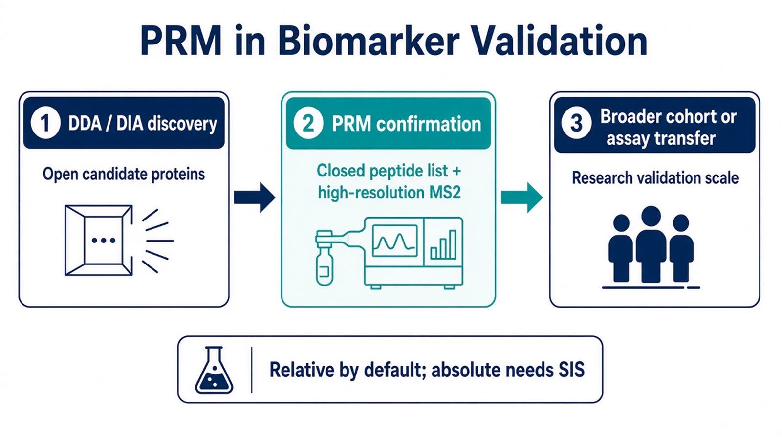
Figure 1. PRM supports biomarker validation as the targeted confirmation stage between open discovery and broader follow-up, not as a discovery or clinical-cutoff method.
PRM validates scheduled candidates; it does not invent new proteins outside the inclusion list.
How a PRM Biomarker Confirmation Assay Is Run
A workable confirmation project keeps discovery shortlisting and PRM acquisition as linked but separate steps.
1. Close the candidate peptide list
Select proteotypic peptides from discovery data, spectral libraries, or literature. Prefer unique sequences with usable chromatography and fragment ions. Shared peptides and sparse discovery detections should be flagged before scheduling.
2. Decide relative versus absolute readout
Same-peptide peak areas support relative confirmation when digestion and loading stay comparable. Absolute concentration needs matched SIS peptides, with spike timing recorded. An SIS added after failed digestion cannot reconstruct the lost protein.
3. Schedule precursors and acquire full MS2
Place precursor m/z and retention-time windows on the inclusion list. Isolate each precursor, fragment it, and store the high-resolution product-ion spectrum. Panel size is limited by peak width and MS2 cycle time.
4. Extract fragments and compare cohorts
Integrate quantitative and qualifier fragment chromatograms in software such as Skyline. Report group differences only for peptides that pass peak-shape, interference, and retention-time checks. ROC-style summaries describe discrimination in the studied cohort; they are not diagnostic claims unless a separately validated assay framework is in place.
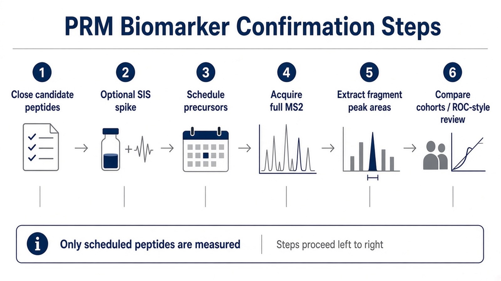
Figure 2. PRM biomarker confirmation locks the peptide list before acquisition and quantifies from extracted high-resolution fragment peak areas.
For operational PRM detail beyond the biomarker application focus, researchers can refer to Parallel Reaction Monitoring (PRM): Principles, Workflow, and Applications.
Judgment Criteria: When PRM Fits Biomarker Validation
Study need |
PRM often fits |
Reassess before scheduling |
|---|---|---|
Candidate status |
Closed shortlist from DDA/DIA or literature |
Open discovery inventory still required |
Evidence need |
High-resolution fragment review for confirmation |
Only a locked unit-resolution transition panel is needed |
Matrix |
Complex digests where accurate-mass fragments help |
Interference remains after isolation-window review |
Claim type |
Relative confirmation; absolute with SIS |
Clinical diagnostic cutoff or IVD decision rule |
Scale |
Tens to low hundreds of peptides within cycle-time limits |
Very large mature panel better suited to MRM transfer |
Prefer PRM when nominated biomarker candidates must be confirmed with post-run fragment choice. Prefer continued DDA or DIA while candidates are still open. Prefer MRM when transitions are already locked and high multiplex on a triple quadrupole is the limiting need. Researchers comparing those targeted options can refer to PRM vs. MRM: Comparative Analysis in Quantitative Proteomics.
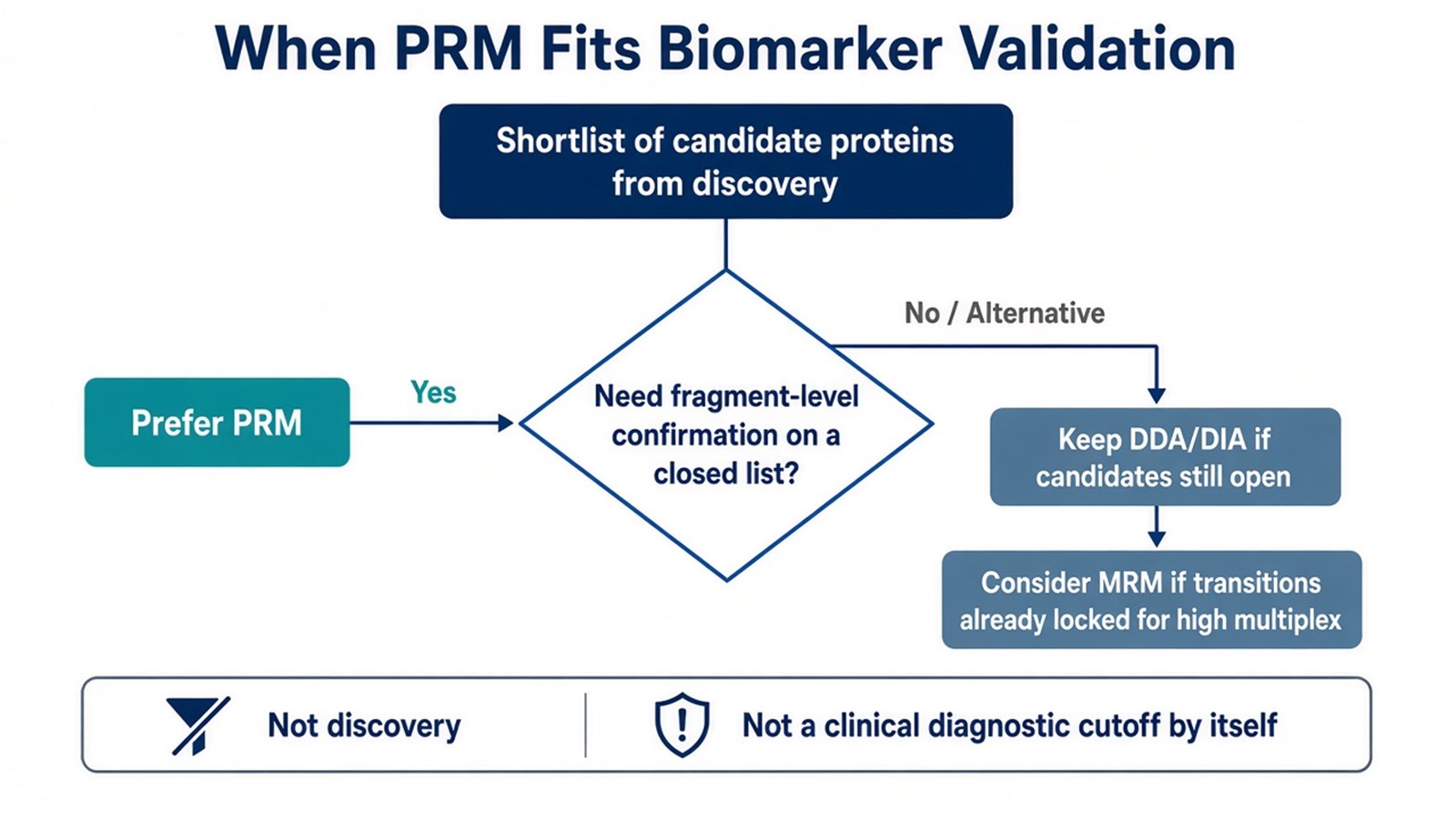
Figure 3. PRM fits biomarker validation when a closed shortlist needs fragment-level confirmation; keep discovery open, or consider MRM, when those conditions are not met.
Limits and Application Boundaries
PRM cannot recover proteins that were never scheduled. Co-isolation within the precursor window can still admit interfering ions. Saying PRM is always highly sensitive, always within a fixed CV, or always clinically translatable overstates what method class alone decides. Performance depends on peptide choice, chromatography, fill time, matrix, and QC.
Typical applications are plasma or tissue candidate confirmation after discovery, pathway-protein shortlists that need fragment-level checks, pharmacodynamic monitoring of selected targets, and selected modified-peptide assays when site-discriminating fragments can be defined. Diagnostic cutoffs, patient triage rules, and regulated clinical reporting sit outside ordinary research PRM unless a separately validated assay framework is in place.
Once the shortlist, relative-versus-absolute design, and cohort comparisons are defined, the project can be evaluated as a PRM biomarker confirmation study. Researchers can review the MtoZ Biolabs Parallel Reaction Monitoring (PRM) Service for sample evaluation, panel feasibility, and workflow planning. For discovery preceding confirmation, see the Deep DIA Proteomics Service.
Frequently Asked Questions
1. Does PRM discover new biomarker candidates?
No. PRM measures only scheduled precursors. Discovery still requires DDA or DIA, or another open acquisition strategy.
2. Does successful PRM confirmation equal a clinical biomarker?
No. Confirmation supports a research quantitative claim for the studied cohorts. Clinical use needs separate analytical and clinical validation frameworks.
3. Is SIS required for every biomarker PRM study?
No. Relative confirmation is the default without SIS. Absolute amount requires matched SIS and a calibration model.
4. How does PRM differ from MRM for biomarker work?
PRM stores a full high-resolution fragment spectrum and allows post-run ion selection. MRM monitors predefined transitions on a triple quadrupole and often fits mature high-multiplex panels.
5. Can PRM replace ELISA in biomarker projects?
Not automatically. ELISA and related immunoassays remain common for later-scale measurement of individual proteins. PRM is a mass spectrometry confirmation route for a defined peptide panel.
Reference
- A.C. Peterson, J.D. Russell, D.J. Bailey, M.S. Westphall, J.J. Coon (2012). Parallel reaction monitoring for high resolution and high mass accuracy quantitative, targeted proteomics. Mol. Cell. Proteomics, 11, 1475-1488. https://doi.org/10.1074/mcp.O112.020131
- V. Lange, P. Picotti, B. Domon, R. Aebersold (2008). Selected reaction monitoring for quantitative proteomics: a tutorial. Mol. Syst. Biol., 4, 222. https://doi.org/10.1038/msb.2008.61
