






How Mitochondrial Proteomics Supports Disease Mechanism Research
Mitochondrial proteomics supports disease mechanism research by showing which proteins in mitochondrial or mitochondria-enriched material change between disease-relevant states, then ranking candidates for follow-up. In practical terms, mitochondrial protein analysis can connect a phenotype you already observe to a protein-level shortlist, without claiming that the proteome alone proves causality.
That distinction keeps the method useful. Mechanism work usually needs both comparative protein evidence and orthogonal functional tests. Proteomics supplies the comparative map; respiration, membrane potential, ROS, genetics, or imaging answer different parts of the mechanism question.
Planning a disease-mechanism study? Contact MtoZ Biolabs and share your model, comparison groups, sample type, and the phenotype you want to connect with protein-level changes with us. We can help assess whether mitochondrial proteomics fits the question and what should be defined before sample submission.
Where Proteomics Fits in a Mechanism Workflow
Disease mechanism research often starts from a phenotype: energy failure, stress sensitivity, tissue injury, treatment response, or genotype-linked dysfunction. Mitochondrial proteomics enters when you need to know which proteins in the mitochondrial preparation differ across those states.
It is especially useful when:
- A disease or stress model already shows a mitochondrial-linked phenotype.
- You need an unbiased or broad protein shortlist rather than one predetermined target.
- Sensitive versus disease, treated versus untreated, or genotype contrasts can be designed with biological replicates.
- Follow-up assays are planned for the top candidates after ranking.
It is less useful as a stand-alone mechanism proof. A differential protein list can support a hypothesis about mitochondrial remodeling; it cannot, by itself, establish that a protein caused the disease phenotype.
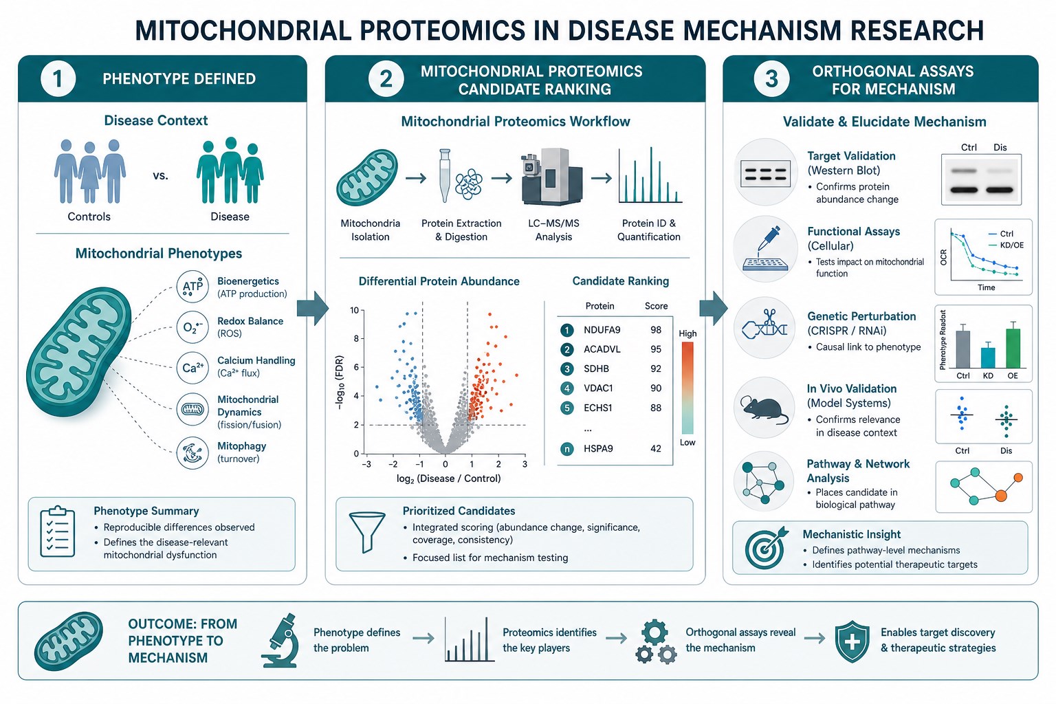
Figure 1. Use proteomics to rank mitochondrial-fraction candidates after a phenotype is defined, then confirm mechanism with orthogonal assays.
What Protein Evidence Can Contribute
For mechanism-oriented studies, mitochondrial proteomics can provide:
- Identification of proteins recovered from cells, tissue, or extracted mitochondrial preparations under disease-relevant conditions.
- Quantitative comparisons across designed groups when DDA or DIA paths are matched to the cohort.
- Ranked candidate lists that focus follow-up on proteins with the strongest, most coherent changes.
- A report package with raw files and result tables that collaborators can review.
Acquisition options for this service line include Orbitrap Exploris 480, timsTOF Pro, and Orbitrap Astral. DDA data are commonly processed with MaxQuant or Proteome Discoverer; DIA data with Spectronaut or DIA-NN. Choose mode from the comparison you need, not from a hoped-for protein total. No fixed identification count should be assumed for every disease model.
Sample planning still decides whether the mechanism contrast is measurable. Planning references are about 5×10^7 cells, about 200 mg animal tissue, or extracted mitochondrial protein of at least about 50 µg with about 80-100 µg commonly planned. Keep amounts matched across disease and reference arms so technical input gaps do not imitate mechanism.
Study Designs That Support Mechanism Questions
Mechanism-oriented mitochondrial protein analysis works best when the biological contrast is explicit.
Useful designs include:
- Disease model versus matched reference under identical handling.
- Genotype contrasts that isolate the pathway under study.
- Treatment or stress challenge versus vehicle in the same background.
- Time points after an insult when mitochondrial remodeling is expected to unfold.
Across these designs, proteomics answers “what changed in the mitochondrial preparation.” Parallel assays answer “what that change does.” Keep those lanes separate in the project brief so the proteomics deliverable is not mistaken for a bundled functional workup.
Functional and imaging readouts such as oxidative phosphorylation panels, membrane potential, ROS, respiratory-chain enzyme tests, calcium or permeability-transition assays, toxicity or injury panels, electron microscopy, and high-content imaging are outside this proteomics service scope. They remain valuable orthogonal tools and should be planned explicitly when the mechanism claim depends on them.
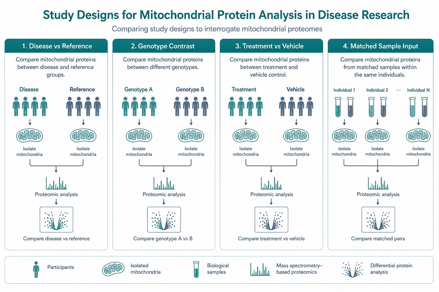
Figure 2. Build clear disease-relevant contrasts first, then keep sample input and acquisition matched across groups.
From Candidate List to Mechanism Without Overclaiming
The most productive use of mitochondrial proteomics in mechanism research is disciplined interpretation.
Read differentials as ranked candidates associated with the mitochondrial preparation. Do not treat every co-enriched protein as an exclusive mitochondrial resident. Do not convert a fold change into a proven causal step. Do not assume pathway annotation modules are automatic for every project; confirm report contents at kickoff.
A practical sequence looks like this:
- Define the phenotype and the mechanism question in one sentence.
- Design groups and biological replicates that isolate that contrast.
- Confirm sample amounts and whether mitochondria are already extracted.
- Run mitochondrial proteomics to rank protein changes.
- Move priority proteins into orthogonal assays that test function or localization.
Planning guidance for the proteomics segment is about 4 weeks, with timing still dependent on sample condition and project complexity. Borderline samples should be reviewed case by case before a mechanism timeline is promised.
|
Mechanism need |
What proteomics contributes |
What to add outside proteomics |
|---|---|---|
|
Map remodeling in disease vs reference |
Quantitative candidate ranking |
Matched phenotype definition |
|
Prioritize follow-up targets |
Differential protein shortlist |
Validation assays for top proteins |
|
Link treatment to mitochondrial-fraction change |
Designed group contrasts |
Dose and exposure metadata |
|
Claim causal mechanism |
Supportive association only |
Functional, genetic, or imaging proof |
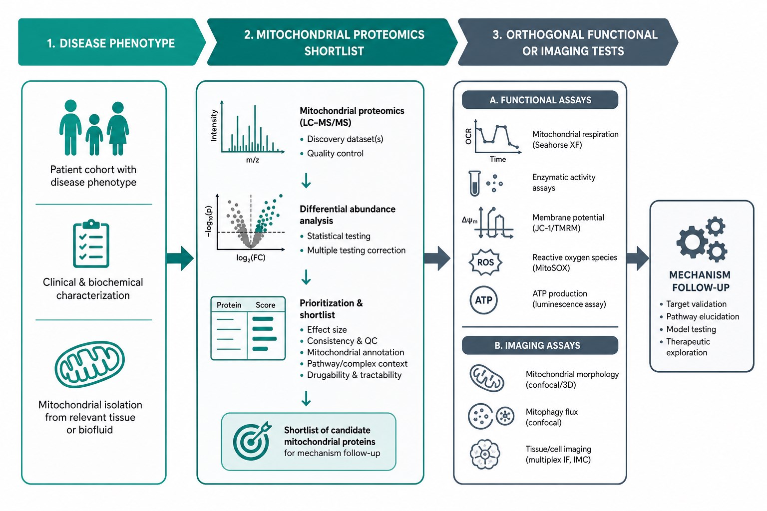
Figure 3. Proteomics narrows candidates; orthogonal assays test whether those candidates explain the phenotype.
When requesting a mechanism-focused plan, send the disease or stress model, the one-sentence question, group map, sample type and amounts, preferred DDA or DIA path if known, and which orthogonal assays will run in parallel. MtoZ Biolabs can align the mitochondrial proteomics package to that mechanism workflow without overstating what protein data alone can prove.
Related Services
Teams connecting disease-model proteomics to broader organelle options can review the services below while scope is still open.
Mitochondrial Proteomics Service
The main route for mitochondrial proteomics when disease-mechanism studies need protein identification and quantitative comparison.
Subcellular Proteomics Service
Use this when the mechanism question spans multiple subcellular fractions rather than a mitochondria-focused design alone.
Subcellular Structure and Organelle Proteomics Service
A broader organelle proteomics option when mitochondrial changes are interpreted alongside other compartments.
Frequently Asked Questions
1. How does mitochondrial proteomics support disease mechanism research?
It ranks proteins that differ in mitochondrial preparations across disease-relevant states, giving a candidate map for follow-up.
2. Can proteomics prove a disease mechanism by itself?
No. It provides associative protein evidence. Causal claims need orthogonal functional, genetic, or imaging support.
3. What study design works best?
Clear disease versus reference, genotype, or treatment contrasts with matched handling and biological replicates.
4. Are mitochondrial function assays included?
No. Membrane potential, ROS, respiration, enzyme activity, and related phenotype assays are outside this proteomics scope and should be planned separately if needed.
5. How much sample is typically required?
Planning references are about 5×10^7 cells, about 200 mg tissue, or about 50 µg protein minimum for extracted mitochondria, with about 80-100 µg commonly planned.
6. What should I send to start planning?
The mechanism question, model details, group map, sample amounts, acquisition preference if known, and parallel assay plans.
Conclusion
Mitochondrial proteomics supports disease mechanism research by turning disease-relevant contrasts into ranked protein evidence from mitochondrial preparations. That evidence is most powerful when paired with orthogonal assays rather than asked to carry the full causal load alone.
If the phenotype, group design, and follow-up plan are clear before kickoff, mitochondrial protein analysis becomes a focused mechanism tool instead of an open-ended data dump. Teams ready to align that workflow can review the model and data needs with MtoZ Biolabs before the project begins.
How to order?




