






Histone Methylation: Site-Specific Marks and Proteomics Analysis
Histone methylation involves the addition of methyl groups to specific lysine and arginine residues on histone proteins and is associated with chromatin regulation. Sites such as H3K4 and H3K27 refer to lysine positions on histone H3, while me1, me2, and me3 represent different lysine methylation states. Arginine methylation includes monomethylation and symmetric or asymmetric dimethylation. Histone methylation analysis identifies these site-specific forms and can compare their relative abundance across biological conditions. Interpretation depends on the modified residue, methylation state, and cellular context.
For histone methylation identification, site localization, and relative quantification, researchers can review the MtoZ Biolabs Histone Methylation Analysis Service for sample suitability and study planning.
What Is Histone Methylation?
Histone methylation involves the enzyme-catalyzed transfer of methyl groups from S-adenosylmethionine (SAM) to lysine or arginine residues on histone proteins. Lysine methylation occurs in mono-, di-, and trimethylated forms (me1, me2, and me3), whereas arginine methylation occurs as monomethylation or symmetric or asymmetric dimethylation (Bedford and Clarke 2009). Histone lysine methylation is regulated by methyltransferases and demethylases, while arginine methylation is catalyzed by protein arginine methyltransferases (PRMTs).
Histone methylation is a residue-specific modification whose biological associations depend on the modified site, methylation state, and cellular context. Different methylation patterns can be associated with distinct chromatin states, making the identity of the modified residue important for interpreting histone methylation profiles (Kouzarides 2007).
Common Histone Methylation Sites and Their Biological Contexts
The histone methylation sites listed below are frequently investigated in chromatin studies. Their biological associations depend on the methylation state and cellular context. LC-MS/MS identifies methylated histone peptides and their modification states, while chromatin immunoprecipitation followed by sequencing (ChIP-seq) provides information about the genomic distribution of specific histone marks.
|
Site |
Methyl forms often compared |
Biological contexts reported in chromatin studies |
|---|---|---|
|
H3K4 |
me1, me2, me3 |
H3K4me1 is associated with enhancers; H3K4me3 is enriched at active promoters; H3K4me2 occurs at promoters and regulatory regions |
|
H3K36 |
me2, me3 |
H3K36me3 is associated with transcriptionally active gene bodies; H3K36me2 shows a broader genomic distribution |
|
H3K9 |
me2, me3 |
H3K9me2 is associated with repressive chromatin domains; H3K9me3 is enriched in heterochromatin and repetitive regions |
|
H3K27 |
me1, me2, me3 |
H3K27me1 can associate with active transcription; H3K27me2 occurs across broad chromatin domains; H3K27me3 is associated with Polycomb-mediated repression |
|
H4K20 |
me1, me2, me3 |
H4K20me1 is linked to cell-cycle and transcriptional regulation; H4K20me2 participates in DNA damage responses; H4K20me3 is associated with heterochromatin |
|
H3R2, H3R8, H3R17, H4R3 |
me1, me2 |
Histone arginine methylation is associated with context-dependent transcriptional regulation; symmetric and asymmetric dimethylation require distinct assignment |
How Do Mono-, Di-, and Trimethylation Differ at the Same Histone Site?
Methylation States on Lysine Residues
Each added methyl group introduces a mass shift of approximately 14.016 Da. Lysine monomethylation (me1), dimethylation (me2), and trimethylation (me3) produce mass increases of +14.016 Da, +28.031 Da, and +42.047 Da, respectively. These represent distinct methylation states of the same lysine residue and can be distinguished by their mass differences when other peptide modifications are accounted for. Database searches should include the relevant methylation states as variable modifications to enable their identification.
Histone methylation sites are reported according to the modified residue and its methylation state. For example, H3K27me1, H3K27me2, and H3K27me3 represent distinct modifications at lysine 27 of histone H3. Accurate assignment requires mass measurements that distinguish the methylation states and MS/MS fragment-ion evidence supporting localization to the specified residue.
Symmetric and Asymmetric Arginine Dimethylation
Arginine monomethylation introduces a mass shift of +14.016 Da, while both symmetric dimethylarginine (SDMA) and asymmetric dimethylarginine (ADMA) produce a mass increase of +28.031 Da. Because these dimethylation forms have identical precursor masses, their differentiation requires diagnostic fragment ions or characteristic neutral-loss evidence. When the available MS/MS spectra cannot distinguish SDMA from ADMA, the modification should be reported as arginine dimethylation without assigning its symmetry. Additional analysis using validated isomer-specific antibodies or suitable mass spectrometry methods may be considered when distinguishing these forms is essential to the research objective.
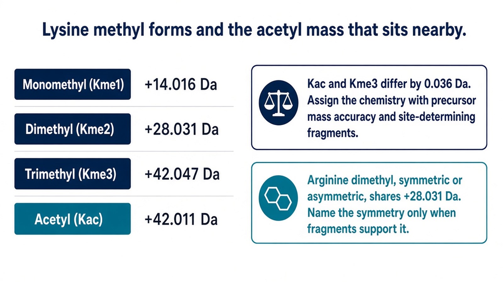
Figure 1. Lysine me1, me2, and me3 masses, the 0.036 Da gap between trimethylation and acetylation, and the shared mass of the two arginine dimethyl isomers.
How Is Histone Methylation Analyzed by Mass Spectrometry?
Histone Peptide Preparation for Methylation Analysis
Histone tails are rich in lysine and arginine. Unrestricted trypsin cuts after both residues and yields very short pieces that are hard to retain and to assign. One published route derivatizes unmodified and monomethyl lysines with propionic anhydride before trypsin, so cleavage is directed to arginine, while di- and trimethyl lysines remain underivatized at the amine and keep their methyl masses (Garcia et al. 2007; Huang et al. 2015). Monomethyl lysine picks up the propionyl group and is searched as that derivative.
Propionylation before trypsin digestion produces Arg-C-like histone peptides suitable for LC-MS/MS analysis. However, chemically introduced propionyl groups can complicate the identification of endogenous propionylation, while propionylated monomethyllysine can be indistinguishable by mass from butyryllysine. Isotopically labeled derivatization reagents or alternative sample preparation methods can be considered when these endogenous acylations are also analytical targets (Huang et al. 2015). Histone recovery before this chemistry is a separate sample step and can be planned with the Histone Isolation and Enrichment Service.
LC-MS/MS Detection of Methylated Histone Peptides
Histone peptides are separated by liquid chromatography and analyzed using high-resolution tandem mass spectrometry, such as an Orbitrap-based LC-MS/MS platform. The precursor mass corresponds to me1, me2, or me3. Fragment ions have to cover the modified residue before the site is localized. On tails with several lysines, a methyl mass that cannot be placed on one residue is reported as a peptide-level methyl peptide.
Database searches specify the relevant lysine and arginine methylation states as variable modifications, together with propionyl derivatives when chemical derivatization is used. The absence of a particular methylation state from the results may reflect low abundance, incomplete peptide detection, insufficient spectral evidence, or search settings. Search parameters and modification-specific fragment evidence should be reviewed when interpreting these results.
What Can Histone Methylation Proteomics Reveal?
Methylated Histone Peptides and Site-Level Results
Histone methylation proteomics reports may include identified histone proteins, methylated peptides, modification sites, and methylation states, together with supporting peptide-identification and site-localization evidence. Histone variants can be assigned separately when the detected peptide sequences or complementary evidence distinguish them, while shared peptides may not support a unique variant assignment. When multiple modifications are confidently localized within the same peptide, their co-occurrence can be reported at the peptide level without inferring the modification pattern across the intact histone molecule.
These site-specific assignments provide candidate histone marks, such as H3K4me3, for subsequent chromatin profiling or functional investigation.
Interpreting Methylation-State Assignments
Histone methylation results can be interpreted by examining the protein or variant assignment, peptide sequence, modified residue, methylation state, and supporting localization evidence. Multiple methylation states, such as me1, me2, and me3, may be detected at the same lysine within a sample, representing different histone molecules in the analyzed population. Their identification establishes the presence of these forms but does not determine their relative proportions.
For quantitative comparisons of histone methylation states across experimental groups, researchers can review the MtoZ Biolabs Histone PTMs Quantitative Analysis Service for project-specific study planning.
Frequently Asked Questions
Is histone methylation proteomics the same experiment as DNA methylation analysis?
No. Histone methylation proteomics measures methyl groups on lysine and arginine of histone proteins. DNA methylation measures 5-methylcytosine in DNA. The two assays use different extraction methods and return different readouts.
An H3K27me3 antibody blot is already in hand. What does a methylation site table add?
An H3K27me3 Western blot detects the corresponding methylation epitope, whereas LC-MS/MS can identify additional methylation states, such as H3K27me1 and H3K27me2, and modifications at other histone residues, including H3K9 and H3K36. The detected forms and their site assignments depend on peptide coverage and supporting MS/MS evidence. Western blot is suitable for examining a predefined methylation mark, while mass spectrometry provides broader information about histone methylation states and sites.
Can me1, me2, and me3 at the same lysine appear in one result?
Yes. Different histone molecules within the same sample can carry me1, me2, or me3 at the same lysine residue. When supported by peptide-identification and site-localization evidence, these methylation states can be reported as separate results.
Conclusion
Histone methylation proteomics identifies site-specific lysine and arginine methylation states through LC-MS/MS analysis. Distinguishing different methylation forms and evaluating their localization confidence are important for interpreting histone modification profiles. Combined with relevant chromatin or gene-expression evidence, these results can support further investigation of histone methylation in transcriptional regulation and chromatin organization.
For histone methylation identification and analysis, researchers can review the MtoZ Biolabs Histone Methylation Analysis Service for sample evaluation and project planning.
Reference
- T. Kouzarides (2007). Chromatin modifications and their function. Cell, 128, 693-705. https://doi.org/10.1016/j.cell.2007.02.005
- A. Barski, S. Cuddapah, K. Cui, T.Y. Roh, D.E. Schones, Z. Wang, G. Wei, I. Chepelev, K. Zhao (2007). High-resolution profiling of histone methylations in the human genome. Cell, 129, 823-837. https://doi.org/10.1016/j.cell.2007.05.009
- K. Zhang, P.M. Yau, B. Chandrasekhar, R. New, R. Kondrat, B.S. Imai, M.E. Bradbury (2004). Differentiation between peptides containing acetylated or tri-methylated lysines by mass spectrometry: an application for determining lysine 9 acetylation and methylation of histone H3. Proteomics, 4, 1-10. https://doi.org/10.1002/pmic.200300503
- B.A. Garcia, S. Mollah, B.M. Ueberheide, S.A. Busby, T.L. Muratore, J. Shabanowitz, D.F. Hunt (2007). Chemical derivatization of histones for facilitated analysis by mass spectrometry. Nat. Protoc., 2, 933-938. https://doi.org/10.1038/nprot.2007.106
- M.T. Bedford, S.G. Clarke (2009). Protein arginine methylation in mammals: who, what, and why. Mol. Cell, 33, 1-13. https://doi.org/10.1016/j.molcel.2008.12.013
- H. Huang, S. Lin, B.A. Garcia, Y. Zhao (2015). Quantitative proteomic analysis of histone modifications. Chem. Rev., 115, 2376-2418. https://doi.org/10.1021/cr500491u
How to order?