






Histone Lactylation: Site Identification and Proteomics Analysis
Histone lactylation is a post-translational modification (PTM) associated with cellular metabolism and epigenetic regulation. Since its discovery, histone lactylation has attracted interest in studies of inflammation, cancer, cellular differentiation, and metabolic reprogramming.
Mass spectrometry-based proteomics enables the identification of lactylated histone peptides and the localization of modification sites. However, reliable analysis requires distinguishing lactylation from other modifications with similar mass characteristics and obtaining sufficient evidence to assign specific modified residues.
For researchers investigating histone lactylation, explore MtoZ Biolabs Histone Lactylation Analysis service to learn more about the available analytical options. Histone modifications encompass a range of PTMs with distinct roles in chromatin regulation. Their major types and biological functions are discussed in Exploring Histone Post-Translational Modifications.
What Is Histone Lactylation?
Histone lactylation is a type of lysine acylation in which a lactyl group is attached to the ε-amino group of a lysine residue. Currently, reported histone lactylation primarily refers to lysine lactylation (Kla). It has been identified on core histones, including H2A, H2B, H3, and H4, with a characteristic mass shift of approximately +72.021 Da. Histone lactylation is associated with cellular lactate metabolism. Lactate produced through glycolysis has been investigated as a source of lactyl groups for histone modification, providing a link between cellular metabolic conditions and chromatin-associated protein modifications.
A landmark study by Zhang et al. (2019) identified histone lactylation in human and mouse cells and investigated its association with gene expression during macrophage polarization. This discovery has encouraged further research into histone lactylation in inflammation, cancer metabolism, cellular differentiation, and other biological processes.
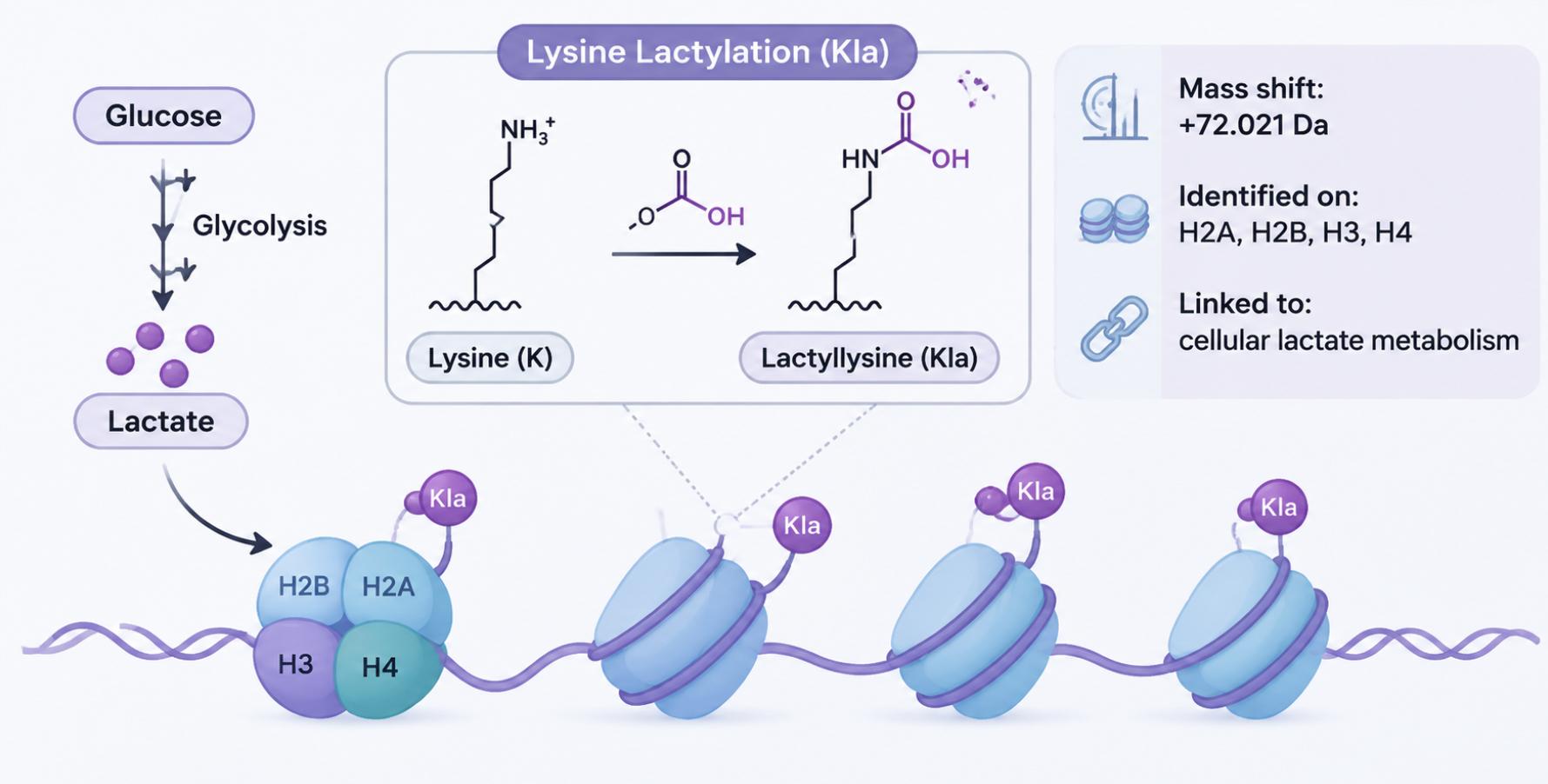
Figure 1. Overview of histone lactylation and its link to cellular metabolism.
Reported Histone Lactylation Sites
Mass spectrometry-based studies have identified lysine lactylation across the four core histones: H2A, H2B, H3, and H4. H3K18la is one of the most extensively investigated sites, while additional lactylation sites have been reported across different histones and biological samples. The following table summarizes representative sites identified in human and mouse samples.
| Histone | Human (HeLa cells) | Mouse (BMDMs) |
|---|---|---|
| H2A | K11, K13, K115 | K11, K115 |
| H2B | K5, K11, K15, K16, K20, K23, K43, K85, K108, K116, K120 | K5, K11, K15, K16, K20, K85, K108 |
| H3 | K9, K18, K23, K27, K79 | K14, K18, K23, K27, K56 |
| H4 | K5, K8, K12, K16, K31, K77, K91 | K8, K12, K31, K91 |
H3K18la has received particular attention because of its association with gene expression during macrophage polarization. Other reported sites, including H3K23la, H3K27la, and H4K12la, have also been investigated in different biological contexts.
The reported site distributions are not identical between human and mouse samples. Differences may reflect species, cell type, biological conditions, and analytical coverage. These reported sites provide useful references for histone lactylation research but do not represent an exhaustive list of all possible modification sites. Additional candidate sites may be identified when sufficient peptide coverage and site-localization evidence are available.
Histone Lactylation Site Identification by LC-MS/MS
Mass spectrometry-based histone lactylation analysis involves histone preparation, modified peptide identification, and site localization. Each step contributes to the identification of lactylated histone peptides and the assignment of modification sites to specific lysine residues.
Histone Preparation and Lactylated Peptide Detection
Histone analysis requires appropriate sample preparation to isolate histones and obtain sufficient peptide coverage for modification analysis.
A typical analytical workflow involves:
1. Histone Extraction: Isolate nuclei and extract histones from the biological sample.
2. Histone Purification: Remove interfering components and obtain histone-enriched fractions.
3. Enzymatic Digestion: Use suitable proteases to generate peptides for mass spectrometry analysis. Multiple proteases may improve sequence coverage and help detect modified regions that are not adequately represented by a single digestion strategy.
4. Peptide Enrichment: When appropriate, modification-specific enrichment can improve the representation of lactylated peptides for subsequent analysis. Histone enrichment before digestion can be arranged through the Histone Isolation and Enrichment Service.
5. LC-MS/MS Analysis: Separate peptides by liquid chromatography and acquire tandem mass spectra for peptide identification and modification analysis.
The resulting mass spectra provide information about peptide sequences and potential modification sites. Accurate mass measurements and database searches support the identification of candidate lactylated peptides.
Distinguishing Lactylation from Other Modifications
Accurate modification assignment is important because different lysine acylations produce characteristic mass shifts. Their mass differences help distinguish modification types during high-resolution mass spectrometry analysis.
| Modification | Mass shift |
|---|---|
| Acetylation | +42.011 Da |
| Crotonylation | +68.026 Da |
| Lactylation | +72.021 Da |
| Succinylation | +100.016 Da |
However, lactylation also occurs in different stereochemical forms, including L-lactyllysine and D-lactyllysine. These forms have identical molecular masses and cannot be distinguished by conventional LC-MS/MS based on mass measurements and routine fragmentation alone. Additional approaches, such as chiral derivatization combined with chromatographic separation, are required when stereochemical identification is a research objective.
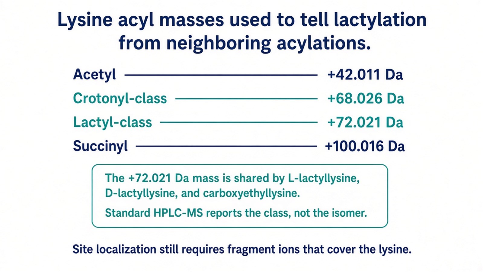
Figure 2. Precursor mass shifts that distinguish lactyl-class lysine from acetyl, crotonyl-class, and succinyl lysine, together with the isomers that share the +72.021 Da mass.
MS/MS Evidence for Lactylation Site Localization
After lactylated peptides are identified, tandem mass spectrometry provides fragment-ion evidence for determining which lysine residue carries the modification. During MS/MS fragmentation, peptide bonds are broken to generate b- and y-type ions. Fragments containing a modified lysine retain the corresponding modification mass, while fragments that do not include the modified residue remain unmodified. Comparing these fragment ions helps distinguish candidate modification positions.
This distinction is particularly important for peptides containing multiple lysine residues. When sufficient site-determining ions are available, the lactylation site can be localized to a specific lysine residue.
What Can Histone Lactylation Analysis Reveal?
Mass spectrometry-based histone lactylation analysis provides information about modification sites and their relative abundance across biological samples. The resulting data can support several research objectives:
-
Lactylation Site Identification: Identify histone proteins and lysine residues carrying lactylation-associated modifications.
-
Modification Pattern Analysis: Investigate the distribution of lactylation sites across H2A, H2B, H3, and H4.
-
Comparative Analysis: Compare relative lactylated peptide abundance between control and treatment groups or different biological conditions.
-
Candidate Site Discovery: Identify previously unreported candidate sites for further investigation.
Together, these findings provide molecular evidence for studying histone lactylation patterns and their relationships with cellular metabolism, chromatin regulation, and biological responses.
Frequently Asked Questions
Can I use a gel band containing histones for lactylation site identification?
Yes. A gel band containing a histone protein can be used for lactylation site identification, provided that sufficient target protein is recovered. After enzymatic digestion, LC-MS/MS analysis can identify lactylated peptides and localize modification sites on the recovered histone protein. Histone immunoprecipitates can also be considered as input materials.
Can a Western blot identify the specific histone lactylation site?
Not with a pan-lactylation antibody alone. A pan-lactylation immunoblot indicates the presence of antibody-reactive lactylated proteins but does not identify the modified lysine residue. Site-specific antibodies validated against individual histone lactylation sites can provide more targeted information. Alternatively, LC-MS/MS can identify modified peptides and localize lactylation sites using site-determining fragment ions. Distinguishing L-lactylation, D-lactylation, and Nε-carboxyethyllysine requires additional analytical methods beyond routine immunoblotting or conventional LC-MS/MS (Zhang et al., 2024).
Does increased lactylation indicate increased cellular lactate levels?
Not necessarily. Histone lactylation is associated with cellular lactate metabolism, but its abundance is also influenced by lactylation and delactylation activities, histone turnover, and other cellular regulatory processes. Increased histone lactylation alone does not establish an increase in intracellular lactate concentration. Additional metabolite measurements are needed to investigate this relationship.
Conclusion
Histone lactylation proteomics enables the identification of lactylated histone peptides, localization of modification sites, and comparison of lactylation patterns across biological conditions. By combining appropriate sample preparation, high-resolution LC-MS/MS, and reliable site-localization evidence, researchers can investigate histone lactylation across different core histones and explore its potential relationships with cellular metabolism and epigenetic regulation. These findings provide a foundation for further studies of histone lactylation and its biological significance.
If your research involves histone lactylation site identification or comparing lactylation changes across experimental conditions, MtoZ Biolabs provides Histone Lactylation Analysis using mass spectrometry-based approaches. Contact our team for a free consultation to discuss your sample requirements and research objectives.
References
- B.R. Sabari, D. Zhang, C.D. Allis, Y. Zhao (2017). Metabolic regulation of gene expression through histone acylations. Nat. Rev. Mol. Cell Biol., 18, 90-101. https://doi.org/10.1038/nrm.2016.140
- D. Zhang, Z. Tang, H. Huang, G. Zhou, C. Cui, Y. Weng, et al. (2019). Metabolic regulation of gene expression by histone lactylation. Nature, 574, 575-580. https://doi.org/10.1038/s41586-019-1678-1
- D. Zhang, J. Gao, Z. Zhu, Q. Mao, Z. Xu, P.K. Singh, et al. (2024). Lysine l-lactylation is the dominant lactylation isomer induced by glycolysis. Nat. Chem. Biol., 21, 91-99. https://doi.org/10.1038/s41589-024-01680-8
How to order?