


High Content Imaging-Based Mitochondrial Phenotype Analysis Service
- Disease Mechanism Research
- Drug Screening and Mechanism of Action Studies
- Aging and Age-Related Disease Research
- Cellular Stress Response Research
High Content Imaging-Based Mitochondrial Phenotype Analysis Service combines high-throughput imaging technology with automated image analysis software to comprehensively assess mitochondrial morphology and its dynamic changes under various physiological and pathological conditions. This service offers researchers an efficient and precise solution for monitoring mitochondrial phenotypic alterations, while enabling large-scale sample processing and analysis through an automated imaging platform.
Mitochondria serve as the cellular powerhouses, playing essential roles in metabolism, energy production, and cellular signaling regulation. Their functional state is closely tied to overall cell health. Notably, changes in mitochondrial morphology are strongly associated with their functional status. Studying mitochondrial phenotypic variations is therefore critical for understanding cellular health, stress responses, and the mechanisms underlying disease development.
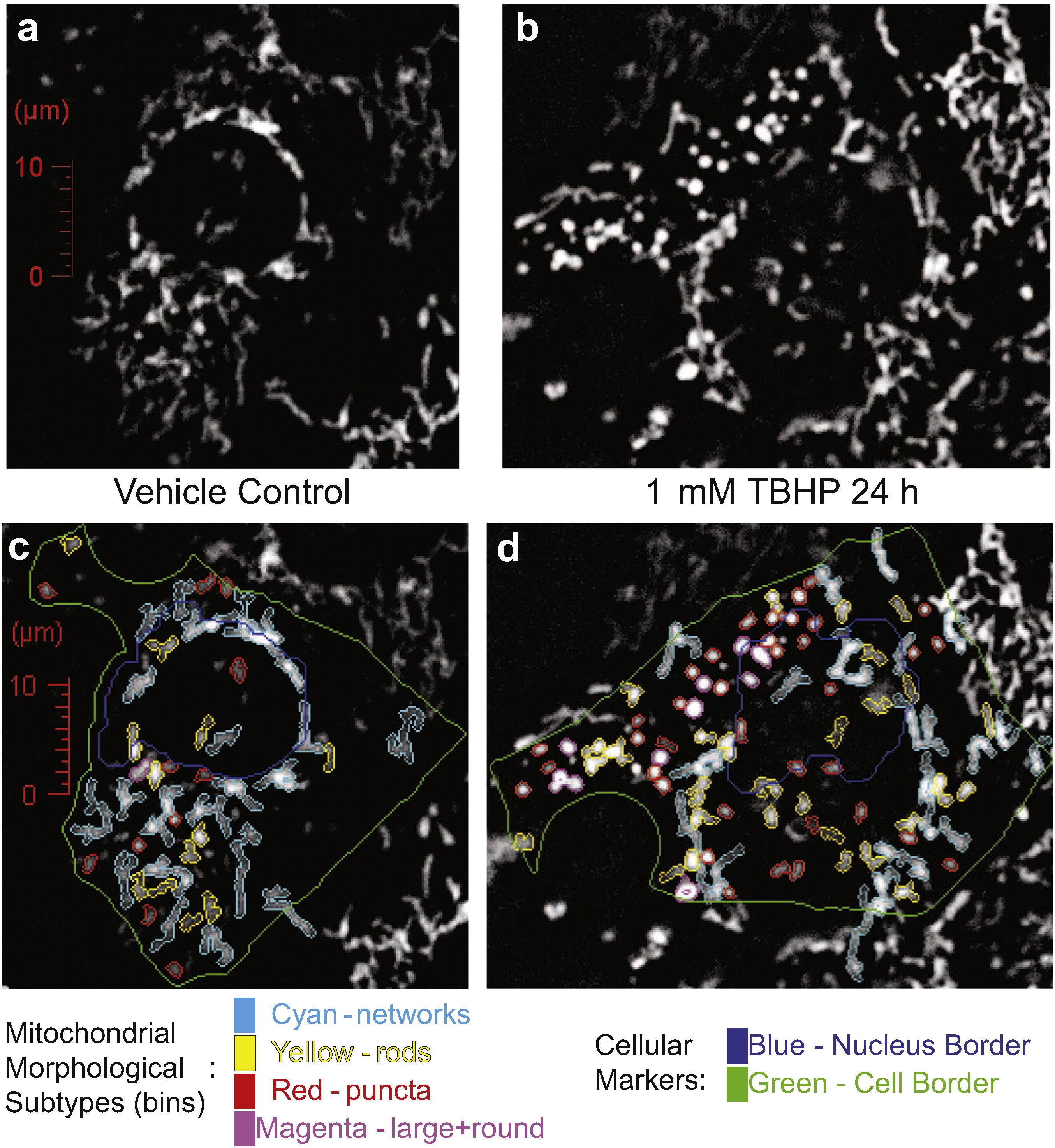
Leonard AP. et al. Biochim Biophys Acta. 2015.
Figure 1. Representative images of t-butyl hydroperoxide (TBHP)-induced damage to mitochondria.
High Content Imaging-Based Mitochondrial Phenotype Analysis is an advanced method that integrates high-resolution fluorescence imaging with automated image analysis to dynamically monitor mitochondrial morphology, distribution, and function at the cellular level. By labeling mitochondria with specific fluorescent dyes and utilizing high-throughput imaging systems such as confocal microscopy, researchers can acquire large volumes of high-quality image data. These images are then quantitatively analyzed using specialized software to reveal mitochondrial phenotypic changes under various physiological, pathological, and drug treatment conditions.
Utilizing high-throughput confocal microscopy and automated image analysis platforms, MtoZ Biolabs offers High Content Imaging-Based Mitochondrial Phenotype Analysis Service to assess mitochondrial morphology, spatial distribution, and dynamic changes within cells. This service provides critical insights into the role of mitochondria in cellular metabolism, disease mechanisms, and drug discovery.
Analysis Workflow
The general process of High Content Imaging-Based Mitochondrial Phenotype Analysis Service is as follows:
1. Sample Preparation
Cells are cultured or tissue sections are prepared to ensure sample quality suitable for high content imaging analysis.
2. Staining and Labeling
Mitochondria-specific fluorescent dyes are used to stain the samples, enabling clear visualization of mitochondrial structures.
3. Image Acquisition
High-resolution confocal microscopy is used to capture images across multiple fields and channels, generating comprehensive mitochondrial image datasets.
4. Image Analysis
Automated image analysis software processes and analyzes the imaging data to quantitatively assess mitochondrial morphological features (e.g., shape, size), membrane potential, spatial distribution, and dynamic changes within the cells.
5. Data Processing and Reporting
Image data are statistically analyzed to generate a quantitative report, including graphs and representative image outputs.
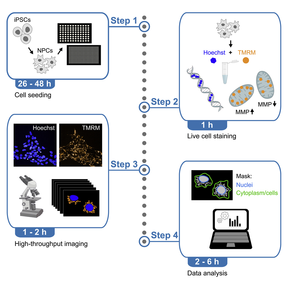
Zink A. et al. STAR Protoc. 2022.
Figure 2. High-content screening of mitochondrial polarization.
Service Advantages
High-Resolution Imaging: Provides high-resolution mitochondrial images to capture subtle morphological changes and detailed distribution patterns.
High-Throughput Analysis: Automated image analysis systems enable large-scale sample processing and efficient data analysis, saving time and enhancing research productivity.
Non-Invasive Live-Cell Imaging: Utilizes real-time, non-destructive live-cell imaging techniques that preserve the natural state of cells without causing damage.
Multichannel Imaging: Supports multichannel imaging, allowing simultaneous visualization of multiple cellular structures and revealing spatial relationships between mitochondria and other organelles.
Customized Experimental Design: Offers tailored High Content Imaging-Based Mitochondrial Phenotype Analysis Services to meet specific research needs, ensuring data relevance and experimental efficiency.
Sample Submission Suggestions
Sample Types: Both cell and tissue samples are acceptable.
Storage and Shipping: Samples should be as fresh as possible. After fixation, they can be stored at 4 °C and shipped using cold chain logistics to prevent quality degradation.
Additional Notes: We recommend contacting us prior to sample submission for detailed and tailored sample preparation guidance.
Applications
Examples of applications of High Content Imaging-Based Mitochondrial Phenotype Analysis Service:
Investigate the role of mitochondria in neurodegenerative and metabolic diseases, and uncover the relationship between mitochondrial morphological changes and disease progression.
Evaluate the impact of candidate drugs on mitochondrial function by observing changes in mitochondrial morphology and dynamics before and after treatment, identifying potential therapeutic compounds.
Examine mitochondrial functional decline during the aging process and explore potential interventions to delay aging and related conditions.
Study changes in mitochondrial morphology and function under environmental stress conditions (e.g., oxidative stress, hypoxia) to reveal the role of mitochondria in cellular stress responses.
Deliverables
1. Comprehensive Experimental Details
2. Materials, Instruments, and Methods
3. High-Resolution Images
4. Quantitative Analysis Report
5. Data Statistics and Graphs
6. Experimental Result Interpretation
Related Services
Mitochondrial Phenotype Analysis Service
How to order?

