


Fluorescent Dyes-Based Exosomes Labeling Service
Fluorescent dyes-based exosomes labeling is a technical service that enables the visual labeling of exosomes using fluorescent dyes, aiming to achieve real-time tracking and localization of exosomes in both in vivo and in vitro environments. This service employs fluorescent dyes with membrane affinity or chemical reactivity and labels fluorescent signals onto the surface or interior of exosomes through methods such as physical adsorption, covalent conjugation, or bioorthogonal click chemistry, while preserving the biological function and structural integrity of the exosomes.
Fluorescent dyes-based exosomes labeling service is widely applied in various fields such as in vivo distribution studies of exosomes, analysis of drug delivery mechanisms, tracking of cellular uptake pathways, and construction of disease diagnostic models. It is particularly suitable for visual verification of tumor targeting, immunotherapy, and nano-delivery systems. With the aid of high-sensitivity imaging platforms, researchers can dynamically monitor exosome behavior, providing critical data support for both basic research and clinical translation of exosomes.
González, M I. et al. Biomedicines, 2021.
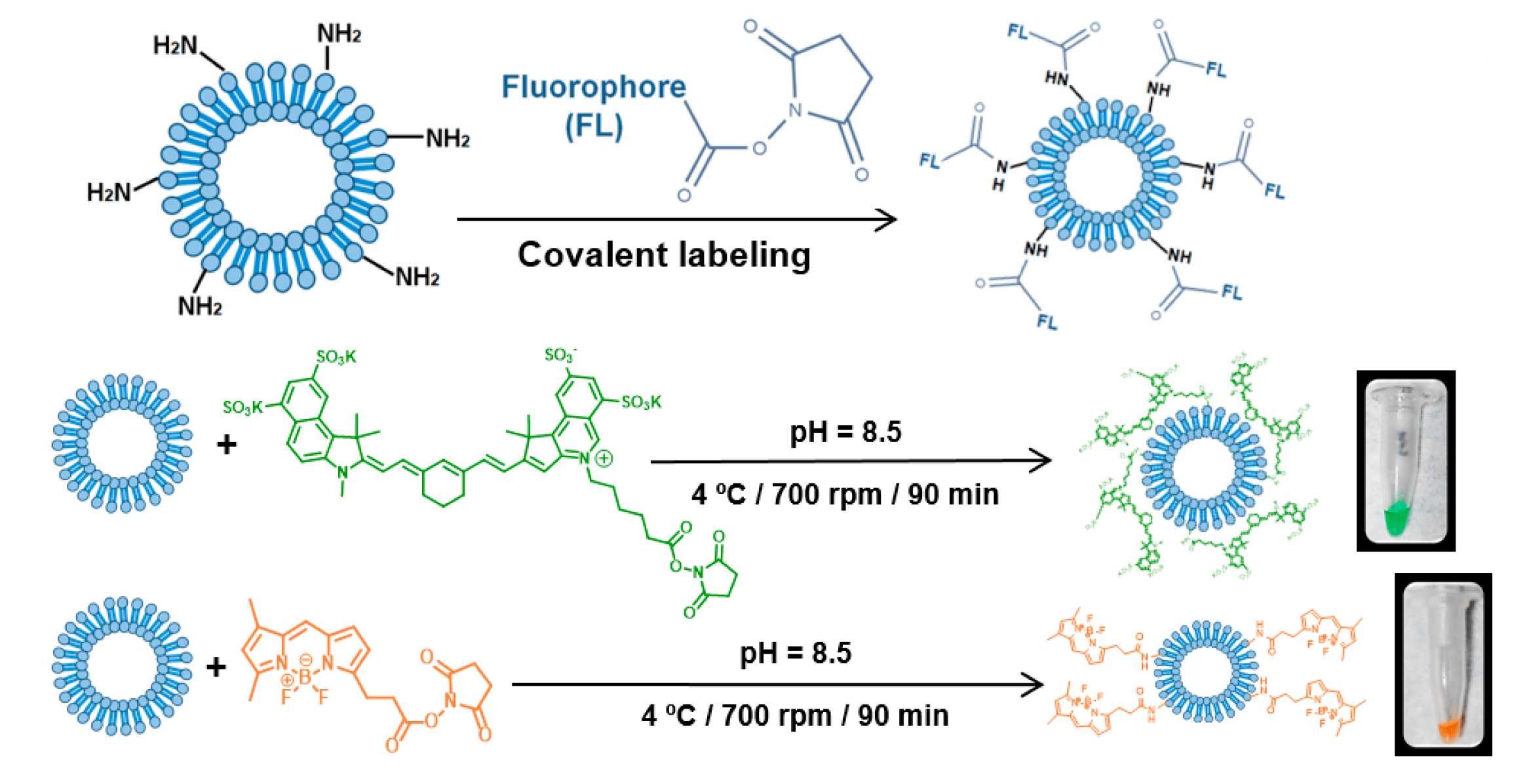
Figure 1. Chemical Strategies for Labeling Exosomes with Fluorescent Dyes.
Services at MtoZ Biolabs
Based on advanced platforms such as transmission electron microscopy (TEM), nanoparticle tracking analysis (NTA), and flow cytometry (FACS), the fluorescent dyes-based exosomes labeling service provided by MtoZ Biolabs utilizes high-affinity membrane dyes or covalent-binding dyes to evaluate the size, morphology, and fluorescence intensity of labeled exosomes, ensuring structural integrity, uniform labeling, and excellent photostability. Fluorescent dye-based exosome labeling services can be divided into the following categories according to the chemical characteristics and labeling methods of the dyes, each with different binding mechanisms and application advantages, and the application of fluorescent dyes in the treatment of cancer, it is suitable for imaging tracking, uptake analysis and functional study of exosomes:
1. Lipophilic Carbocyanine Dyes for Exosomes
Carbocyanine dyes label exosome membranes efficiently by inserting their hydrophobic alkyl chains into the lipid bilayer, without disrupting exosome structure. Common dyes such as DiI, DiO, DiD, and DiR offer diverse emission colors, high fluorescence intensity, and excellent photostability. These dyes are ideal for confocal imaging and in vivo fluorescence tracking experiments, particularly useful for studying the biodistribution and dynamic transport of exosomes in living organisms.
2. PKH Lipophilic Dyes for Exosomes
PKH series dyes are lipophilic and readily incorporate into exosome membranes, producing long-lasting fluorescent signals. With simple labeling procedures and strong signal output, these dyes are widely used in exosome uptake studies, intercellular communication pathway research, and fluorescence imaging analysis.
3. Covalent Labeling Dyes for Exosomes
These dyes, typically NHS esters, form covalent bonds with amino groups on the exosome membrane surface, enabling stable and uniform fluorescent labeling. They are well-suited for long-term imaging, membrane protein localization, and quantitative analysis. With high labeling specificity and minimal fluorescence drift, these dyes are ideal for high-resolution tracking experiments.
4. Bioorthogonal Reactive Dyes for Exosomes
By employing click chemistry strategies, functional groups such as azide or alkyne are first introduced onto the exosome surface, followed by rapid and specific labeling with bioorthogonal fluorescent probes. This mild and biocompatible approach preserves exosome function and enables in situ imaging and real-time tracking in complex biological systems. It is especially advantageous for precise targeting studies both in vitro and in vivo.
Service Advantages
1. Wide Selection of Dyes
A variety of commonly used fluorescent dyes are available, including the PKH series, DiI/DiO/DiR, Alexa Fluor, and NHS ester dyes, covering different emission wavelengths and compatible with various fluorescence imaging systems.
2. Good Preservation of Structure and Function
Experimental conditions are optimized to maximally preserve the structural integrity and biological activity of exosomes, ensuring smooth progress in downstream functional experiments.
3. Robust Data Validation
Using platforms such as TEM, NTA, and FACS, labeled exosomes are comprehensively characterized, with complete experimental data provided on particle size, fluorescence intensity, and labeling efficiency.
4. Exceptional Technical Support
MtoZ Biolabs offers personalized experimental design and full-process technical guidance through its experienced technical team, ensuring that labeling results align with research objectives.
Applications
1. In Vivo Biodistribution and Homing Studies of Exosomes
Fluorescent dyes-based exosomes labeling service can be used for real-time tracking in small animal models to study the distribution patterns and tissue-specific homing abilities of exosomes in organs such as the liver, spleen, lungs, and tumor tissues, providing data to support their potential in drug delivery and tissue targeting.
2. Cellular Uptake and Intracellular Trafficking Mechanism Analysis
In in vitro experiments, fluorescently labeled exosomes co-cultured with cells allow observation of their uptake process and intracellular localization (e.g., endosomal and lysosomal pathways), enabling investigation of entry mechanisms, transport routes, and roles in regulating cellular behavior.
3. Drug Delivery and Carrier Performance Validation
In studies where exosomes are used as delivery carriers, the fluorescent dyes-based exosomes labeling service can be utilized to validate their efficiency in delivering RNA, proteins, or small molecule drugs. It allows for tracking the delivery pathways and drug release profiles, further optimizing the design and functional evaluation of the delivery system.
4. Exosome Functional Validation
In conjunction with disease models, labeled exosomes can be used to investigate their specific functions in immune modulation, inflammatory responses, tumor metastasis, or tissue regeneration. This helps to elucidate the biological effects mediated by exosomes and validate strategies for functional modifications.
5. Imaging and Visualization Analysis
The fluorescent dyes-based exosomes labeling service can be used in flow cytometry, confocal microscopy, and in vivo imaging systems to enable the visualization and tracking of exosomes in both in vitro and in vivo experiments. This allows researchers to intuitively obtain key information such as localization, quantity, and dynamic changes, enhancing experimental efficiency and data quality.
Case Study
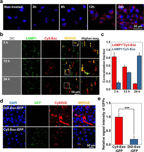
1. In Situ One-Step Fluorescence Labeling Strategy of Exosomes via Bioorthogonal Click Chemistry for Real-Time Exosome Tracking in Vitro and in Vivo
This study explored methods for labeling exosomes using different fluorescent dyes, aiming to develop an efficient and stable fluorescence labeling strategy for real-time tracking of exosomes. The study focused on the application of fluorescence labeling via exosomes, particularly using bioorthogonal click chemistry to label exosomes. In terms of methodology, the researchers used various fluorescent dyes for one-step click chemistry labeling on the surface of exosomes, with the dyes reacting with chemical groups on the exosome surface. The results showed that this method effectively labeled exosomes, and different fluorescent dyes provided stable and intense signals after labeling, making them suitable for dynamic tracking in both in vitro and in vivo studies. The conclusion suggests that using different fluorescent dyes for exosome labeling provides higher labeling efficiency and tracking sensitivity, avoids potential interference with exosome function from traditional labeling methods, and offers broad application potential, especially in drug delivery and disease research.
Song, S. et al. Bioconjugate Chemistry, 2020.
Figure 2. In Vitro Tracking of Cy5-Exo and DiD-Exo to Study Intracellular Behavior.
FAQ
Q1: Will the Labeling Affect the Structure or Function of the Exosomes?
A1: We have optimized dye concentration, incubation time, and washing procedures to preserve the structural integrity and biological activity of exosomes as much as possible, ensuring no interference with subsequent functional experiments or cell uptake assays.
Q2: How Stable Is the Fluorescence of the Labeled Exosomes?
A2: We select dyes with strong photostability and minimize exposure to strong light during the procedure. The fluorescence signal remains stable in the short term, making it suitable for imaging and quantitative analysis.
Q3: Are Exosome Samples Provided by Customers Supported?
A3: Yes, we support customer-provided exosome samples. Customers can provide exosome extracts or exosome source cells, and we will assess the labeling strategy based on the sample condition and proceed with the necessary operations and validation.
How to order?

