


Exosome Fluorescent Labeling Service
Exosome Fluorescent Labeling Service refers to the efficient and specific fluorescent labeling of exosomes through fluorescent probes, fluorescent proteins or fluorescent conjugated antibodies, so that they can be visualized and analyzed in intracellular uptake, tissue distribution and in vivo tracing studies.
Exosomes are small extracellular vesicles secreted by cells that play a crucial role in intercellular communication and have significant applications in disease diagnosis, biomarker research, and targeted drug delivery. Fluorescent labeling is one of the most commonly used visualization techniques, allowing researchers to analyze exosome origin, distribution, uptake mechanisms, and biological functions. By utilizing fluorescence microscopy, flow cytometry, and in vivo imaging systems, fluorescent labeling technology enables the real-time tracking of exosomes, providing precise monitoring of their behavior and dynamics in both in vitro and in vivo studies.
González MI. et al. Biomedicines. 2021.
The core principle of exosome fluorescent labeling is the selective binding of fluorescent probes to the exosome membrane structure or internal contents, making them detectable under fluorescence imaging systems. Depending on the labeling strategy, fluorescent labeling techniques can be categorized into membrane fluorescence labeling, protein fluorescence labeling, and cargo fluorescence labeling. Each method is suited for different research objectives, and selecting the optimal approach depends on the specific experimental requirements.
MtoZ Biolabs offers Exosome Fluorescent Labeling Service can be applied to exosome tracking, functional studies, drug delivery research, and disease biomarker identification, supporting both basic and clinical research.
Analysis Workflow
Our Exosome Fluorescent Labeling Service follows a standardized, high-precision workflow to ensure high-quality and reproducible results:
1. Exosome Isolation and Quality Assessment
Exosomes are isolated and purified, followed by quality control using transmission electron microscopy (TEM), Western blot, and other techniques to ensure purity and integrity.
2. Selection of Appropriate Fluorescent Labeling Method
Based on the research objective, the most suitable fluorescent probe is selected. Dye concentration, incubation temperature, and time are optimized to ensure high labeling efficiency and specificity.
3. Exosome Fluorescent Labeling and Purification
Exosomes are incubated with the fluorescent probe in an appropriate buffer system. Free dye is removed using ultracentrifugation, size-exclusion chromatography (SEC), or dialysis to ensure a clean labeling process.
4. Quality Control and Delivery
Fluorescence microscopy and other techniques are used to confirm successful exosome labeling, evaluate labeling efficiency, and verify exosome integrity post-labeling. The labeled exosomes are then delivered to the client for further analysis.
Applications
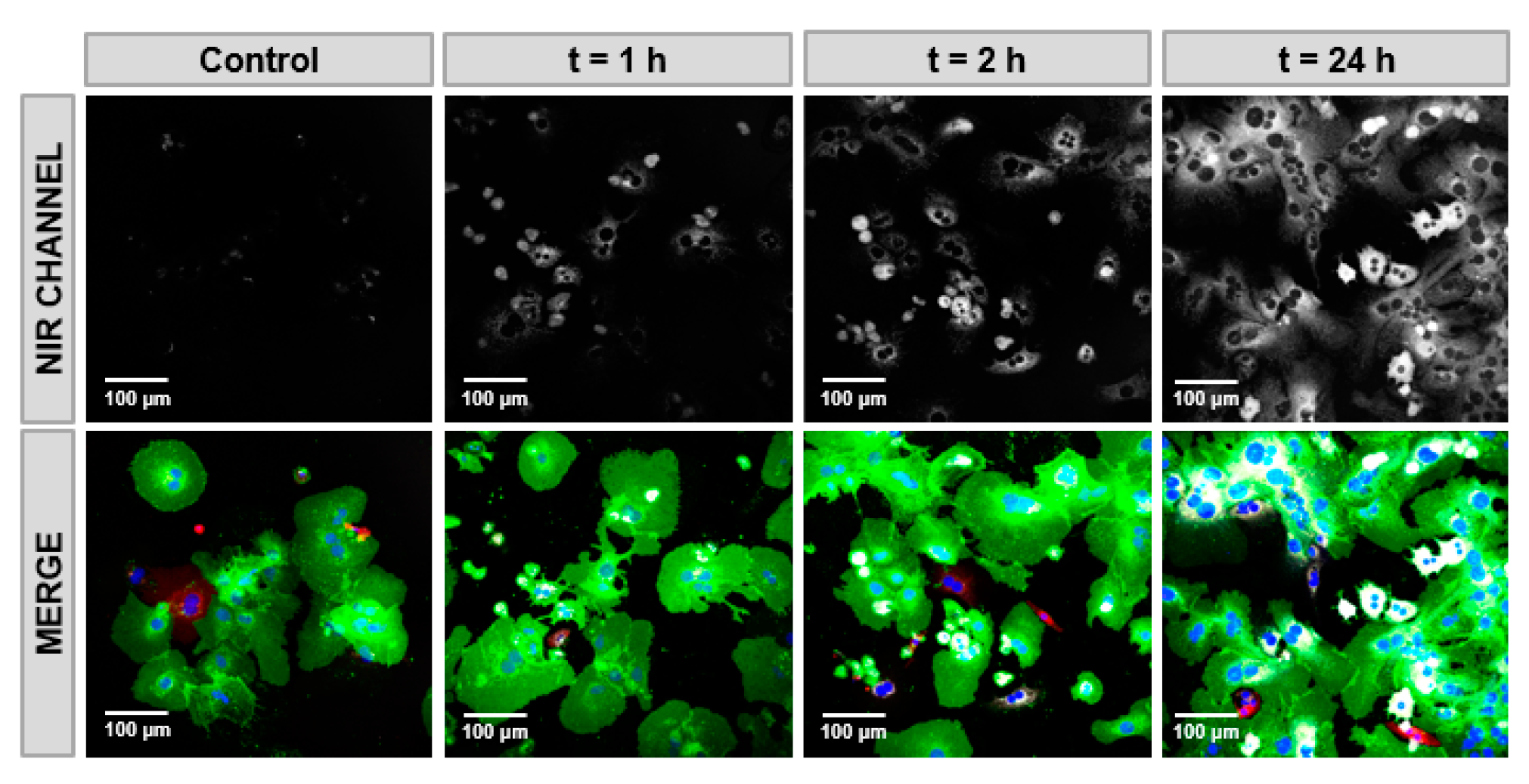
Exosome Uptake Studies
Investigate how exosomes are taken up by target cells and elucidate their delivery mechanisms.
Exosome Tracing and In Vivo Distribution
Track the movement and distribution of exosomes in both in vitro and in vivo environments.
Exosome Functional Studies
Label specific RNAs or proteins to study the role of exosomes in disease progression and intercellular communication.
Exosome-Based Drug Delivery Systems
Assess whether fluorescently labeled exosomes successfully deliver targeted drugs or gene-editing tools.
Service Advantages
1. High-Efficiency and Precise Fluorescent Labeling Strategies: Utilizing a combination of lipid dyes, protein labeling, and genetic fluorescence fusion techniques, we customize the optimal labeling strategy based on experimental needs.
2. Advanced Quality Control System: Comprehensive validation using TEM, NTA, Western blot, and flow cytometry ensures the purity and integrity of labeled exosomes.
3. Comprehensive One-Stop Exosome Analysis Services: Beyond fluorescent labeling, we provide exosome isolation, proteomics, and transcriptomics services to support in-depth exosome functional research.
FAQ
Q. Does fluorescent labeling affect the biological function of exosomes?
Optimized fluorescent labeling strategies can minimize the impact on the biological functions of exosomes. Lipophilic fluorescent probes (e.g., PKH67) primarily integrate into the exosome membrane without altering its main components. However, excessive labeling may lead to membrane structural changes or aggregation. Therefore, it is essential to optimize probe concentration and incubation time, and use ultracentrifugation or size-exclusion chromatography (SEC) to remove unbound dyes. Additionally, genetically encoded fluorescent fusion proteins (e.g., GFP-CD63) can be used to label exosomes without affecting their natural function, although this method requires cell transfection. To ensure that labeled exosomes retain their biological activity, it is recommended to perform cell uptake experiments and functional assays before proceeding with downstream applications.
Case Study
This study employed exosome fluorescent labeling technology to track the distribution and function of mesenchymal stem cell (MSC)-derived exosomes in an acute liver injury (ALI) model. The results demonstrated that exosomes secreted by MSCs can deliver SLC7A11, inhibit ferroptosis, and mitigate pathological changes in ALI. These findings suggest that MSCs counteract ferroptosis through exosome-mediated stabilization of SLC7A11, highlighting their potential therapeutic value in ALI treatment.
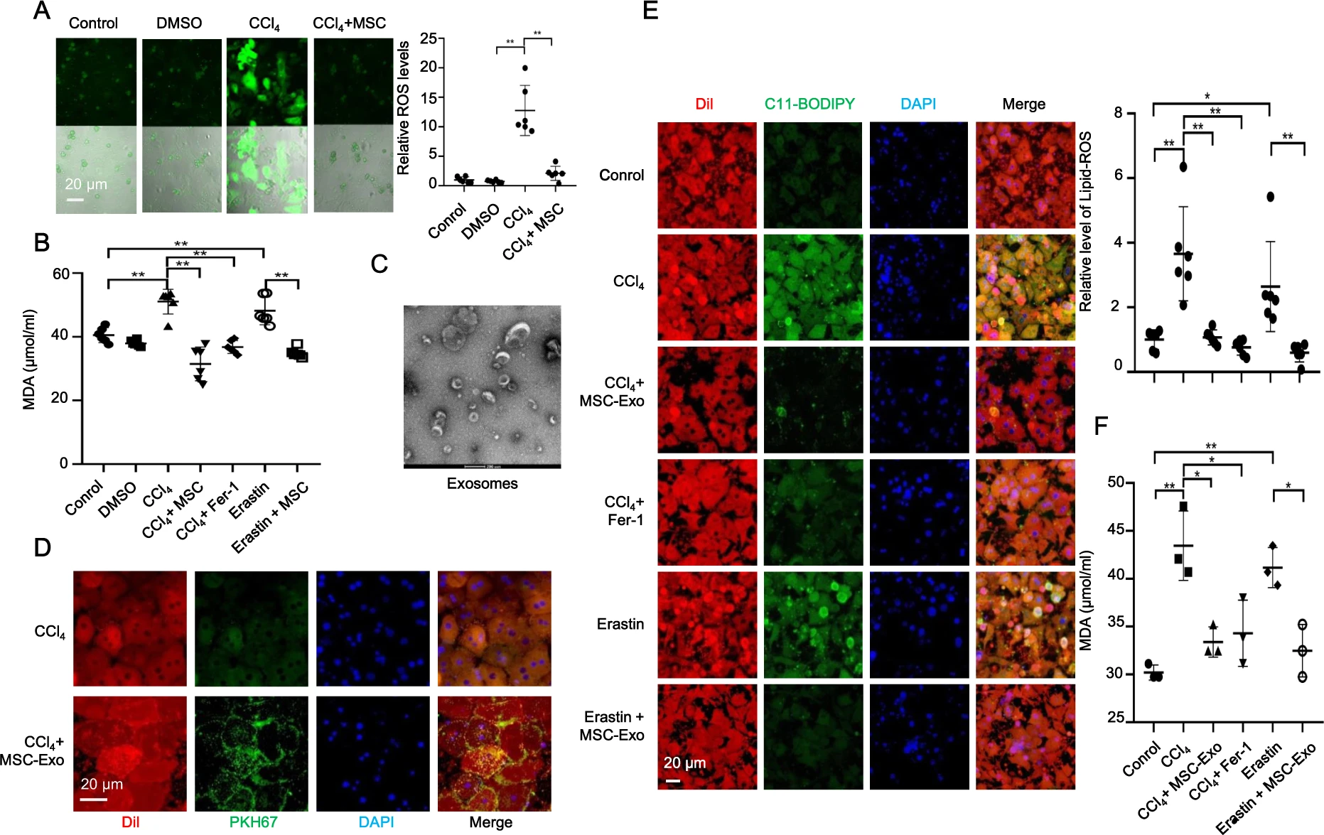
Lin F. et al. Cell Death Dis. 2022.
How to order?

