



Data-independent acquisition (DIA) quantitative proteomics is often applied in oncology when unlabeled digests from tumor tissue, adjacent normal, plasma, serum, or other research matrices must be compared across predefined cancer groups on separate liquid chromatography-tandem mass spectrometry (LC-MS/MS) injections (Gillet et al. 2012). Windowed or staggered DIA schedules support relative candidate lists when the contrast is declared before acquisition, not after files are grouped post hoc. The guide below maps DIA application choices for cancer biomarker discovery from matched prep through extraction. It is not an early-detection promise, not a multi-marker panel product page, and not a generic DDA versus DIA theory article.
Oncology samples combine clinical heterogeneity with matrix-specific prep risk. Formalin-fixed paraffin-embedded (FFPE) blocks, fresh frozen tissue, and depleted plasma each change digestion yield and missing-value patterns. DIA acquisition does not remove stroma composition differences or hemolysis tracked to case labels. Matched pretreatment, a frozen contrast, and unified false discovery rate (FDR) control remain the application gate before candidates are named (Rosenberger et al. 2017).
In MtoZ Biolabs offerings, oncology discovery cohorts are often acquired as label-free DIA or SWATH-style schedules, with extraction and contrast testing documented after prep and acquisition records are fixed.
Cancer Biomarker Application Contexts Where DIA Fits
Five oncology discovery patterns appear most often in project scoping.
Tumor versus matched normal or adjacent tissue. Relative contrasts need pathologist-confirmed regions and matched extraction so stroma shifts are not prep artifacts.
Stage or grade contrasts within one histology. Early versus advanced, or low versus high grade, need inclusion rules frozen before search thresholds move.
Liquid biopsy-oriented plasma or serum discovery. Case versus control or stage-defined arms fit DIA when depletion and collection SOPs are equal across groups and the contrast is not relabeled after acquisition.
Treatment response or resistance contrasts. Pre- and post-therapy biopsies, or sensitive versus resistant lines in research collections, need synchronized timing metadata.
FFPE retrospective cohorts with declared input rules. Archival blocks fit when extraction chemistry is uniform and contrast arms share fixation history where possible.
Cancer DIA biomarker application value is a contrast-tested relative matrix on matched digests, not a validated diagnostic panel by default.
High-file cohort mechanics are covered in High-throughput SWATH-MS Quantitative Proteomics for Biomarker Discovery. Broader clinical research framing appears in Typical Applications of DIA-Based Quantitative Proteomics in Clinical Research.
Study Designs for Oncology DIA Cohorts
Application quality follows cancer-specific design choices.
Contrast and histology lock. Tumor type, stage class, and arm labels should be frozen before extractor tuning. Merging arms after acquisition inflates candidate lists.
Matched pretreatment across arms. Lysis, depletion, digestion, and cleanup must apply equally to tumor and control fluids. Arm-specific prep creates false oncology markers.
Tumor content and cellularity metadata. Bulk tissue contrasts should record necrosis, stroma, and cellularity where available so interpretive claims stay bounded.
Library or library-free extractor declared upfront. Library-based SWATH-style extraction needs provenance-matched evidence. Library-free direct DIA searches under FDR control need interference review (Demichev et al. 2020).
Discovery versus validation intent. Open DIA fits relative candidates for a predefined contrast. Clinical utility, companion diagnostics, and screening claims need independent cohorts and often parallel reaction monitoring (PRM) or immunoassay stages outside the discovery matrix (Gerber et al. 2003).
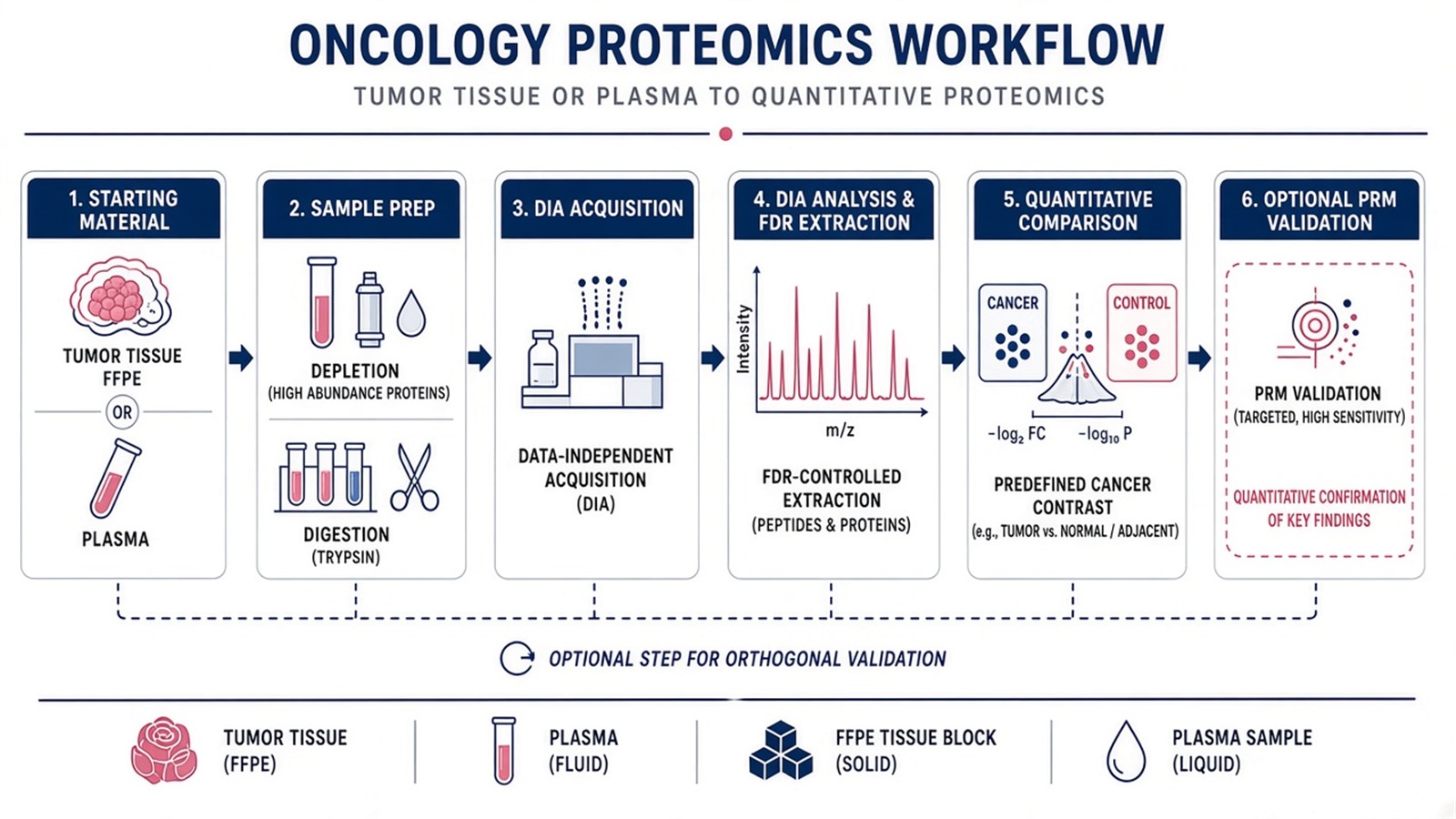
Figure 1. Cancer biomarker DIA applications depend on matched prep, a frozen oncology contrast, and FDR-controlled extraction before candidate lists.
Pathway overlay belongs after contrast QC. Mechanism wording for drug contrast projects can cross-check Quantitative Proteomics in Disease and Drug Discovery. Extraction statistics belong with DIA Data Analysis Methods in Proteomics.
Shared Workflow From Cancer Sample to DIA Matrix
A practical oncology biomarker DIA spine usually runs as follows.
- Confirm matrix, histology, contrast, and relative claim.
- Collect or section samples under documented SOP; record fixation, storage, and tumor content notes.
- Prepare digests with one declared route; apply depletion or FFPE recovery equally across arms when used.
- Run pilot injections when the oncology matrix or prep route is new.
- Acquire DIA or SWATH-style windows under a locked plan across the cohort.
- Extract peptides under FDR and interference rules with one declared extractor version.
- Test only the predefined contrast; document missing-value policy.
- Optional targeted PRM or stable isotope-labeled internal standard (SIS) follow-up on a short list after QC.
Steps 2 through 4 dominate oncology failure modes. Step 6 cannot repair unequal FFPE extraction or plasma depletion applied to cases only. Missing values in oncology fluids often track abundance and prep loss, not only acquisition chance (Lazar et al. 2016).
Parameters and Limits in Cancer Biomarker DIA Applications
Parameter |
Why it matters in oncology work |
Failure mode if ignored |
|---|---|---|
Histology and stage in contrast map |
Bounds interpretive claims |
Mixed tumor types in one arm |
Matched depletion and digestion |
Keeps peptide yield comparable |
False fluid markers from prep |
Tumor cellularity or macrodissection |
Separates biology from stroma mix |
Stroma shifts read as regulation |
Window plan locked to cohort |
Keeps extraction valid |
Mid-study method changes |
Unified FDR across files |
Controls peptide error rate |
Per-run cutoffs that drift |
Missing-value policy |
Handles fluid dropout |
Imputed differences without evidence |
Follow-up on short lists |
Separates discovery from deployment |
Discovery p-values as clinical test |
Limits follow the same table. DIA does not ensure detection of every low-abundance secreted protein in every stage I plasma sample. Machine-learning panels built on the same discovery matrix without holdout cohorts overfit. Retrospective reanalysis still needs a declared extractor and contrast. Early detection, prognosis scores, and therapy selection claims need validation designs orthogonal to the initial DIA table.
Isobaric tagging may fit a closed multi-arm tumor set labeled the same day. Open biobank enrollment with staggered plasma draws more often stays on label-free DIA separate injections.
Researchers who need three-axis route selection can refer to Protein Quantification Method. Targeted confirmation trade-offs appear in Advantages and Disadvantages of Targeted Proteomics.
How to Choose DIA for the Next Oncology Biomarker Project
Use matrix, enrollment, and follow-up goal together.
Project need |
DIA application plan that often fits |
Recheck before committing |
|---|---|---|
Plasma case-control, open enrollment |
Label-free DIA with locked extractor |
Depletion on cases only |
Tumor versus adjacent normal |
Matched macrodissection prep |
Different extraction routes by arm |
FFPE retrospective stage contrast |
Uniform FFPE digestion pilot |
Full cohort without yield QC |
Multi-center plasma biobank |
Site metadata plus batch covariate plan |
Site tracks cancer type |
Short list before assay transfer |
DIA discovery then PRM panel |
Panel longer than duty cycle |
Screening or companion diagnostic claim |
Separate validation cohort design |
Discovery matrix as final test |
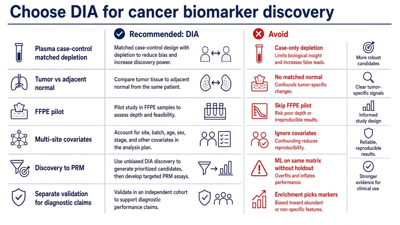
Figure 2. Choose cancer DIA when matched prep and a frozen oncology contrast fit; diagnostic deployment and ML panels need validation beyond one discovery matrix.
Teams that already have matrix, histology contrast, cohort design, and follow-up goal can review the MtoZ Biolabs Label-Free Quantitative Proteomics Service for oncology DIA biomarker planning.
Frequently Asked Questions
1. Does cancer DIA discovery equal a blood test for early detection?
No. DIA yields research candidates for predefined contrasts. Screening assays need separate validation and regulatory paths.
2. Can pathway enrichment pick diagnostic markers?
No. Enrichment interprets a contrast-tested list. Marker choice still needs statistical and analytical follow-up.
3. Is FFPE compatible with DIA biomarker discovery?
Often yes with a declared FFPE prep route and pilot yield QC. Fixation history should be balanced across arms where possible.
4. Must candidates be validated by PRM?
Not always, but remeasurement on a closed peptide list is common before assay transfer. Discovery intensities alone are not concentration claims.
5. Does deeper protein identification mean stronger cancer markers?
No. Identification depth and contrast quality differ. Replicate structure and follow-up design decide candidate strength.
MtoZ Biolabs can support oncology DIA biomarker application planning when sample type, histology contrast, cohort metadata, and the intended relative claim are available.
If further evaluation of plasma depletion, FFPE prep, multi-center batch plans, or discovery-to-targeted staging is needed, project details can be submitted for a feasibility review.
Reference
- S.A. Gerber, J. Rush, O. Stemman, M.W. Kirschner, S.P. Gygi (2003). Absolute quantification of proteins and phosphoproteins from cell lysates by tandem MS. Proc. Natl. Acad. Sci. U.S.A., 100, 6940-6945. https://doi.org/10.1073/pnas.0832254100
- L.C. Gillet, P. Navarro, S. Tate, H. Rost, N. Selevsek, L. Reiter, R. Bonner, R. Aebersold (2012). Targeted data extraction of the MS/MS spectra generated by data-independent acquisition: a new concept for consistent and accurate proteome analysis. Mol. Cell. Proteomics, 11, O111.016717. https://doi.org/10.1074/mcp.O111.016717
- V. Demichev, C. Messner, S. Vernardis, K.S. Lilley, M. Ralser (2020). DIA-NN: neural networks and interference correction enable deep proteome coverage in high throughput. Nat. Methods, 17, 41-44. https://doi.org/10.1038/s41592-019-0638-x
- C. Lazar, L. Gatto, M. Ferro, C. Bruley, T. Burger (2016). Accounting for the multiple natures of missing values in label-free quantitative proteomics data sets to compare imputation strategies. J. Proteome Res., 15, 1116-1125. https://doi.org/10.1021/acs.jproteome.5b00981
- G. Rosenberger, S. Liu, J. Röst, H.L. Ludwig, C. Buil, B. Borchers, R. Aebersold, L. Collins (2017). Statistical control of peptide and protein error rates in large-scale targeted data-independent acquisition analyses. Nat. Commun., 8, 14224. https://doi.org/10.1038/ncomms14224
