



Data-independent acquisition mass spectrometry (DIA-MS) on mouse tissue digests is often chosen when unlabeled peptides from liver, brain, heart, kidney, muscle, or other organs must stay comparable across genotypes, treatments, or time points on separate liquid chromatography-tandem mass spectrometry (LC-MS/MS) injections (Gillet et al. 2012). DIA window plans sit after injection-ready digests exist. The guide below focuses on mouse tissue sample handling and pretreatment through the DIA handoff. It is not a mouse model catalog, not an instrument parameter sheet, and not a generic DDA versus DIA theory page.
Mouse tissues differ in lipid content, connective matrix, membrane protein load, and blood contamination when perfusion is incomplete. Those differences change lysis yield and cleanup needs before any DIA schedule is tested. DIA acquisition does not remove lipids left in the digest or correct prep batches that track genotype labels. Matched extraction and digestion across arms remain the gate for organ contrast studies (Wisniewski et al. 2009).
In MtoZ Biolabs offerings, mouse tissue digests are commonly acquired as label-free DIA or SWATH-style windowed schedules after pretreatment is documented, with extraction and contrast testing fixed after acquisition records are locked.
Mouse Tissue Matrices Used for DIA-MS Quantification
Common mouse DIA inputs include perfused or flash-frozen organs, dissected subregions, and pooled aliquots from cohort culls. Each matrix sets the first prep bound.
Liver and kidney often carry high metabolic enzyme load and variable blood residue when perfusion is skipped. Brain needs thorough disruption with lipid-aware cleanup when myelin and membrane fractions dominate. Heart and skeletal muscle carry abundant contractile proteins and connective material that benefit from consistent homogenization depth. White adipose and brown adipose need lipid management so digestion and desalting recover peptides comparably across groups.
Mouse DIA quantification starts when digests are clean, desalted, and loaded under matched rules, not when frozen tissue enters the homogenizer without a contrast map.
Bulk Bradford, bicinchoninic acid (BCA), Lowry, and 280 nm assays help match total protein before digestion. They do not replace peptide-level DIA extraction after LC-MS acquisition.
Pretreatment Steps That Keep Mouse DIA Digests Comparable
Label-free DIA comparability depends on matched prep across genotypes and treatments, not on window width alone.
Homogenization and lysis. Groups should share buffer family, bead-mill or dounce protocol, and extraction temperature. Organ-specific routes are allowed when each contrast arm uses the same route for that organ.
Reduction, alkylation, and digestion. Shared enzyme-to-substrate ratio and digestion time limit missed cleavages that track groups. Filter-aided sample preparation and solid-phase cleanup help remove detergents and chaotropes that harm LC and fragment extraction.
Cleanup and recovery. StageTip-style or cartridge desalting should recover peptides equally across arms (Rappsilber et al. 2007). Group-specific losses create missing values that track treatment, not biology.
Desalting and injection load. DIA needs stable chromatography across the cohort. Unequal dry-down recovery or mismatched peptide loads per injection distort extracted ion traces before FDR-controlled extraction runs.
No isobaric tagging step appears in ordinary label-free mouse DIA prep. Digests proceed to LC when cleanup is complete. Stable isotope-labeled internal standard (SIS) peptides belong to targeted follow-up on named peptides, not to open organ discovery prep by default.
Researchers who need general label-free SWATH pretreatment framing can refer to SWATH-MS Label-Free Protein Quantification Technology Analysis. For LC injection discipline shared across routes, see Quantitative Analysis of Proteins by Liquid Chromatography.
Study Designs for Mouse Tissue DIA Cohorts
Application quality follows mouse-specific design choices.
Genotype, treatment, and organ lock. Mixing organs in one contrast arm without declaration creates false regulation lists. Cull time and fasting state should be fixed in the contrast map.
Matched prep batches interleaved with injection order. Sacrifice day, dissector, and extractor version should not track treatment unless that tracking is intentional.
Sex and age as declared factors. Cohort tables should list sex and age so contrasts are not confounded with unlabeled arm merges.
Pilot tissue when the organ or prep route is new. Brain lipid, adipose yield, or fibrous muscle routes often need a pilot digest before the full DIA batch is scheduled.
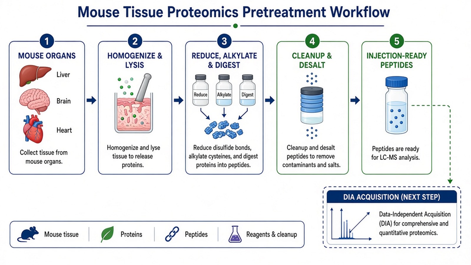
Figure 1. Mouse DIA-MS depends on matched organ prep and injection-ready digests before windowed acquisition and extraction.
Enrollment checks before scaling appear in Exploring Quantitative Analysis in Proteomics. Extraction, FDR control, and matrix statistics belong with DIA Data Analysis Methods in Proteomics after DIA-ready injections exist.
Shared Workflow From Mouse Tissue to DIA-Ready Injection
A practical mouse tissue DIA prep spine usually runs as follows.
- Confirm strain, organ, contrast, and relative claim across separate injections.
- Harvest with matched perfusion or flash-freeze rules; record cull batch and storage.
- Homogenize and extract proteins with one declared organ-compatible route.
- Reduce, alkylate, and digest under shared rules; remove debris and lipids that interfere with LC.
- Clean and desalt peptides to LC-ready loads shared across arms.
- Run pilot injections when the organ or prep route is new.
- Acquire DIA or SWATH-style windows under a locked plan across the cohort.
- Hand files to the declared extractor with frozen contrast and missing-value policy.
Steps 1 through 6 define this page. Step 7 crosses into acquisition. Step 8 belongs with analysis SOPs. Order errors show up as alignment failure or abundance-dependent missingness (Lazar et al. 2016). Pathway enrichment after search cannot repair unequal cleanup or confounded sacrifice batches.
Researchers who need the full lysis-to-digest spine can refer to Quantitative Protein Analysis.
Parameters and Limits in Mouse Tissue DIA Prep
Parameter |
Why it matters for mouse DIA prep |
Failure mode if ignored |
|---|---|---|
Organ and dissection map |
Sets valid contrast |
Mixed tissues read as regulation |
Perfusion or blood residue control |
Stabilizes plasma protein carryover |
Blood proteins track genotype |
Matched input (weight or total protein) |
Stabilizes yield |
Load bias mimics biology |
Lipid-aware cleanup for brain or adipose |
Protects digestion and LC |
Missing peptides by arm |
Equal cleanup recovery |
Limits group-specific dropout |
Missingness that tracks treatment |
Injection load per run |
Stabilizes DIA peak groups |
Saturation or weak alignment |
Pilot before cohort |
Catches bad organ routes early |
Full batch repeat homogenization |
Limits follow the same table. Mouse DIA prep does not label channels. Late culls cannot join prior injections as hidden replicates without a new prep record. DIA does not report absolute concentration from open discovery intensities without SIS on named peptides. Window width, library choice, and extractor version are acquisition and analysis parameters. They must be declared with the matrix, but they do not replace matched pretreatment.
Isobaric tagging may fit a freezable multi-arm mouse set harvested for one labeling day. Open colonies with staggered culls more often stay on label-free DIA separate injections.
How to Choose Pretreatment for the Next Mouse DIA Project
Use organ, enrollment, and follow-up goal together.
Project need |
Pretreatment plan that often fits |
Recheck before committing |
|---|---|---|
Genotype contrast, one organ |
Matched homogenization and digest, separate DIA injections |
Sacrifice day equals genotype |
Treatment versus vehicle, liver |
Synchronized cull, interleaved prep |
Different cleanup routes by arm |
Multi-organ panel within one mouse |
Paired prep rules per organ, separate contrasts |
One contrast merges unlike organs |
Brain or adipose discovery |
Lipid-aware cleanup pilot |
Full cohort without LC pilot |
Time course with open culls |
Interleaved prep and injection order |
All late time points in one batch |
Short list after DIA discovery |
Same cleanup rules, then targeted panel |
Open DIA stats as confirmation |
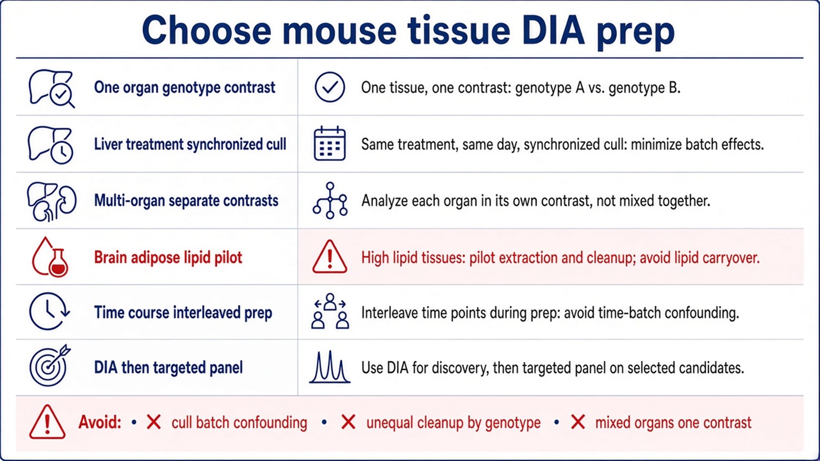
Figure 2. Choose mouse DIA prep from organ and enrollment rules; unequal cleanup, confounded cull batches, and amount-without-SIS requests need repair before acquisition.
Teams that already have strain, organ map, contrast, and relative claim can review the MtoZ Biolabs Label-Free Quantitative Proteomics Service for mouse tissue DIA pretreatment and acquisition planning. For route selection against TMT or targeted follow-up, see Protein Quantification Method.
Frequently Asked Questions
1. Is DIA-MS a different labeling method from label-free quantification?
No. Label-free names the chemistry. DIA names a window acquisition schedule on unlabeled digests.
2. Must every mouse DIA project use the same lysis buffer for all organs?
No. Organ-specific buffers are common. Contrast arms within one organ still need matched chemistry.
3. Can prep differ by genotype group?
Only when prep is an explicit experimental factor. Otherwise matched lysis and cleanup are required across groups.
4. Does successful DIA extraction prove pretreatment was matched?
No. Extraction can return IDs from uneven prep. Matrix QC and contrast design still decide whether ratios are trustworthy.
5. Should perfusion be planned for all mouse organs?
Perfusion reduces blood protein carryover in vascular organs. The same perfusion rule should apply to every arm in the contrast.
MtoZ Biolabs can support mouse tissue DIA pretreatment and acquisition planning when strain, organ type, study design, and the intended relative claim are available.
If further evaluation of organ-specific lysis, lipid cleanup, injection loads, or DIA versus TMT enrollment is needed, project details can be submitted for a feasibility review.
Reference
- L.C. Gillet, P. Navarro, S. Tate, H. Rost, N. Selevsek, L. Reiter, R. Bonner, R. Aebersold (2012). Targeted data extraction of the MS/MS spectra generated by data-independent acquisition: a new concept for consistent and accurate proteome analysis. Mol. Cell. Proteomics, 11, O111.016717. https://doi.org/10.1074/mcp.O111.016717
- C. Lazar, L. Gatto, M. Ferro, C. Bruley, T. Burger (2016). Accounting for the multiple natures of missing values in label-free quantitative proteomics data sets to compare imputation strategies. J. Proteome Res., 15, 1116-1125. https://doi.org/10.1021/acs.jproteome.5b00981
- J. Rappsilber, M. Mann, Y. Ishihama (2007). Protocol for micro-purification, enrichment, pre-fractionation and storage of peptides for proteomics using StageTips. Nat. Protoc., 2, 1896-1906. https://doi.org/10.1038/nprot.2007.261
- J.R. Wisniewski, A. Zougman, N. Nagaraj, M. Mann (2009). Universal sample preparation method for proteome analysis. Nat. Methods, 6, 359-362. https://doi.org/10.1038/nmeth.1322
- G. Rosenberger, S. Liu, J. Röst, H.L. Ludwig, C. Buil, B. Borchers, R. Aebersold, L. Collins (2017). Statistical control of peptide and protein error rates in large-scale targeted data-independent acquisition analyses. Nat. Commun., 8, 14224. https://doi.org/10.1038/ncomms14224
