






Cell Proteomics Data Analysis: From Protein Identification to Functional Interpretation
Cell Proteomics data analysis is used to identify stable protein changes from protein identification and quantitative results and to determine whether altered proteins converge on specific functional categories, molecular pathways, or protein relationship networks. In comparisons such as treatment versus control, different cellular states, or distinct genetic backgrounds, interpretation also requires consideration of protein coverage, replicate consistency, and overall quantitative patterns.
Protein-level data are typically evaluated from multiple aspects, including which proteins are detected, whether their abundance differs between experimental groups, and whether the changed proteins are associated with specific biological functions or pathways. Through these analyses, Cell Proteomics results can provide evidence for understanding cellular protein changes and support further biological investigation.
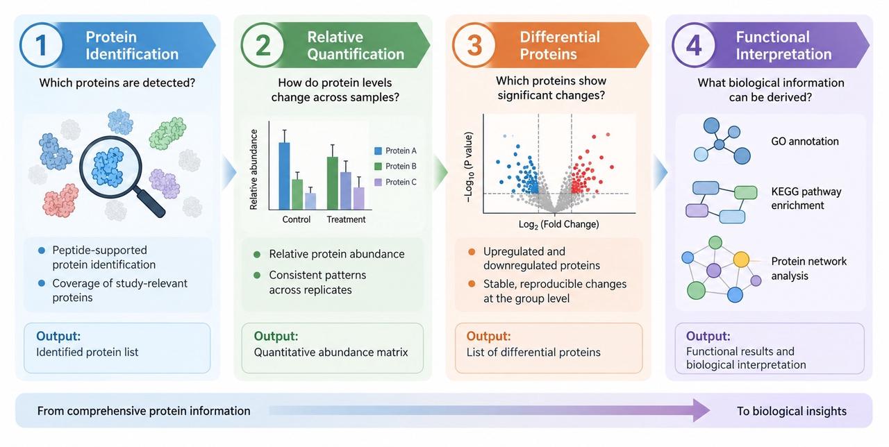
What Data Are Generated from Cell Proteomics?
Cell Proteomics results can be organized into four data levels: protein identification, relative protein quantification, differential proteins, and functional analysis. The first two describe the detectable protein range and abundance information across samples, whereas the latter two focus on between-group changes and organize the functional relationships among altered proteins.
| Data Level | Typical Result Format | Primary Interpretation Focus |
| Protein Identification | Identified protein list with peptide evidence | Whether study-relevant proteins are covered; whether identification ranges are comparable across samples |
| Relative Protein Quantification | Protein × sample relative abundance matrix | Whether biological replicates are consistent; whether between-group abundance changes are stable |
| Differential Proteins | Set of altered proteins associated with a predefined comparison | Which proteins account for the main between-group changes; whether changes are supported across multiple samples |
| Functional Analysis | GO, pathway enrichment, and protein relationship networks | Whether altered proteins converge on shared biological processes, pathways, or protein modules |
Understanding Differential Protein Changes
Cell Proteomics quantitative results can reveal differences in protein abundance between samples or experimental groups. These changes describe differences in protein abundance between specific conditions and provide information about protein-level variation between compared groups. Differential protein analysis further summarizes proteins with distinct abundance patterns between comparison groups, helping identify protein changes that may be associated with the studied conditions.
1. Protein Abundance Changes
Changes in the abundance of an individual protein should be interpreted together with both the direction of change and consistency across biological replicates. When multiple samples in an experimental group show a similar increase or decrease relative to the control group, the protein change has stronger within-group consistency. When a change is driven mainly by one or a few samples, the apparent between-group difference may be influenced by sample-to-sample variation.
Interpretation of protein abundance changes also depends on the original comparison. Differences between control and treatment groups reflect protein changes associated with the treatment condition, whereas differences between baseline and post-treatment samples reflect relative changes across time or state. An abundance change alone does not explain the cause of the change.
2. Differential Proteins
Differential proteins are proteins that show clear abundance differences in a predefined comparison. Compared with reviewing proteins individually, a differential protein set is more suitable for determining whether proteome changes concentrate within particular protein classes or shared functional themes. Differential protein lists usually include both the direction and relative magnitude of change and provide the input for subsequent functional analysis.
The number of differential proteins should not be used alone to judge the strength of a result. More important considerations include whether the changes are consistent across biological replicates, whether they correspond to the study variable, and whether multiple differential proteins converge on a coherent functional pattern.
Functional Analysis of Cell Proteomics Results
Functional analysis evaluates whether differential proteins show concentrated functional patterns. Interpretation can be divided into two broad categories: enrichment analysis based on functional annotation and pathway databases, and network analysis based on known protein relationships. These approaches address two different questions: which functions or pathways contain concentrated protein changes, and whether the altered proteins form connected protein modules.
1. Functional Annotation and Pathway Enrichment
GO functional annotation organizes differential proteins across three dimensions: Biological Process, Molecular Function, and Cellular Component. Interpretation should focus on whether multiple differential proteins converge on the same or related biological processes, molecular functions, or cellular components, and whether the major terms are consistent with the experimental condition and cellular background.
KEGG pathway enrichment evaluates whether differential proteins are jointly represented in known molecular pathways. KEGG results should be interpreted together with the actual differential proteins mapped to each pathway, the direction of protein changes, and the original comparison. When multiple altered proteins converge on the same KEGG pathway or on related pathways, the result supports a coordinated pathway-level pattern. KEGG enrichment reflects concentration of a protein set within annotated pathways and cannot by itself establish pathway activation or inhibition.
2. Protein Relationships and Network Analysis
Protein relationship networks organize differential proteins using known interaction or functional association information. Network analysis can show whether altered proteins form connected modules, whether highly connected nodes are present, and whether multiple differential proteins cluster within the same region of a protein relationship network. Compared with isolated proteins, network modules are more useful for identifying protein sets that may participate in the same biological process.
Connections in a network are derived from protein relationships supported by databases or published literature and do not indicate that direct physical interactions were measured in the current samples. The main value of network analysis is therefore to identify known relationship structures and candidate protein modules among altered proteins.
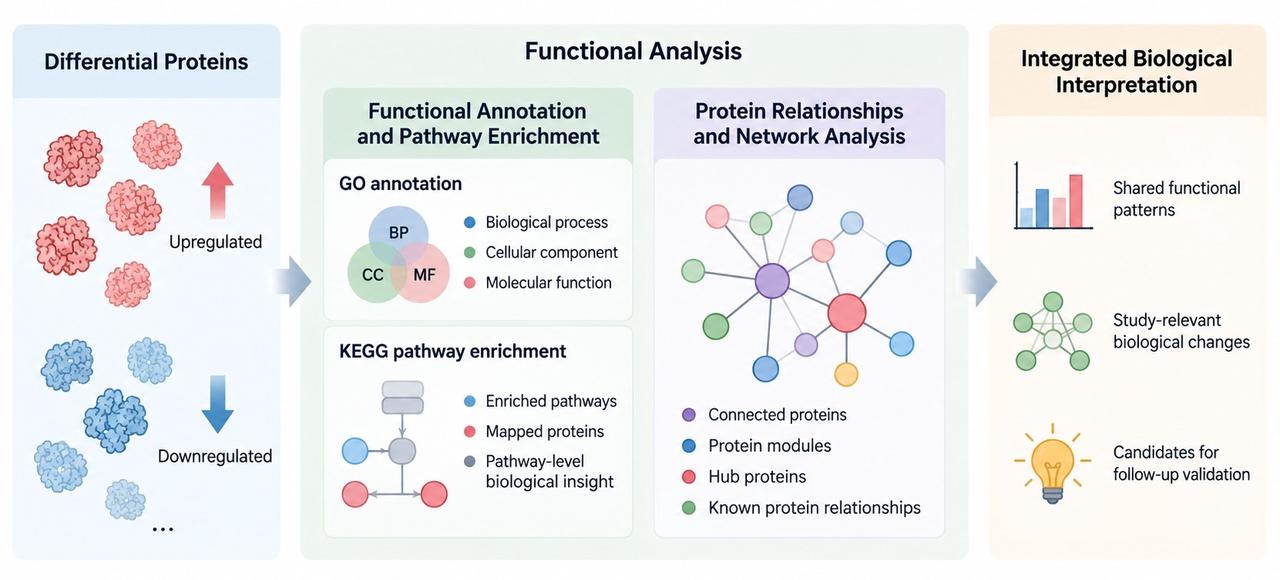
Connecting Proteomics Results with Biological Questions
Biological interpretation requires quantitative patterns, differential proteins, and functional analysis to be related back to the original comparison. The key question is not whether a single result is significant, but whether different levels of evidence consistently support the same protein changes.
| Interpretation Level | Question to Address | Main Evidence |
| Sample and Group Level | Are protein abundance patterns consistent within groups, and are between-group differences stable? | Quantitative patterns across biological replicates; relative abundance differences between groups |
| Protein-Set Level | Do differential proteins converge on shared protein classes or functional themes? | Differential protein list, direction of change, shared alteration patterns |
| Functional Level | Do multiple altered proteins converge in GO, pathways, or PPI networks? | Functional enrichment, pathway mapping, protein network modules |
| Research-Question Level | Do proteome-level changes correspond to cell type, experimental condition, and predefined comparison? | Alignment between experimental context and the preceding proteomics evidence |
1. Biological Interpretation
Biological interpretation should evaluate whether multiple levels of results point to the same direction of change. When differential proteins show stable patterns across biological replicates and also converge on related GO terms, pathways, or protein networks, the functional interpretation is stronger than interpretation based on an isolated protein change. When different analyses show poor agreement, less weight should be placed on individual findings.
Cell type and experimental condition define the biological context for interpreting protein changes. The same protein pattern can have different research implications in different cell models or treatment conditions, so functional results should always be interpreted in relation to the original experimental comparison.
2. Research Hypothesis Generation
Proteomics results can be used to prioritize directions for follow-up validation. Higher-priority candidates generally show stable between-group abundance changes and are consistent with major protein sets, pathways, or network modules identified in functional analysis. Follow-up studies can then focus on candidate proteins, co-varying protein modules, or functional themes to test whether the proteomics findings support a more specific biological hypothesis. The broader applications of Cell Proteomics in cellular research are discussed in Applications of Cellular Proteomics.
Considerations When Interpreting Cell Proteomics Results
Cell Proteomics results should be interpreted by considering data completeness, quantitative consistency, and biological variation. Protein identification, abundance changes, and functional analysis results provide different types of evidence, but the reliability of interpretation depends on how well the detected data represent the analyzed samples and whether observed differences are consistent with the experimental design.
1. Protein Coverage
Protein coverage refers not only to the total number of identified proteins, but also to whether study-relevant proteins are detected and whether coverage is comparable across samples. If one group contains substantially fewer quantifiable proteins than another, subsequent between-group comparisons may be affected by differences in data completeness.
Coverage should therefore be evaluated by examining both detection of key study targets and the range of proteins shared across samples. A high total protein count cannot replace protein evidence that is directly relevant to the research question.
2. Missing Values
Missing values occur when quantitative information for a protein is unavailable in one or more samples. An isolated missing value in a single sample, concentrated missingness within one group, and consistently low detection across multiple samples represent different data patterns and should not be interpreted in the same way.
Proteins with extensive missing values require cautious interpretation in between-group comparisons. It is important to determine whether the apparent difference is supported by genuine abundance changes or is driven mainly by incomplete detection. A missing quantitative signal should not be interpreted directly as protein absence.
3. Biological Variation
Biological variation represents differences in protein abundance patterns between biological samples within or between experimental groups. Such variation is an important factor when evaluating whether observed protein changes are associated with the studied condition or reflect natural differences between samples. Interpretation of Cell Proteomics results requires consideration of biological variation, experimental design, and data quality to distinguish meaningful protein patterns from sample-to-sample differences.
Frequently Asked Questions
1. Should functional analysis use all identified proteins or only differential proteins?
It depends on the analytical objective. All identified proteins are useful for describing the overall functional composition of a dataset, whereas differential proteins are more appropriate for evaluating functional changes associated with a predefined comparison. The two analyses represent different interpretation levels.
2. Do two proteins with similar relative changes necessarily have the same biological significance?
No. Proteins may differ in function, cellular localization, pathway assignment, and relationships with other altered proteins. Similar relative changes can therefore have different research significance.
3. How should a protein be interpreted when the direction of change is inconsistent across biological replicates?
First determine whether the apparent difference is driven by individual samples, and evaluate the result together with within-group variation and data completeness. Proteins with clearly inconsistent replicate patterns should not be interpreted strongly on the basis of the group mean alone.
4. Why can the same differential proteins appear in multiple GO terms or pathways?
Functional annotations are hierarchical and overlapping, and one protein can participate in multiple biological processes or pathways. Interpretation should examine the core proteins shared among related terms and avoid treating highly overlapping results as independent findings.
5. Which proteins or functional themes should be prioritized for follow-up validation?
Priority can be given to candidates with stable changes across biological replicates, complete quantitative information, and consistency with the major GO, pathway, or protein network results.
Conclusion
Cell Proteomics data interpretation connects protein identification, relative abundance patterns, differential protein changes, functional analysis, and biological interpretation. Together, these results allow researchers to evaluate cellular protein changes from protein identification and quantitative patterns to differential proteins, functional analysis, and biological interpretation.
The interpretation of Cell Proteomics results requires consideration of both quantitative findings and biological context. For a broader overview of Cell Proteomics principles, workflow, and applications, refer to Cell Proteomics: Principles, Workflow, and Applications.
For researchers planning cell proteomics projects and requiring evaluation of expected data outputs or result interpretation strategies, MtoZ Biolabs provides Cellular Proteomics Service support. Further project information is available through Contact Us.
How to order?




