






Can PhIP-Seq Detect Conformational or Modified Autoantigens?
- Paired modified and unmodified peptide clones for direct comparison
- Disease-relevant PTM site selection based on prior biology or mapping data
- Sufficient clone representation across the library
- Input library normalization to distinguish true enrichment from clone abundance bias
- Peptide array validation tests shortlisted linear or modified peptides under controlled spot-format conditions
- Recombinant protein or domain binding assays evaluate whether peptide enrichment translates to folded antigen recognition
- Glycoprotein or glycopeptide-focused workflows become relevant when carbohydrate moieties contribute to epitope structure
- PTM mass spectrometry supports site-level confirmation of modification states on purified antigen or immunoprecipitated targets
- Enriched peptide list normalized against input library and background controls
- Case-control or longitudinal comparison summaries
- Protein or tiled-region mapping when annotation supports coordinate assignment
- Modified versus unmodified peptide comparison when PTM variant libraries are used
- Prioritized candidate list flagged for validation follow-up
Introduction
Autoimmune serology often depends on detecting antibodies that recognize self-antigens in native, modified, or structurally folded forms. A rheumatology cohort may show reactivity against citrullinated proteins, yet peptide screens built only from unmodified sequences fail to explain the signal. A neurology study may implicate a multimeric autoantigen whose epitope spans non-adjacent regions. Before patient sera are committed to full library sequencing, teams need to know whether a high-throughput screen can detect conformational or modified autoantigens relevant to the disease model.
PhIP-Seq (phage immunoprecipitation sequencing) supports broad autoantibody profiling by linking phage-displayed peptide libraries with immunoprecipitation and next-generation sequencing. Serum antibodies bind displayed peptides, captured phage are sequenced, and enrichment read counts reveal recognized targets relative to input library representation and background controls. Detection scope is defined by how antigens are represented on phage rather than by full native protein structure alone.
Understanding that boundary early helps teams design libraries, interpret enrichment results, and plan validation before samples are submitted at scale.
What the Platform Detects in Autoantibody Screening
In autoantibody screening, the workflow reports peptide-level enrichment rather than direct measurement of every autoantigen in its native form. Each phage particle displays a short peptide while carrying the DNA sequence that encodes it. Autoantibodies in serum or plasma bind displayed peptides that resemble recognized epitopes or motifs. Immunoprecipitation captures antibody-phage complexes, and sequencing maps read counts back to peptide identity.
The output is a library-defined reactivity profile. Peptides enriched above input and control thresholds are interpreted as candidate autoantigen regions within the peptide space represented in the library. Screening is most direct when the relevant epitope can be approximated by a contiguous linear peptide displayed on phage. When the biological autoantigen depends on three-dimensional folding, subunit assembly, or a modification absent from the library, detection becomes indirect or unsupported unless the study design compensates for that gap.
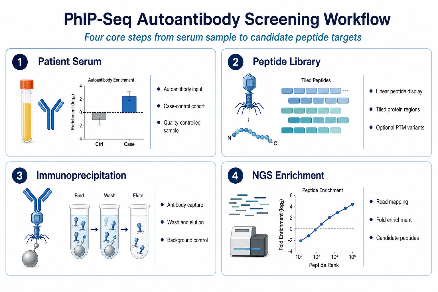
Figure 1. Four-panel workflow summary for autoantibody screening from patient serum through peptide library immunoprecipitation to NGS enrichment.
Conformational Autoantigens: Detection Limits
Conformational autoantigens expose epitopes formed by amino acid residues that are not adjacent in primary sequence. These epitopes often depend on protein folding, domain pairing, or quaternary structure. Multimeric receptors, enzyme complexes, and structurally constrained protein domains may carry diagnostically relevant conformational epitopes that short linear peptides cannot reconstruct.
Standard phage display presents peptides as linear sequences on phage coat proteins. A 7 to 15 amino acid peptide may capture a local linear motif, but the peptide generally does not preserve the three-dimensional arrangement required for many conformational epitopes. Discontinuous epitopes, in which binding requires residues from separate sequence segments brought together by folding, are particularly difficult to represent in conventional peptide libraries.
Tiled peptide libraries can still localize antibody reactivity to protein regions that later guide structural follow-up. Cyclic peptide libraries, constrained peptide designs, or larger displayed scaffolds may approximate some structural features better than unstructured linear peptides. Even so, the default format remains peptide-linear display, and conformational claims require orthogonal validation on folded antigen formats.
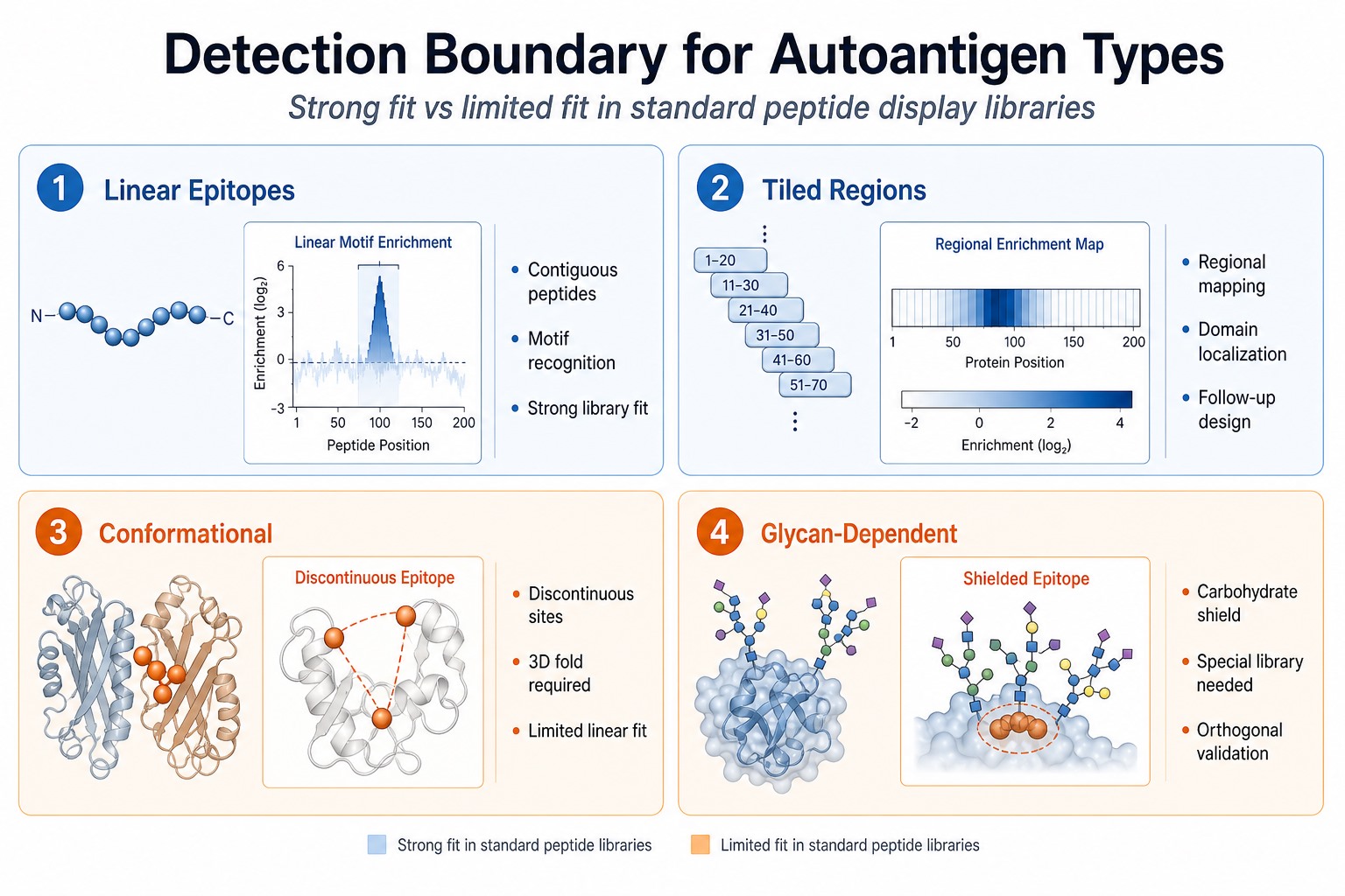
Figure 2. Four-panel detection boundary summary showing strong fit for linear and tiled epitopes and limited fit for conformational and glycan-dependent autoantigens.
Modified Autoantigens: Library Design Determines Coverage
Modified autoantigens carry post-translational modifications (PTMs) that create neo-epitopes or alter antibody recognition. Examples include citrullination in rheumatoid arthritis-related serology, phosphorylation in signaling-associated autoimmunity, acetylation in nuclear antigen responses, and carbamylation in inflammatory settings. Whether modified autoantigens are detected depends primarily on whether the modification is built into the displayed peptide library.
If a library contains only unmodified peptide sequences, the assay cannot report enrichment against a modified epitope that exists only in the PTM-bearing form. If the library includes explicit modified variants, such as citrullinated arginine-containing peptides paired with unmodified controls, the workflow can detect autoantibody reactivity against that defined modified sequence. The readout remains peptide-specific enrichment, not automatic confirmation that the full-length modified protein is the in vivo autoantigen.
Useful library design elements include the following points.
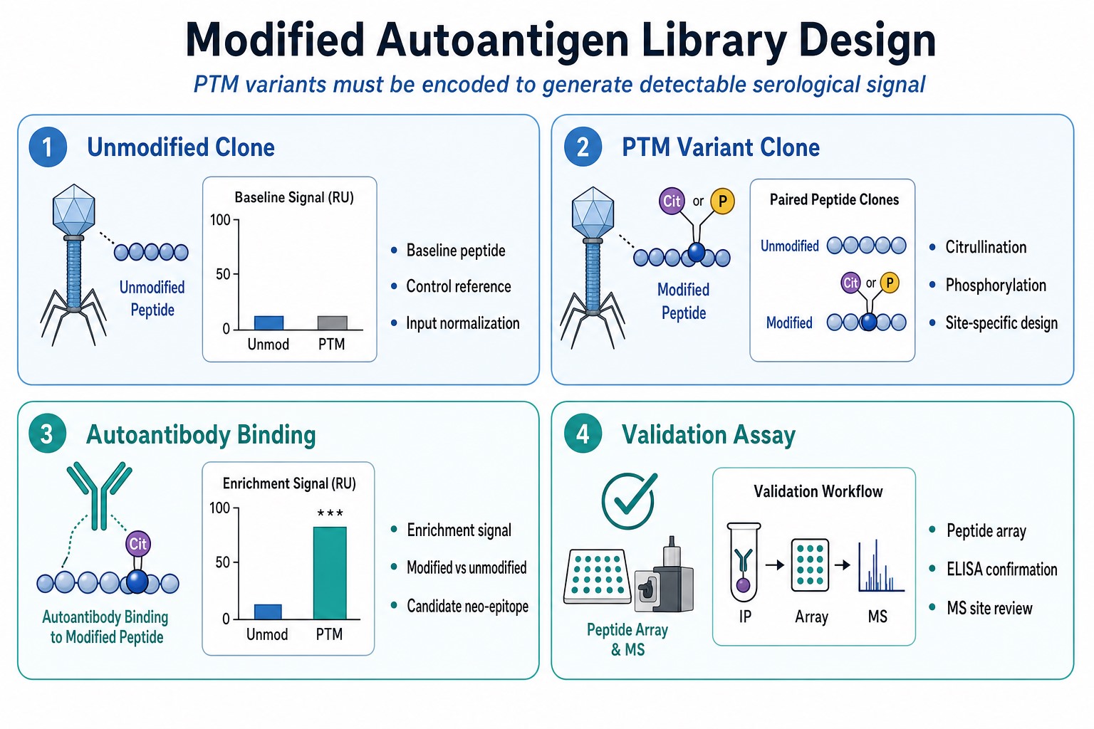
Figure 3. Four-panel library design summary from unmodified and PTM variant clones through autoantibody binding to validation assays.
Related Services
PhIP-Seq Antibody Analysis Service
Antibody Epitope Mapping Service
Peptide Array-Based Epitope Mapping Service
Post-translational Modification Mass Spectrometry Service
Researchers planning autoantibody profiling can consult MtoZ Biolabs to review library scope, modified peptide inclusion, and the validation path suited to conformational or PTM-focused autoantigen questions.
Detection Scope by Autoantigen Type
The table below summarizes how detection performance varies by autoantigen feature. The summary supports project planning but does not replace study-specific feasibility review.
|
Autoantigen Feature |
Detection Potential |
Main Interpretation Constraint |
|---|---|---|
|
Linear contiguous epitope |
Strong when peptide is in library |
Peptide reactivity may not equal full protein binding |
|
Tiled protein region |
Strong for regional mapping |
Requires adequate tiling density |
|
Discontinuous conformational epitope |
Limited in standard linear libraries |
Folded antigen follow-up required |
|
Multimeric or quaternary antigen |
Limited on phage display peptides |
Native complex validation required |
|
PTM-defined modified epitope |
Strong only if modified peptide is encoded |
Modified and control clones both needed |
|
Glycan-dependent epitope |
Limited without glycopeptide library design |
Glycan structure often absent on phage |
This scope summary explains why the answer to conformational or modified autoantigen detection is conditional. The assay can detect what the library presents in a binding-competent format under the experimental conditions used.
Core Technical Advantages and Current Limitations
Core Technical Advantages
Parallel screening across large autoantigen peptide spaces.
Many peptide targets can be tested in one workflow rather than through repeated single-antigen immunoassays.
Quantitative enrichment for cohort comparison.
Sequencing read counts support normalization, replicate review, and case-control or longitudinal comparison across autoimmune sample groups.
Explicit PTM variant testing when libraries are designed accordingly.
Modified and unmodified peptide pairs allow direct comparison of reactivity against defined post-translational modification states.
Regional mapping through tiled libraries.
Overlapping peptides can localize autoantibody reactivity to protein domains or sequence windows for follow-up design.
Discovery-stage candidate generation at scale.
Enriched peptides provide a prioritized list for peptide array, ELISA, or orthogonal binding confirmation.
Current Limitations
Library-defined visibility boundary.
Only peptides represented in the library can generate signal. Unrepresented modified forms, folds, or domains remain outside the search space.
Linear display bias.
Standard phage display peptides do not fully reconstruct many conformational, discontinuous, or glycan-dependent epitopes.
Enrichment is not autoantigen confirmation.
Peptide enrichment indicates candidate recognition under library conditions, not definitive disease specificity without validation.
Background and sample quality sensitivity.
Nonspecific binding, bead background, hemolysis, and storage variability can distort enrichment if controls are insufficient.
Modified epitope coverage depends on prior knowledge.
PTM site selection requires biological hypothesis or prior mapping because untargeted PTM discovery is not inherent to standard peptide libraries.
Applications in Autoimmune Serology Research
The platform is used in autoimmune serology when the research question requires broad peptide-level screening rather than testing one predefined autoantigen at a time.
Case-control autoantibody discovery compares enrichment patterns between patient and matched control sera across human proteome or disease-focused peptide libraries. Modified autoantigen screening applies when citrullinated, phosphorylated, or other PTM peptide sets are included to test neo-epitope hypotheses. Regional autoantigen mapping uses tiled libraries to localize reactivity before protein-level validation. Longitudinal treatment studies compare baseline and follow-up sera within one library framework. Candidate prioritization converts large enrichment lists into shortlists for peptide array, ELISA, or mass spectrometry-supported confirmation.
Project suitability still depends on cohort definition, library composition, control design, and whether conformational or modified autoantigen hypotheses are encoded in the library before screening begins.
Complementary Approaches When Native Autoantigen Biology Matters
When conformational or modified autoantigen biology cannot be captured adequately by standard peptide libraries, complementary methods strengthen interpretation.
A practical program often uses phage immunoprecipitation sequencing in phase 1 for broad candidate discovery, then shifts in phase 2 to targeted validation on modified peptides, folded proteins, or orthogonal immunoassays matched to the autoantigen hypothesis. For programs that move from peptide enrichment to site-level modification review, MtoZ Biolabs can help align discovery screening with PTM analysis and epitope mapping follow-up.
Expected Outputs and Validation Path
A useful autoantibody screening report should include more than raw read counts. Typical deliverables include the following items.
Validation depth should match the intended use. Exploratory autoimmune discovery may accept a ranked peptide list with limited follow-up. Mechanistic or translational projects usually require independent sample validation, modified peptide confirmation, and folded protein binding evidence before an enriched peptide is treated as a conformational or modified autoantigen target.
Frequently Asked Questions
1. Can PhIP-Seq detect conformational autoantigens directly?
The method can detect antibody reactivity against peptides displayed on phage, but standard linear peptide libraries do not fully represent most conformational or discontinuous epitopes. Conformational autoantigen claims usually require follow-up on folded antigen formats.
2. Can PhIP-Seq detect modified autoantigens such as citrullinated peptides?
Yes, when the modified peptide variant is included in the library and enrichment is compared against appropriate unmodified controls. The method does not detect modifications that are absent from the displayed peptide set.
3. Does peptide enrichment prove a protein is the true autoantigen?
No. Enrichment indicates candidate peptide recognition under library conditions. Protein-level binding, modified antigen confirmation, and independent cohort validation are usually required.
4. What controls are needed for modified autoantigen screening?
Typical controls include input library normalization, no-antibody immunoprecipitation controls, healthy donor background, paired modified and unmodified peptide clones, and technical replicates where sample volume allows.
5. When should teams combine peptide display serology with PTM or epitope mapping services?
Combine approaches when the research question involves modified neo-epitopes, glycan-dependent recognition, or conformational epitopes that standard linear peptide libraries may not capture. Discovery screening supports candidate generation, while PTM analysis and epitope mapping support site-level and structural confirmation.
Conclusion
PhIP-Seq can detect autoantibody reactivity against conformational or modified autoantigens only within the limits of library representation and peptide display format. Parallel screening of linear epitopes, tiled protein regions, and explicitly encoded PTM variant peptides remains a core strength of the workflow. The method is limited for native three-dimensional folds, discontinuous epitopes, and glycan-dependent autoantigens unless the study design includes specialized library formats or complementary validation.
The most reliable outcomes come from matching library design to the autoantigen hypothesis, interpreting enrichment as discovery-stage evidence, and validating shortlisted peptides on modified or folded antigen formats suited to the disease model. Researchers evaluating phage immunoprecipitation sequencing for conformational or modified autoantigen screening can contact MtoZ Biolabs to review library composition, control design, and the validation workflow before patient sera are submitted.
How to order?




