






What to Consider Before Starting a Mitochondrial Proteomics Project
Variation introduced during sample collection, mitochondrial enrichment, storage, or protein preparation can persist through LC-MS/MS analysis and cannot be assumed to disappear after normalization. In mitochondrial proteomics, these pre-analytical differences affect which proteins are recovered, how consistently they are quantified, and whether group-associated patterns reflect biology or sample handling.
Project preparation should therefore begin with the intended protein-level evidence and work backward to the experimental design, starting material, enrichment strategy, buffer system, and quality-control plan. This sequence reduces the risk that an otherwise technically successful analysis produces results that are difficult to interpret.
Define the Comparison Before Sample Preparation
1. Clarify the Protein-Level Objective
(1) Identification Versus Quantitative Comparison
An identification-focused study asks which proteins are supported by peptide evidence in the analyzed mitochondrial preparation. A quantitative study asks whether relative protein abundance differs reproducibly across predefined groups. These objectives require different emphasis on replication, batch balance, missing-value control, and statistical design. A project combining both objectives should still identify which output is primary because maximizing identification depth and maximizing cross-sample consistency are related but not identical goals.
(2) Match the Evidence to the Biological Question
Mitochondrial protein analysis is appropriate when the missing evidence concerns protein composition, abundance changes, or pathway-associated patterns. It does not directly measure respiration, ATP production, membrane potential, reactive oxygen species, morphology, or mitophagy flux. Protein-level changes may support hypotheses about these functions, but direct functional conclusions require measurements designed for the relevant phenotype.
2. Establish the Experimental Design
(1) Controls, Biological Replicates, and Comparisons
Matched controls and independently generated biological replicates are central to quantitative interpretation. Replicates should represent biological variation rather than repeated injections of the same preparation. Treatment duration, dose, genotype, tissue region, age, sex, or other study variables should be defined before collection when they could influence mitochondrial composition. Planned contrasts should also be specified so that sample numbers and group structure support the intended comparisons.
(2) Randomization and Batch Structure
Collection order, mitochondrial enrichment batches, digestion batches, and LC-MS/MS acquisition order can become confounded with biological groups. When all control samples are processed before all treated samples, technical drift may resemble a biological effect. Balanced processing and randomization, where feasible, reduce this risk. Batch identifiers should be documented even when the study is small because they may be needed during data review and statistical modeling.
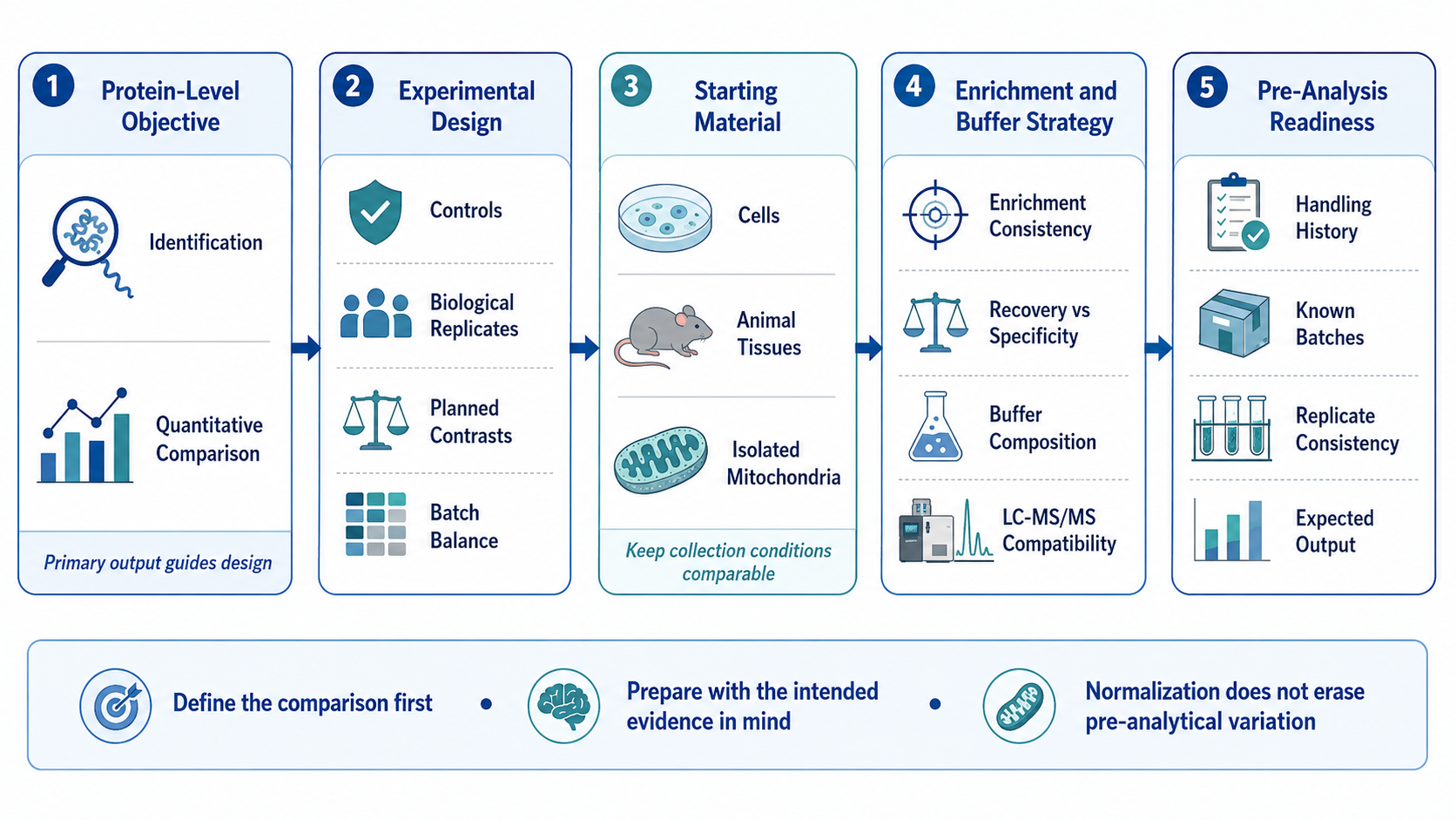
Figure 1. Pre-Analysis Planning for Mitochondrial Proteomics
Evaluate the Starting Material and Enrichment Strategy
1. Assess Sample Readiness
(1) Cells and Animal Tissues
Cells and animal tissues differ in mitochondrial abundance, biological heterogeneity, lipid content, connective tissue, and susceptibility to degradation. These properties affect homogenization, organelle recovery, protein extraction, and sample complexity. Collection conditions should remain consistent across groups, including washing, dissection, handling time, freezing, and storage. Unequal blood content or tissue-region selection may also alter the apparent protein profile independently of the experimental variable.
(2) Isolated Mitochondria and Enriched Fractions
Isolated mitochondrial material should be accompanied by the source sample, isolation procedure, buffer composition, protein amount and concentration, storage conditions, freeze-thaw history, and preparation date. Quantitative comparisons are difficult to interpret when samples were isolated with different protocols or handled under unequal conditions. Documentation is especially important when detergents, stabilizers, carrier proteins, or inhibitors were added during preparation.
2. Interpret Enrichment Quality Carefully
(1) Recovery Versus Enrichment Specificity
An enrichment procedure that maximizes mitochondrial recovery does not necessarily provide the strongest separation from other cellular components. More selective preparation may reduce total yield or alter recovery of fragile mitochondrial subpopulations. The appropriate balance depends on the research question, available material, and required analytical depth. Consistency across comparison groups is often more important than pursuing an undefined standard of absolute purity.
(2) Co-Isolated and Mitochondria-Associated Proteins
Cytosolic, nuclear, endoplasmic reticulum, lysosomal, and other proteins may remain in mitochondrial-enriched fractions through incomplete separation, organelle contact sites, surface association, or cellular debris. Marker proteins and subcellular annotations can support technical assessment, but they do not prove exclusive localization. A detected protein should be interpreted as part of the analyzed preparation unless additional evidence establishes its mitochondrial residence.
Protect Protein Quality and Analytical Compatibility
1. Control Handling and Buffer Variables
(1) Collection, Storage, and Freeze-Thaw History
Delayed processing, inconsistent temperatures, repeated freezing and thawing, and unequal storage durations may change protein recovery or increase degradation. Such effects become especially problematic when handling history aligns with experimental groups. Sample records should capture collection time, preservation method, storage temperature, transfer events, and thawing history. Comparable handling does not remove all variation, but it makes biological comparisons more defensible.
(2) Salts, Detergents, Inhibitors, and Other Additives
Buffer components influence protein solubilization and preservation, yet some salts, detergents, carrier proteins, and inhibitors interfere with cleanup, digestion, labeling, chromatography, or electrospray ionization. Compatibility depends on the selected downstream route, so the complete buffer composition should be available before sample preparation is finalized. Undocumented additives may cause sample loss during cleanup or introduce variable ion suppression.
2. Account for Unequal Protein Accessibility
(1) Membrane Proteins and Hydrophobic Components
Mitochondrial membrane proteins may be underrepresented when extraction and digestion conditions do not adequately address hydrophobicity and membrane embedding. Transporters and respiratory-chain components can produce fewer accessible peptides than soluble matrix proteins. Stronger solubilization is not automatically preferable because reagents must remain compatible with digestion and LC-MS/MS. Recovery should be evaluated as a workflow-dependent outcome rather than a fixed platform property.
(2) Low-Abundance Proteins and Detection Limits
Detectability depends on protein abundance, peptide sequence, digestion efficiency, sample complexity, loading capacity, chromatographic separation, acquisition depth, and data-processing criteria. Low-abundance regulatory proteins may remain below the practical detection range even when present. A missing identification therefore does not establish biological absence, and comparisons should avoid treating nondetection as equivalent to a confirmed decrease without supporting quantitative evidence.
Anticipate Data Quality and Interpretation Limits
1. Connect Preparation Quality With Data Quality
(1) Replicate Consistency and Missing Values
Inconsistent enrichment, extraction, digestion, or sample loading can increase quantitative dispersion and missingness across replicates. These patterns reduce statistical power and make group differences difficult to separate from technical variation. Missing values also have different causes, including low abundance, stochastic sampling, interference, and processing thresholds. Their distribution should be examined before normalization or imputation is used.
(2) Process-Level Quality Assessment
Data quality should be evaluated across the analytical sequence rather than by total protein count alone. Relevant evidence may include enrichment-associated marker patterns, peptide and protein identification confidence, chromatographic stability, signal distributions, digestion performance, replicate correlations, missingness, and sample-level outliers. The most informative metrics depend on whether the primary objective is identification depth, quantitative comparison, or analysis of a defined protein subset.
2. Complete a Pre-Analysis Readiness Check
(1) Information to Document Before Analysis
A preparation record should include the research objective, sample type, preparation status, available material, buffer composition, collection and storage history, experimental groups, controls, biological replicates, known batches, and intended protein-level output. This information allows the analytical route to be matched to the actual samples and identifies limitations before processing begins rather than after data acquisition.
(2) Separate Technical Suitability From Biological Interpretation
A technically suitable sample may produce reproducible identification or quantitative data without confirming mitochondrial function, exclusive localization, pathway activation, or causal mechanism. Technical quality assessment addresses whether the measurement is reliable under the selected workflow. Biological interpretation requires integration with the experimental model and, where appropriate, orthogonal localization, functional, temporal, or perturbation evidence.
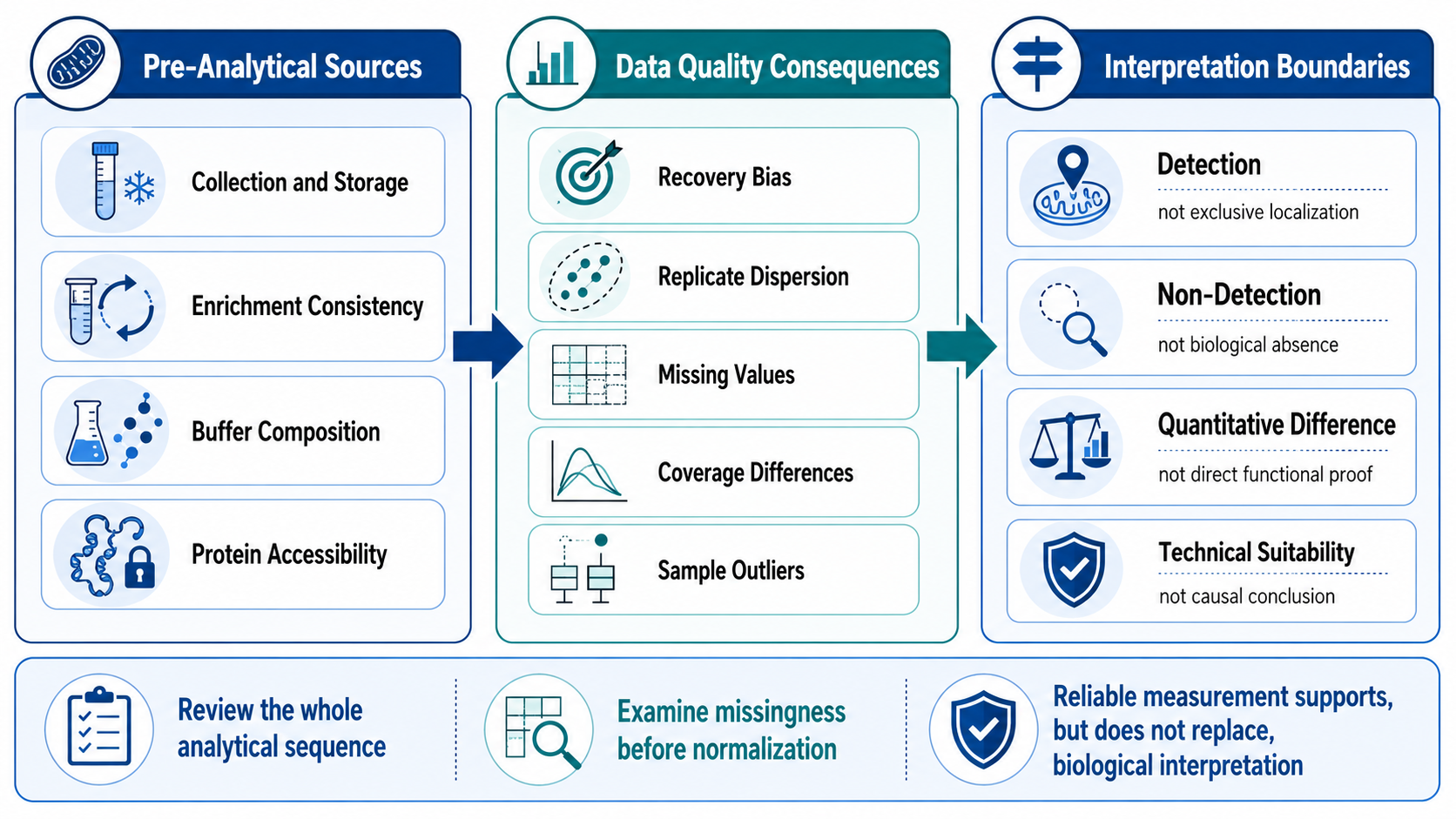
Figure 2. Pre-Analytical Variation and Interpretation Boundaries
Reliable mitochondrial proteomics begins with a defined comparison, consistent sample handling, documented enrichment conditions, and a preparation route compatible with the intended LC-MS/MS analysis. MtoZ Biolabs supports mitochondrial protein identification and quantitative comparison from cells, animal tissues, and isolated mitochondrial material; project evaluation can consider sample status, buffer information, group design, biological replicates, and expected protein-level outputs. Submit your inquiry below for project evaluation.
MtoZ Biolabs, an integrated chromatography and mass spectrometry (MS) services provider.
Related Services
How to order?




