



Chemical proteomics can nominate proteins that took a covalent or photoaffinity mark from a ligand-derived probe. It does not reveal protein–ligand interactions, and it is not a powerful tool for characterizing those interactions across a proteome. A labeled accession is a protein that survived enrichment after that probe. It is not a target, an off-target, or a native contact of the unlabeled parent compound.
Ligands in the original opening can include drugs, metabolites, lipids, and metal ions as study backgrounds. This workflow is built for an organic probe. A metal ion is not installed as a biotinylated photoaffinity analog by default. Affinity chromatography and a biochemical pair assay remain usable later formats. They are not failed high-throughput alternatives that chemical proteomics replaces. Researchers who already have a probe or a parent scaffold and need a labeled-protein table can review the MtoZ Biolabs Chemical Proteomics Analysis Service for project-specific enrichment and LC-MS/MS planning.
A Probe Table Is Not an Interaction Map
The original definition remains usable if it is read as probe capture, not as ligand-target identification. A small-molecule probe carries a reactive or photoreactive group and a handle, often biotin or a clickable reporter. The probe is incubated with live cells or a lysate. Labeled proteins are enriched, digested, and named by liquid chromatography–tandem mass spectrometry (LC-MS/MS). That chain profiles proteins that were modified by the probe. It does not profile proteins that interact with a specific ligand in the native environment of cells or tissues.
Live-cell incubation requires that the probe enter the cell. A lysate incubation is not an in situ physiological context. Enrichment on streptavidin and a subsequent digest do not preserve native conformations or microenvironments, and they do not minimize interaction loss during purification. The non-covalent complex is usually already gone. What remains is a covalent mark.
LC-MS/MS here is the reader of peptides from the enriched pellet. It is not, by itself, a binding detector. For when a covalent mark, a selection cut, or an intact-mass shift can support a binding sentence, Can the Binding of Small Molecules to Proteins Be Detected by LC-MS/MS is the complementary yes-or-no page.
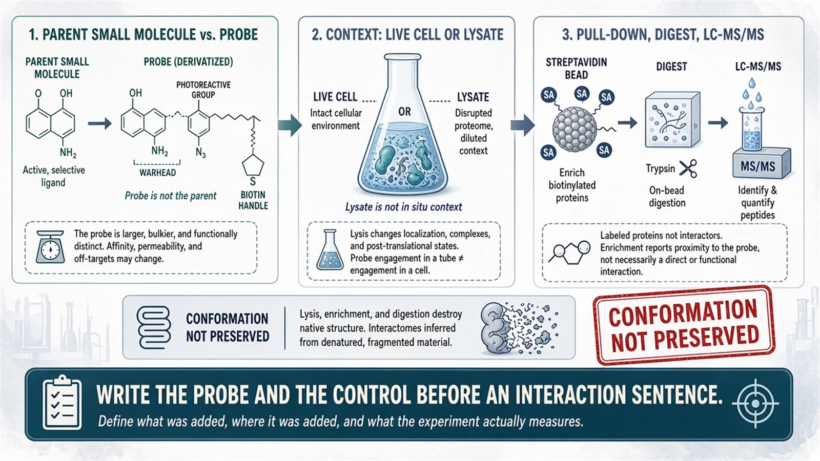
Figure 1. The probe is not the parent ligand. Enrichment names labeled proteins. Native conformation is not preserved through streptavidin and digest.
Write the probe structure, the parent compound, and the control arm before any sentence that the table revealed an interaction.
Three Strategies, Three Files
Activity-based protein profiling
Activity-based protein profiling (ABPP) uses an electrophilic warhead on a scaffold plus a reporter. The warhead covalently modifies a nucleophilic residue in an active site when that residue is available. The classic use is serine hydrolases. Hydrolases and oxidoreductases are not a granted enzyme-class coverage. SILAC or TMT can rank a written contrast between two probe conditions. That ratio is not high-throughput analysis of protein–ligand interactions.
Photoaffinity labeling
A photoaffinity probe generates a short-lived intermediate under ultraviolet light and can form a covalent bond with a nearby residue. Aryl azides, benzophenones, and diazirines are the usual photoreactive groups. Diazonium salts are not treated here as a standard photo-crosslinking moiety. The file names proteins that were close enough to react. Transient or weak binding of the parent is a later interpretation, not a capture proof. For the photo-capture arm, Photocrosslinking Mass Spectrometry Service is the complementary covalent-neighborhood page.
Competition with the unlabeled parent
Excess unlabeled ligand can reduce probe labeling at sites the two molecules share. Competed accessions are probe-and-parent overlapping IDs. They are not validated true targets, and the leftover list is not a nonspecific-binder filter by itself. A no-UV, no-warhead, or scavenger arm still has to travel with the file.
Drug-target identification, natural-product mapping, phenotypic-hit deconvolution, and toxicology remain allowable study backgrounds. Chemical proteomics does not rapidly and accurately identify molecular targets, comprehensively map natural-product interactions, accelerate phenotype-to-mechanism translation, or systematically assess functional perturbations. A long ID list from a toxin-treated lysate is still a labeled-protein list.
Label-free stability methods such as drug affinity responsive target stability (DARTS) or a thermal-shift proteome sit outside this probe page. They report protection or melting behavior, not a covalent handle.
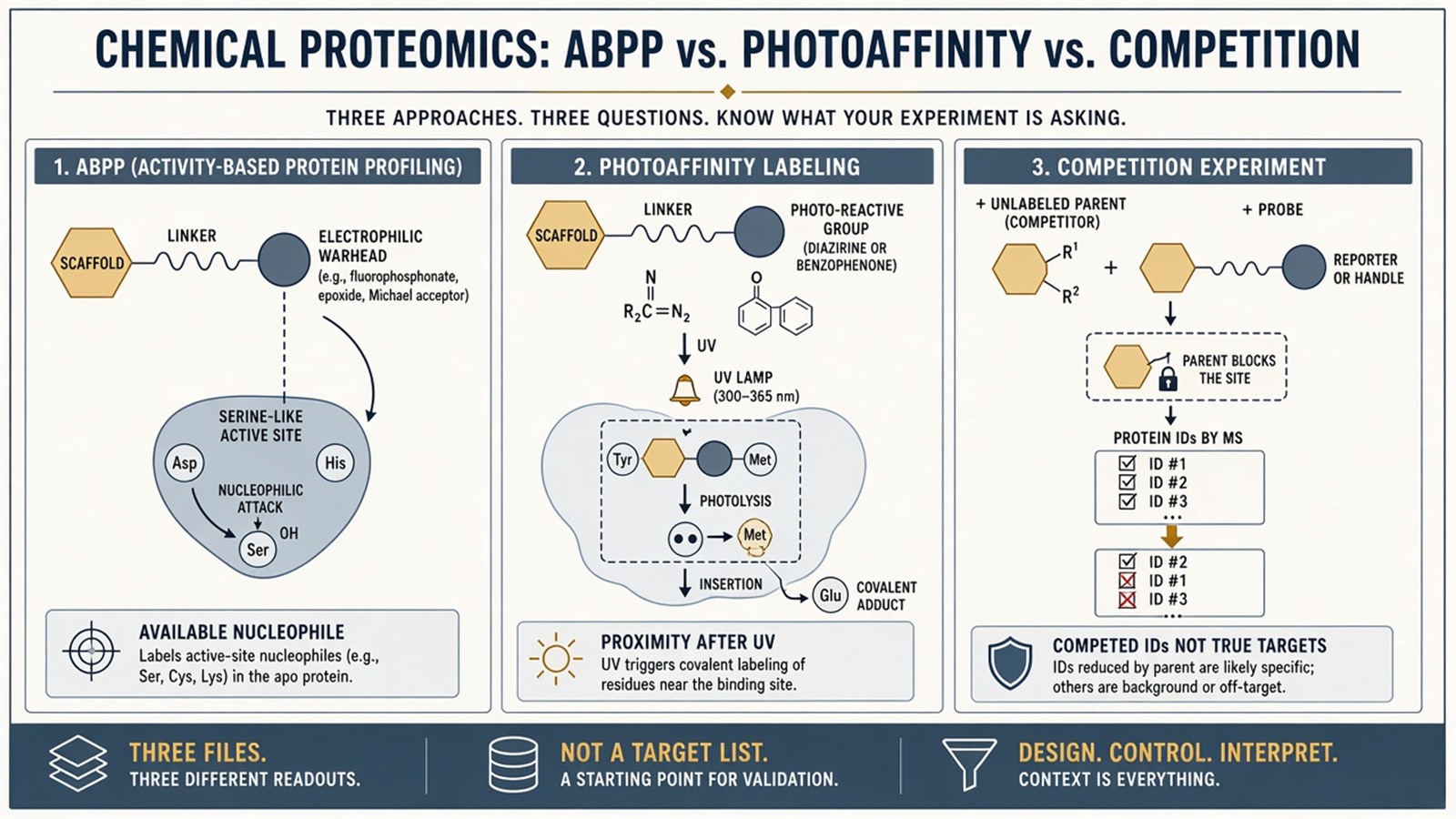
Figure 2. ABPP marks an available active-site nucleophile. Photoaffinity marks proximity after UV. Competition ranks IDs that lost probe signal. None of those tables is a target list.
Experimental Limits
Hundreds of potential targets in a single experiment is a slogan for a long accession list. In situ applicability does not preserve native cellular interaction context through enrichment. Optimized probe structures do not grant high specificity. Pathway annotation and function terms are later overlays. They do not make data interpretation complete.
Probe design still has to balance affinity, reactivity, and permeability. Background still needs a control and a threshold. Those challenges remain. They are why the table is a nomination, not a decoder of protein–ligand interactions. Biomarker discovery, pathway modeling, and an end-to-end essential tool in modern drug discovery sit outside ordinary research reporting unless a separately validated assay is already named.
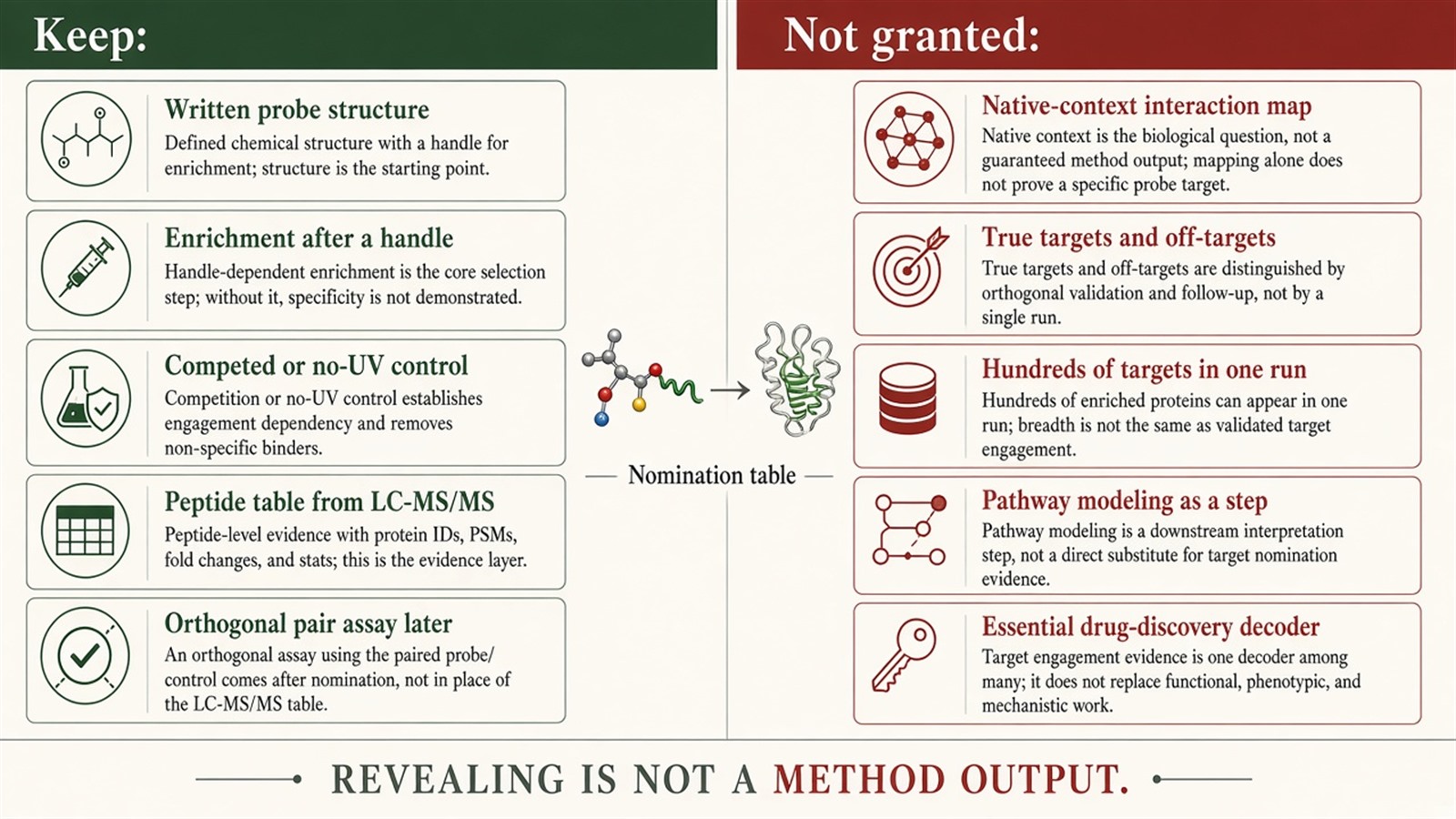
Figure 3. Keep: a written probe, an enrichment, a competed contrast, and a peptide table. Not granted: native-context capture, true-target validation, hundreds of targets, or pathway modeling as a step.
Project Planning and Service Scope
A usable request names the parent ligand, whether a covalent or photoaffinity probe already exists, live-cell versus lysate, the handle that will be used for enrichment, and the competition or no-UV arm that will travel with the digest. MtoZ Biolabs scopes probe-enrichment LC-MS/MS on Orbitrap-class instruments when that arm matches the written question. Interpretability still follows the probe and the control, not a high-throughput heading. Probe synthesis is in scope only when the scaffold and the handle are already specified.
Once the next sentence is an orthogonal pair file for a shortlisted accession, this probe page should hand off. Researchers who need that pair file can review the MtoZ Biolabs Protein Ligand Binding Assay Service for project-specific sample evaluation, feasibility assessment, and workflow planning.
Frequently Asked Questions
1. Does chemical proteomics profile proteins that interact with a ligand in cells?
It profiles proteins labeled by the probe. The parent ligand is a different molecule.
2. Does enrichment preserve native conformation?
No. Streptavidin capture and digest report a covalent mark, not a preserved complex.
3. Does competition distinguish true targets from nonspecific binders?
It ranks IDs whose probe signal dropped. True target is a later claim.
4. Are aryl azides and diazonium salts the standard photo-groups?
Aryl azides, benzophenones, and diazirines are the usual set. Diazonium is not treated as standard here.
5. Does one experiment identify hundreds of drug targets?
It can name hundreds of accessions. Targets are not granted by list length.
Reference
- Cravatt B.F., Wright A.T., Kozarich J.W. (2008). Activity-based protein profiling: from enzyme chemistry to proteomic chemistry. Annu. Rev. Biochem., 77, 383-414. https://doi.org/10.1146/annurev.biochem.75.101304.124125
- Parker C.G., Pratt M.R. (2020). Click chemistry in proteomic investigations. Cell, 180, 605-632. https://doi.org/10.1016/j.cell.2019.12.029
- Smith E., Collins I. (2015). Photoaffinity labeling in target- and binding-site identification. Future Med. Chem., 7, 159-183. https://doi.org/10.4155/fmc.14.152
- Weerapana E., Wang C., Simon G.M., et al. (2010). Quantitative reactivity profiling predicts functional cysteines in proteomes. Nature, 468, 790-795. https://doi.org/10.1038/nature09472
