


Raman-fluorescence Spectroscopy Analytical Service
Raman-fluorescence spectroscopy is a spectroscopic analysis method that combines Raman scattering with molecular fluorescence signals. Its basic principle is that when molecules are excited by laser irradiation, part of the photons produce Raman scattering, reflecting molecular vibrations and chemical bond information; another part is emitted in the form of fluorescence, revealing molecular environments and energy level transition characteristics. By simultaneously obtaining Raman spectra and fluorescence spectra, multidimensional analysis of molecular structures, functional groups, and microenvironments can be achieved. This technique combines the high molecular specificity of Raman spectroscopy with the high sensitivity of fluorescence spectroscopy and is widely applied in biomarker detection, studies of drug–target interactions, and molecular structure and dynamic analysis of complex biological systems.
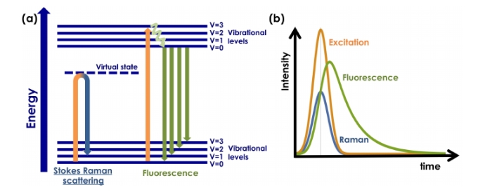
De Luca, A. C. et al. Sensors, 2015.
Figure 1. Energy Level Diagram and Time Variation of Raman Scattering and Fluorescence Emission
Services at MtoZ Biolabs
Based on an advanced Raman-fluorescence spectroscopy platform, MtoZ Biolabs has launched a Raman-fluorescence spectroscopy analytical service to simultaneously collect Raman scattering and fluorescence emission signals under laser excitation. This service can analyze molecular vibration modes, chemical bond characteristics, as well as energy level transitions and microenvironmental changes, making it suitable for the detection of various samples. The final results include complete Raman and fluorescence spectral data, along with qualitative and semi-quantitative analyses of molecular structures, functional group characteristics, and interactions, providing reliable support for biomedical research and drug development.
Analysis Workflow
1. Sample Preparation
Pretreat samples such as cells, tissues, proteins, or drug molecules to ensure uniformity and representativeness.
2. Laser Excitation
Under irradiation with a specific wavelength laser, sample molecules generate Raman scattering signals and fluorescence emission signals.
3. Spectral Acquisition
Use a high-resolution Raman-fluorescence spectrometer to simultaneously record scattered and emitted light, obtaining complete spectral data.
4. Data Processing
Perform background subtraction, peak identification, and signal optimization on the spectra to separate Raman signals from fluorescence signals.
5. Result Interpretation
Output Raman and fluorescence spectra, providing analysis reports on molecular structural features, functional group information, and interactions.
Sample Submission Suggestions
1. Sample Type
Suitable for samples such as cells, tissues, proteins, drug molecules, and nanomaterials. The samples should be uniform and free from obvious impurities or contamination to ensure the accuracy of spectral signals.
2. Sample Purity
It is recommended to minimize sources of background fluorescence interference, such as impurities or residual fluorescent dyes, to avoid affecting the interpretation of Raman and fluorescence signals.
3. Sample Storage
Samples should be stored under low-temperature and dark conditions to prevent changes in fluorescence properties or molecular structure damage caused by light exposure or high temperatures.
4. Sample Transport
Samples should be transported in sealed containers, and when necessary, accompanied by cold-chain conditions to ensure stability and integrity before reaching the analytical platform.
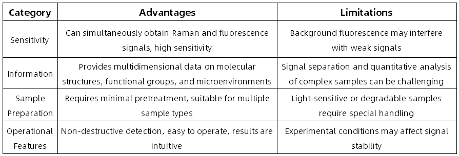
Advantages and Limitations
Applications
1. Cell and Tissue Analysis
By detecting molecular structures and microenvironmental differences in cell and tissue samples, local chemical characteristics can be revealed.
2. Protein and Peptide Research
Raman-fluorescence spectroscopy analytical service can be used to study the conformational changes, interactions, and stability of proteins and peptides.
3. Biological Sample Characterization
Through qualitative and semi-quantitative molecular-level analysis of complex biological samples, structural and compositional information can be obtained.
4. Functional Materials and Nanoprobes
Raman-fluorescence spectroscopy analytical service can be applied to the spectroscopic characterization of biomedical functional materials and nanoprobes, verifying their applicability in biological systems.
FAQs
Q1: Will the Detection Process Cause Damage to the Sample?
A1: This technique is generally considered non-destructive and usually does not alter the properties of the sample. However, for light-sensitive or heat-sensitive samples, excessive laser exposure may cause signal attenuation or structural changes; therefore, optimization of detection conditions is required.
Q2: Is It Suitable for Quantitative Analysis?
A2: Raman-fluorescence spectroscopy is more suitable for qualitative and semi-quantitative studies. Although comparative analysis can be performed through peak intensity or signal ratios, validation with other techniques is still required in complex systems.
How to order?

