


Protein-Nucleic Acid Complexes Structure Characterization Service | Cryo-EM
Protein-nucleic acid complexes (PNACs) play central roles in gene regulation, chromatin remodeling, viral replication, and the discovery of novel drug targets. High-resolution structural analysis of PNACs is essential for understanding molecular mechanisms, guiding rational drug design, and optimizing biologics. However, PNACs are often large, flexible, and heterogeneous, posing significant challenges to traditional crystallography and NMR techniques due to limitations in sample preparation, crystallization, or molecular size.
Cryogenic electron microscopy (Cryo-EM) single-particle analysis has emerged as a mainstream technique for elucidating large and multi-component molecular assemblies, thanks to its ability to image native-state samples without crystallization and to deliver continuously improving resolution. Leveraging an advanced Cryo-EM platform and a team of experienced structural biologists, MtoZ Biolabs offers a comprehensive Protein-Nucleic Acid Complexes Structure Characterization Service Based on Cryo-EM to support both basic research and innovative drug development.
Technical Principles
Cryo-EM captures biomolecular samples rapidly vitrified in a thin amorphous ice layer, thereby avoiding the conformational artifacts introduced by chemical fixation or staining. Under low electron dose conditions, a beam penetrates the sample to generate near-native 2D projection images. These are then processed using statistical image analysis and 3D reconstruction algorithms, combining tens of thousands to millions of particles into a final density map at near-atomic (Å-level) resolution.
Compared to X-ray crystallography, Cryo-EM is more suitable for large, flexible, and heterogeneous complexes. In contrast to NMR, it offers advantages in molecular weight tolerance and data acquisition throughput. When integrated with auxiliary methods such as molecular dynamics simulations, cross-linking mass spectrometry, or hydrogen-deuterium exchange, Cryo-EM can further resolve conformational dynamics and key interaction interfaces.
Zhu, KF. et al. Mil Med Res. 2023.
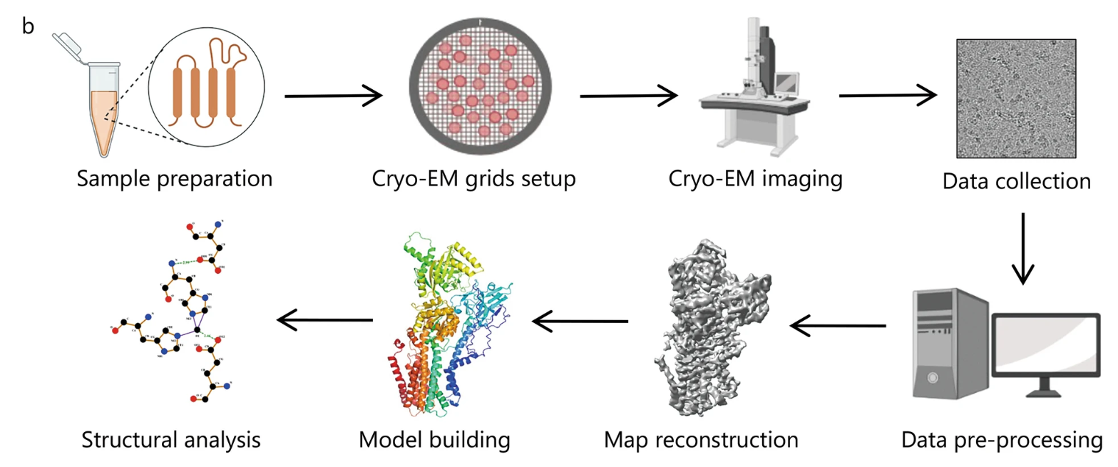
Figure 1. Typical Workflow of Single Particle Cryo-EM for Structural Analysis
Analysis Workflow
1. Sample Pre-Evaluation
Initial assessment of purity, homogeneity, and particle distribution via SDS-PAGE, SEC-MALS, and negative-stain EM.
2. Grid Optimization and Vitrification
Optimization of buffer conditions, glycerol concentration, and ionic strength, followed by plunge-freezing using an automated vitrification system to prepare ultrathin ice layers.
3. High-Throughput Data Acquisition
Automated image acquisition on a 300 kV field-emission Cryo-EM under low-dose mode, collecting thousands of movie stacks with drift correction and focus monitoring.
4. Image Processing and 3D Reconstruction
Motion correction → CTF estimation → Particle picking → 2D classification → Initial model → 3D iterative refinement. For heterogeneous samples, multi-body or continuous heterogeneity classification strategies are applied.
5. Model Building and Structural Annotation
Atomic model construction using homologous templates or de novo modeling tools, followed by geometric optimization, interface analysis, and visualization report generation.
6. Data Delivery and Follow-Up Support
Deliverables include raw movies, complete data processing documentation, resolution validation plots, PDB coordinate files, and interpretation reports. Optional services such as molecular docking or hotspot residue prediction are available.
Service Advantages
1. End-to-End In-House Workflow: All processes from sample screening to structural interpretation are completed in-house, minimizing quality variations caused by multi-party handling.
2. Flexible Billing Models: Options to charge per project phase or delivery milestone; feasibility studies available to reduce initial budget pressure.
3. Multidimensional Quality Control: Every step includes traceable QC records. Resolution validation is based on gold-standard FSC and local resolution heatmaps.
4. Cross-Disciplinary Integration: Seamless integration with mass spectrometry, molecular dynamics simulations, AI-based structure prediction, and bioinformatics for deep annotation.
5. Predictable Turnaround Time: Standard workflows are completed within 6–8 weeks; expedited service is available for time-sensitive drug screening projects.
Applications
1. Gene Expression Regulation Studies
Structural insights into transcription factor–DNA or chromatin remodeling complexes to support the design of regulatory modulators.
2. Viral Replication and Packaging Mechanisms
Visualization of viral polymerase–RNA or nucleocapsid–genome RNA complexes for antiviral drug discovery.
3. CRISPR-Cas System Optimization
Structural elucidation of Cas protein–gRNA–target DNA complexes to guide improvements in editing specificity and efficiency.
4. Cocrystal Analysis of Small Molecules or Peptides
Mapping inhibitor binding pockets at protein–nucleic acid interfaces to support hit-to-lead optimization.
5. Ribosome and Translational Control Research
Characterization of translation initiation, elongation, and termination complexes to explore antibiotic resistance mechanisms.
6. Solution-State Conformational Polymorphism
Capture of functional states from a single sample using classification and reconstruction to analyze transitional conformations.
Case Study
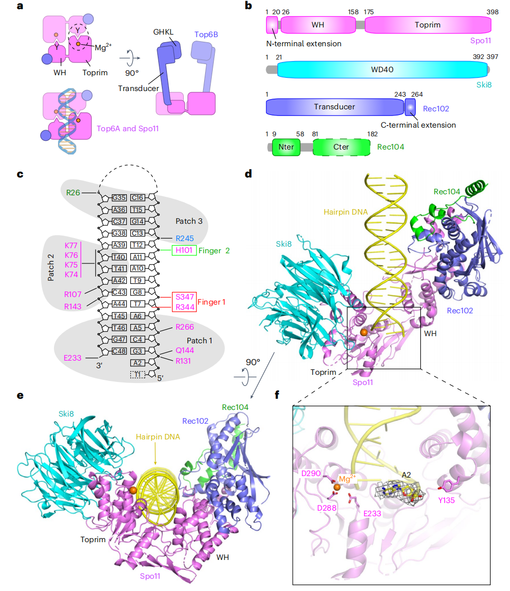
1. Structure Analysis of Spo11 Core Complex Bound to Hairpin DNA by Cryo-EM
This study presents 3.3 Å-resolution cryo-EM structures of the yeast Spo11 core complex bound to DNA, comprising Spo11, Rec102, Rec104, and Ski8. The monomeric complex engages DNA by recognizing the recessed 3′-OH end and 5′ overhang, revealing the molecular basis of DNA end-binding specificity and the role of metal ions in DNA interaction. Structural variation in the Top6BL homolog was uncovered, and key interfaces were validated through functional assays in yeast. Protein-Nucleic Acid Complexes Structure Characterization Service enables the structural resolution of DNA-bound protein complexes, supporting the analysis of conformational states, binding interfaces, and end-recognition modes critical to protein–nucleic acid interactions.
Yu, Y. et al. Nat Struct Mol Biol. 2025.
Figure 2. Cryo-EM Structure of Spo11 Core Complex Bound to Hairpin DNA
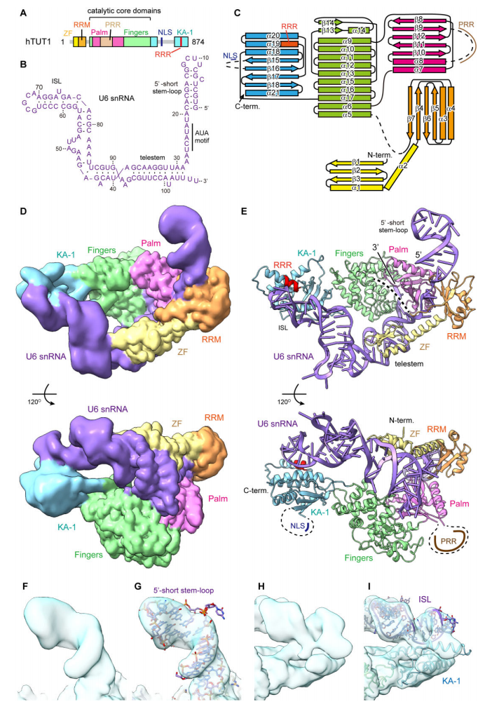
2. Cryo-EM Structure of Human TUT1: U6 snRNA Complex
This study presents the cryo-EM structure of the human TUT1\:U6 snRNA complex, revealing how TUT1 precisely recognizes and stabilizes U6 snRNA through multiple domains. The N-terminal zinc finger and Palm domain clamp the AUA-rich region, the Fingers domain secures the telestem, and the C-terminal KA-1 domain anchors the internal stem-loop at the 3′ end, preventing RNA displacement. The structure demonstrates how TUT1 specifically engages both the sequence and structural features of U6 snRNA for effective oligo-uridylylation. Protein-Nucleic Acid Complexes Structure Characterization Service enables high-resolution imaging of multi-domain protein–noncoding RNA assemblies, supporting structural analysis of sequence recognition, anchoring interactions, and dynamic binding states.
Yamashita, S. et al. Nucleic Acids Res. 2025.
Figure 3. Cryo-EM Analysis of Full-Length hTUT1 in Complex with Full-Length U6 snRNA
FAQs
Q1: What are the sample concentration and volume requirements?
A1: Recommended final concentration: 0.2–1 mg/mL; volume ≥ 50 µL. For limited sample amounts, we offer feasibility evaluation and concentration strategies.
Q2: Are there specific buffer requirements?
A2: Avoid high salt, high glycerol, and strong reducing agents. Standard buffers like 20 mM HEPES/KCl or Tris/KCl are suitable. Special cofactors can be preserved upon discussion.
Q3: Can you handle highly flexible or heterogeneous mega-complexes?
A3: Yes. We apply negative-stain screening, angular limitation, and 3D classification to minimize heterogeneity interference.
Q4: How is data confidentiality ensured?
A4: All client data is stored on isolated internal servers with mutual NDA agreements. Raw data can be deleted upon request after project completion.
Q5: Do you offer downstream functional or drug screening support?
A5: Yes, we can assist with compound docking, molecular simulations, and biochemical assays, but these require separate project discussions.
Through rigorous workflows, quantitative quality control, and application-driven reporting, MtoZ Biolabs’ Protein-Nucleic Acid Complexes Structure Characterization Service provides reliable, actionable structural solutions for both basic research and drug development. For more information or to obtain our sample submission guide, please contact our technical support team.
How to order?

